% !
Rev Saúde Pública 2004;38(5):723-8 www.fsp.usp.br/rsp
Correspondence to: Mario J Avila-Campos Laboratório de Anaeróbios
Departamento de Microbiologia - ICB/ USP Av. Prof. Lineu Prestes, 1374
05508-900 São Paulo, SP, Brasil E-mail: [email protected]
Partially financied by Fundação de Amparo à Pesquisa do Estado de São Paulo (Fapesp - Process n. 00/07785-3 and 00/ 07582-5).
Based on thesis presented to Departamento de Microbiologia, Instituto de Ciências Biomédicas, Universidade de São Paulo, 2002.
Received on 16/6/2003. Reviewed on 26/11/2003. Approved on 5/2/2004.
D etection of pathogens from periodontal
lesions
Detecção de patógenos de lesões periodontais
Veruska de João M alheiros and M ario Julio Avila-Campos
Laboratório de Anaeróbios. Departamento de Microbiologia. Instituto de Ciências Biomédicas. Universidade de São Paulo. São Paulo, SP, Brasil
Keywords
A. actinomycetemcomitans. F. nucleatum. Periodontopathogens. Diagnosis. Periodontal disease. PCR.
D escritores
A. actinomycetemcomitans. F. nucleatum. Periodontopatógenos. Diagnóstico. Doença periodontal. PCR.
Abstract
Objective
To comparatively detect A. actinomycetemcomitans and F. nucleatum from periodontal and healthy sites.
Methods
Subgingival clinical samples from 50 periodontitis adult patients and 50 healthy subjects were analyzed. Both organisms were isolated using a trypticase soy agar-bacitracin-vancomycin (TSBV) medium and detected by PCR. Conventional biochemical tests were used for bacteria identification.
Results
A. actinomycetemcomitans and F. nucleatum were isolated in 18% and 20% of the patients, respectively, and in 2% and 24% of healthy subjects. Among A. actinomycetemcomitans isolates, biotype II was the most prevalent. Primer pair AA was 100% sensitive in the detection of A. actinomycetemcomitans from both subject groups. Primers ASH and FU were also 100% sensitive to detect this organism in healthy subject samples. Primer pair FN5047 was more sensitive to detect F. nucleatum
in patients or in healthy samples than primer 5059S. Primers ASH and 5059S were more specific in the detection of A. actinomycetemcomitans and F. nucleatum, respectively, in patients and in healthy subject samples.
Conclusions
PCR is an effective tool for detecting periodontal pathogens in subgingival samples, providing a faster and safer diagnostic tool of periodontal diseases. The method’s sensitivity and specificity is conditioned by the choice of the set of primers used.
Resumo
Objetivo
Realizar a detecção comparativa de A. actinomycetemcomitans e F. nucleatum de sítios periodontais e sadios.
Métodos
% " Rev Saúde Pública 2004;38(5):723-8 www.fsp.usp.br/rsp
Detection of periodontal pathogens Malheiros VJ & Avila-Campos MJ
INTRODUCTION
It is known that a great number of different micro-organisms exist in the oral cavity but only some species, particularly anaerobic bacteria, have been implicated in the etiology of periodontal disease. Both Actinobacillus actinomycetemcomitans and Fusobacterium nucleatum are Gram-negative bac-teria involved in the pathogenesis of human peri-odontitis but they can also be associated to other endogenous infections.9
A. actinomycetemcomitans is an important periodontopathogen that is involved in the etiology of different forms of periodontal diseases, particu-larly in the localized juvenile and adult periodonti-tis, and also in several extra-oral infections such as endocarditis, pericarditis, pneumonias, septicemias, and abscesses.3,10,17
F. nucleatum has also been considered an impor-tant periodontopathogen for the development of gingivitis and periodontitis and as the most com-mon anaerobic species found in human and animal infections, particularly in the oral cavity.2
The isolation and identif ication of periodontal pathogens characterize an important tool for increas-ing knowledge on periodontal microbiota as well as on the etiology and progression of periodontal in-fections. However, molecular methods have contrib-uted to the detection of putative periodontopatho-gens in several oral or extra-oral infections.15
Even so, there have been some limitations of bac-terial culture such as high cost and time-consuming procedures, besides the fact that it may fails to
Resultados
A. actinomycetemcomitans e F. nucleatum foram isolados em 18 e 20% dos pacientes, respectivamente, e em 2 e 24% dos indivíduos sadios. Entre os isolados de A. actinomycetemcomitans, o biótipo II foi o mais prevalente. O par de iniciadores AA mostrou 100% de sensibilidade na detecção de A. actinomycetemcomitans, em ambos os grupos de indivíduos. Iniciadores ASH e FU foram também 100% sensíveis para detectar esse organismo em amostras de indivíduos sadios. O iniciador FN5047 foi mais sensível para detectar F. nucleatum em amostras de pacientes ou de sadios que o 5059S. Iniciadores ASH e 5059S foram mais específicos na detecção de A. actinomycetemcomitans e F. nucleatum, respectivamente, em amostras de pacientes e de sadios.
Conclusões
PCR constitui-se uma ferramenta efetiva na detecção de patógenos periodontais de espécimes clínicos, fornecendo um diagnóstico rápido e seguro da doença periodontal. Entretanto, esse método depende da escolha dos iniciadores específicos utilizados.
uncultivable organisms. Additionally, cell viabil-ity is necessary for isolation but can be partially lost during transport and in the sampling procedure.6
Several methods for the rapid detection of peri-odontal pathogens have been reported such as im-munologic and immunoenzymatic assays, protein electrophoresis, and DNA-DNA hybridization. However, these methods show different limitations leading to false-positive results as well as cross-reactivity.1,4
Polymerase chain reaction (PCR) is an excellent tool used to identify putative periodontopathogens directly from subgingival samples. Also, it is a fast and efficient method to detect, identify, and differ-entiate periodontal organisms due to its sensitivity and specificity but appropriate standardization is necessary.5
The aim of this study was detect the presence of A. actinomycetemcomitans and F. nucleatum from clini-cal subgingival samples of periodontal patients us-ing two different methods.
M ETH O D S
% #
Rev Saúde Pública 2004;38(5):723-8 www.fsp.usp.br/rsp
Detection of periodontal pathogens Malheiros VJ & Avila-Campos MJ
The Ethics Commission of the Instituto de Ciências Biomédicas. University of São Paulo, approved this study.
Initially, the supragingival plaque was removed and subgingival samples were obtained using three steri-lized paper points inserted in the deepest site of peri-odontal pocket or gingival crevice. After 60 seconds, the paper points were transferred into tubes contain-ing 3.5 ml of the viability maintaincontain-ing microbiostatic medium (VMGA III) transport medium.7
Samples were mixed and diluted 10-fold in VMGA I solution,7 and then plated on trypticase soy agar-bacitracin-vancomycin (TSBV).13 Plates were incu-bated in anaerobiosis (90% N2 + 10% CO2) at 37oC for four days.
To obtain a pure culture, characteristic colonies of both organisms were subcultured on blood agar. Then, they were presumptively identified by Gram-staining, and catalase, H2S, and indole production as well as by their susceptibility to sodium fluoride.14 Definitive identification was achieved using biochemical tests.14
Reference strains of A. actinomycetemcomitans ATCC 29522, ATCC 33384, ATCC 43718, and KC 517 CDC (Centers for Disease Control and Preven-tion, GA, USA) and of F. nucleatum ATCC 10953 were used as controls.
Biotypes of A. actinomycetemcomitans isolates were detected by examining the ability to ferment dex-trose, maltose, xylose, and mannitol.12
VMGA III media containing clinical samples were warmed at 37oC for 10 minutes and then homog-enized. Next 300 µl aliquots were washed three times with sterilized Milli-Q water. Pellets were resuspended in 300 µl water and then boiled for 10 minutes; supernatants were used as templates after centrifuga-tion (14,000 x g, 10 minutes).1
Also, bacterial DNA was extracted from 10 colo-nies of A. actinomycetemcomitans and five colocolo-nies of F. nucleatum. They were suspended in 500 µl ster-ile Milli-Q water, homogenized, and then boster-iled for 10 minutes. Then, after centrifugation, the supernatant (DNA) was used as template.
DNA amplification was performed in volumes of 25 µl containing 8.25 µl sterile Milli-Q water, 2.5 µl 10x PCR buffer, 1 µl MgCl2 (50 mM), 1 µl dNTP (0.2 mM), 1 µl of each primer (0.4 M), 0.25 µl Taq DNA polymerase (0.5 U), and 10 µl DNA.
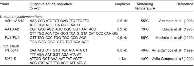
A termocycler Perkin Elmer, Gene Amp PCR Sys-tem 2,400, was programmed to: 1 cycle at 94ºC (5 minutes); 30 cycles at 94ºC (30 seconds), annealing temperature for each specific primer pair (Table 1), and 72ºC (30 seconds); 1 cycle at 72ºC (5 minutes) until the final DNA extension. Specific primers used for identifying A. actinomycetemcomitans and F. nucleatum are listed in Table 1.
PCR products were detected by electrophoresis in 1% agarose gel in 1x TBE at 70 V for 2.5 hours. Gels were stained with an ethidium bromide solution (0.5 µg/ml), and then photographed on a UV transillumi-nator using the Electrophoresis Documentation and the Analysis System 120. A 1 kb DNA ladder was used as molecular mass standard.
To assess concordance between both detection methods (culture and PCR) as well as to determine their sensitivity and specificity, data were analyzed using a Kappa Index (K).
RESU LTS
To detect A. actinomycetemcomitans and F. nucleatum, 50 subgingival samples from adult peri-odontitis patients and 50 from healthy subjects were analyzed. In a trypticase soy agar bacitracin-vanco-mycin (TSBV), nine (18%) clinical samples from
peri-Table 1 - Specific primers used in the detection of A. actinomycetemcomitans and F. nucleatum.
Pri mer O ligonucleotide sequence Amplicon Annealing Reference
(5’→3’) Temperature
A. actinomycetemcomitans
ASH 1/ASH 2 AAA CCC ATC TCT GAG TTC TTC TTC 0.5 kb 55ºC Ashimoto et al1 (1996)
ATG CCA ACT TGA CGT TAA AT
AA1/AA2 CGT GCC AGC AGC CGC GGT AAT ACG 0.2 kb 70ºC Garcia et al7 (1998)
CTT TGC ACA TCA GCG TCA G GTA CAT CCC CAA GG
FU 1/FU 2 GTT TAG CCC TGG TGC CCG AGG 0.5 kb 55ºC Macheleidt et al9 (1999)
TGA CGG GCG GTG TGT ACA AGG F. nucleatum
FN 5047 CAA ATG CTT GTG TCA ATA ATA CT 0.5 kb 40ºC Avila-Campos et al3 (1999)
TTT AGA AAT GGT AGA ATA AT
5059 S ATTGG GCT AAA AAT TAT AGTT 1 kb 40ºC Avila-Campos et al3 (1999)
% $ Rev Saúde Pública 2004;38(5):723-8 www.fsp.usp.br/rsp
Detection of periodontal pathogens Malheiros VJ & Avila-Campos MJ
odontal patients were positive to A. actinomycetem-comitans, with 17 isolates recovered, and ten (20%) were positive to F. nucleatum with 19 isolates. Two A. actinomycetemcomitans were isolated from one (2%) healthy subject and 18 F. nucleatum isolates were recovered from 12 (24%) healthy subjects.
A. actinomycetemcomitans isolates were grouped in five biotypes (II, VI, VIII, IX and X). The most preva-lent was biotype II.
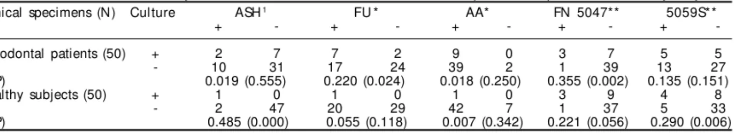
Table 2 shows the sensitivity and specificity of the primer sets used for detecting A. actinomycetem-comitans and F. nucleatum from clinical samples. Primers AA were highly sensitive (100%) to detect A. actinomycetemcomitans from both subject groups. Furthermore, primers ASH and FU were also 100% sensitive to detect this organism in healthy subject samples. Primer FN5047, used to detect F. nucleatum, was more sensitive to detect this organism in pa-tients or healthy samples than primer 5059S. On the other hand, primer ASH and 5059S were more spe-cif ic to detect A. actinomycetemcomitans and F. nucleatum, respectively, in patients and healthy sub-ject samples (Table 2).
All amplified products were compared and the primer pairs produced amplicons of predicted size (data not shown). Table 3 shows a comparison of the bacterial detection between culture method and each
primer used in PCR. It can also be observed that the detection values were as signif icant to detect F. nucleatum in periodontal patients using primer FN5047 (p<0.002) as to detect A. actinomycetem-comitans in healthy subjects using primer ASH (p<0.000).
Statistical analysis showed that primers AA and FU had higher sensitivity, and primers ASH had higher specificity for detecting A. actinomycetemcomitans. However, primer FN 5047 showed higher sensitivity and primer 5059S showed higher specificity for de-tecting F. nucleatum (Table 3).
D ISCU SSIO N
A. actinomycetemcomitans and F. nucleatum are important organisms of both human and animal in-digenous microbiota, and they have been involved in several oral cavity infections. It is well known that improvements in diagnostic methods are useful in the prevention and treatment of periodontal dis-ease and contribute for increasing knowledge on sub-gingival microbiota. Bacterial cultures are used for allowing to recovering cultivable organisms, al-though being a time-and-labor-consuming method. Several molecular tools are often used to identify periodontopathogens but PCR is considered to be an easy and fast detection method even in clinical samples.11
In this study, two methods of detecting A. actino-mycetemcomitans and to F. nucleatum were com-pared. The primary isolation of both organisms stud-ied was performed on selective TSBV medium.13 It is important to mention that although TSBV agar is used as the first choice to isolate A. actinomycetem-comitans, a high recovery rate of F. nucleatum was also observed.
Studies have detected A. actinomycetemcomitans using a PCR method but at different rates in popula-tions with (19%) and without (70%) periodontal dis-ease. These studies have also shown PCR sensitivity and the specificity in comparison to traditional bac-terial culture.8,16
*Sensibility (positive-true) = (N umber of positive samples by PCR and culture/Number of positive samples by culture) x 100
**Specificity (negative-true) = (Number of negative samples by PCR and culture/Number of negative samples by culture) x 100
VMGA: Viability maintaining microbiostatic medium
Table 2 - Sensitivity and specificity of specific primer pairs used to detect A. actinomycetemcomitans and F. nucleatum from clinical samples in VMGA III.
Pri mer Sensitivity* (%) Specificity** (%) Patient H eal thy Patient H eal thy
A. actinomycetemcomitans
ASH 5 100 75 95
AA 100 100 5 15
FU 77 100 59 60
F. nucleatum
FN 5047 50 34 67 87
5059 S 30 25 97 97
Table 3 - Detection of A. actinomycetemcomitans and F. nucleatum strains from periodontal patients and healthy subjects. Clinical specimens (N) Culture ASH1 FU * AA* FN 5047** 5059S**
+ - + - + - + - +
-Periodontal patients (50) + 2 7 7 2 9 0 3 7 5 5
- 10 31 17 24 39 2 1 39 13 27
K (P) 0.019 (0.555) 0.220 (0.024) 0.018 (0.250) 0.355 (0.002) 0.135 (0.151)
Healthy subjects (50) + 1 0 1 0 1 0 3 9 4 8
- 2 47 20 29 42 7 1 37 5 33
K (P) 0.485 (0.000) 0.055 (0.118) 0.007 (0.342) 0.221 (0.056) 0.290 (0.006) *Primers to detect A. actinomycetemcomitans
% %
Rev Saúde Pública 2004;38(5):723-8 www.fsp.usp.br/rsp
Detection of periodontal pathogens Malheiros VJ & Avila-Campos MJ
In the present study, primers AA and FU had higher sensitivity for detecting A. actinomycetemcomitans but primers ASH had higher specificity (Table 3). On the other hand, when compared to culture for detect-ing A. actinomycetemcomitans, primers AA and FU showed similarly sensitivity (100%), which confirms their specificity.
DNA from A. actinomycetemcomitans and F. nucleatum strains were amplified by specific meth-ods and produced predicted bands. It is important to note that both the annealing temperature and magne-sium concentration are critical factors in PCR detec-tion.1 F. nucleatum detection by PCR (primer 5059S) was similar to culture methods; however, when primer FN 5047 was used, PCR showed higher sensitivity
than culture. The study results suggest that PCR is a sensitive and highly specific technique to detect A. actinomycetemcomitans and F. nucleatum in the sub-gingival plaque.
In conclusion, the present study demonstrated the usefulness of specific primers based on the PCR detection of periodontal organisms in subgingival samples. The use of molecular tools in the bacte-rial detection will provide faster and safer diag-nostics of periodontal diseases and PCR reaction may be helpful in detecting putative periodon-topathogens from subgingival samples. A single method could not be ideal, and using both tradi-tional and molecular methods is recommended in the bacterial detection.
REFEREN CES
1. Ashimoto A, Chen C, Bakker I, Slots J. Polymerase chain reaction detection of 8 putative periodontal pathogens in subgingival plaque of gingivitis and advanced periodontitis lesions. Oral Microbiol Immunol 1996;11:266-73.
2. Avila-Campos MJ, Sacchi CT, Whitney AM, Steigerwalt AG, M ayer LW. Arbitrarily primed-polymerase chain reaction for identification and epidemiologic subtyping of oral isolates of F. nucleatum. J Periodontol 1999;70:1202-8.
3. Chen YC, Chang SC, Luh KT, Hsieh WC.
Actinobacillus actinomycetemcomitans endocarditis: a report of four cases and review of the literature. Q J Med 1991;81:871-8.
4. Chen C, Slots J. Microbiological tests for Actinobacillus actinomycetemcomitans and Porphyromonas gingivalis. Periodontology 2000 1999;20:53-64.
5. Doungudomdacha S, Rawlison A, Douglas WI. Enumeration of Porphyromonas gingivalis, Prevotella intermedia and Actinobacillus
actinomycetemcomitans in subgingival plaque samples by a quantitative-competitive PCR method. J Med Microbiol 2000;49:861-74.
6. Lopez NJ. Occurrence of Actinobacillus actinomycetemcomitans, Porphyromonas and gingivalis, Prevotella intermedia in progressive adult periodontitis. J Periodontol 2000;71:948-54.
7. Möller AJR. Microbiological examination of root canals and periapical tissues of human teeth. Odontol Tidskr 1966;74:1-380.
8. Mullally BH, Dace B, Shelburne CE, Wolff LF, Coulter WA. Prevalence of periodontal pathogens in
localized and generalized forms of early-onset periodontitis. J Periodont Res 2000;35:232-41.
9. Paster BJ, Boche SK, Galvin JL, Ericson RE, Lau CN, Levanos VA, Sahasrabudhe A, Dewhirst FE. Bacterial diversity in human subgingival plaque. J Bacteriol 2001;183:3770-83.
10. Preus HR, Haraaszthy VI, Zambon JJ, Genco RJ. Differentiation of strains of Actinobacillus actinomycetemcomitans by arbitrarily primed polymerase chain reaction. J Clin Microbiol 1993;31:2773-6.
11. Riggio MP; Lennon A. Rapid identification of Actinobacillus actinomycetemcomitans, haemophilus aphrophilus, and haemophilus paraphrophilus by restriction enzyme analysis of PCR-amplified. J Clin Microbiol 1997;168:1-8.
12. Slots J, Reynolds HS, Genco RJ. Actinobacillus actinomycetemcomitans in human periodontal disease: a cross-sectional microbiological. Infect Immun 1980;29:1013-20.
% & Rev Saúde Pública 2004;38(5):723-8 www.fsp.usp.br/rsp
Detection of periodontal pathogens Malheiros VJ & Avila-Campos MJ
14. Slots J. Salient biochemical characters of Actinobacillus actinomycetemcomitans. Arch Microbiol 1982;131:60-7.
15. Slots J, Ashimoto A, Flynn MJ, Li G, Chen C. Detection of putative periodontal pathogens in subgingival specimens by 16S ribosomal DNA amplification with the polymerase chain reaction. Clin Infect Dis 1995;20:304-7.
16. Tran SD, Rudney JD. Improved multiplex PCR using conserved and species-specific 16S rRNA gene primer for simultaneous detection. J Clin Microbiol 1999;37:3504-8.
17. van Winkelhoff AJ, Overbeek BP, Pavicic MJAM, van den Bergh JPA, Ernest JPMG, Graaff J. Long standing bacteremia caused by oral Actinobacillus