Inês do Carmo Viegas Baptista
Characterization of TRIB2 following PI3K
inhibition
Mestrado em Oncobiologia:
Mecanismos Moleculares do Cancro
Trabalho efetuado sob a orientação de:
Professor Doutor Wolfgang Link
Professor Doutor Richard Hill
UNIVERSIDADE DO ALGARVE
Departamento de Ciências Biomédicas e Medicina
2015
I
Characterization of TRIB2 following PI3K
inhibition
Declaração de autoria de trabalho
Declaro ser a autora deste trabalho, que é original e inédito. Autores e trabalhos consultados estão devidamente citados no texto e constam da listagem de referências incluída.
II
Copyright em nome do estudante da UAlg, Inês do Carmo Viegas Baptista
A Universidade do Algarve reserva para si o direito, em conformidade com o disposto no Código do Direito de Autor e dos Direitos Conexos, de arquivar, reproduzir e publicar a obra, independentemente do meio utilizado, bem como de a divulgar através de repositórios científicos e de admitir a sua cópia e distribuição para fins meramente educacionais ou de investigação e não comerciais, conquanto seja dado o devido crédito ao autor e editor respetivos.
III
Acknowledgements
First of all, I must give my gratitude to my supervisors. I would like to thank Dr Wolfgang Link for the opportunity, the guidance and the support throughout the research and writing of my thesis. To Dr Richard Hill, I thank you for your patience, motivation and the knowledge you bestowed on me, I will be eternally in your debt.
I would like to thank all my laboratory colleagues Marta, Neuton, Eduarda and Gisela, for their support and companionship during the last year. You are all crazy but lab work wouldn’t be the same without you there.
I will also thank my friends for their encouragement, nagging and patience for my “social reclusion” during my writing of the thesis.
Finally, my thanks to my family for always believing in me. To my sister Vera, for always pushing me to my limits. To my mother Ana, for always supporting me in my endeavours. Last but not least, to my father Augusto, who is gone but I am sure would be proud.
IV
Abstract
Cancer can be defined as an unbalance between cell proliferation and apoptosis. The PI3K/AKT signalling pathway is one of the most mutated pathways in cancer and is involved in cell growth and survival. The transcription factor FOXO3a is a critical effector of this pathway and its inactivation by AKT prevents the expression of genes involved in cell cycle arrest and apoptosis. The TRIB2 protein was found to be a repressor of FOXO3a, and over expressed in many types of cancer. Importantly, TRIB2confers resistance to PI3K inhibitors which are being tested in clinical trials.
This work shows that following PI3K inhibitor treatment, there is an increase of the TRIB2 protein levels by reducing proteasomal degradation. Furthermore, the TRIB2 COP1 binding domain is critical for AKT activation and subsequent FOXO repression.
Altogether, our results provide insight into the mechanism underlying TRIB2 mediated tumorigenesis and drug resistance implicating the binding and activation of AKT and suggests a role of TRIB2 in acquired resistance against PI3K inhibitors.
V
Resumo
O Cancro é caracterizado por uma proliferação celular descontrolada oriunda de danos e/ou mutações no ADN. Estas alterações surgem na grande maioria de erros durante o processo de replicação, falhas nos mecanismos de reparação, exposição a fatores ambientais ou a agentes cancerígenas. Existem seis alterações essências que uma célula normal submete-se na transformação em célula tumoral: evasão a supressores externos de crescimento celular, sinalização constitutiva de proliferação, evasão à apoptose (morte celular programada), capacidade própria de induzir angiogênese (produção de novos vasos sanguíneos), potencial replicativo ilimitado que torna a célula “imortal” e capacidade metastática de invadir outro tecidos. Recentemente, mais quatro características foram adicionadas, sendo elas: evasão ao sistema imunitário, instabilidade genómica, desregulação do balanço energético celular e capacidade de inflamação indutora de tumores.
O Cancro é uma das maiores ameaças á saúde humana a nível mundial. Dentro dos tipos de cancro mais preocupantes está o cancro de pele, mais especificamente o Melanoma. Esta forma agressiva de cancro de pele desenvolve-se a partir de melanócitos, células especializadas na produção de melanina. Embora este tipo de cancro de pele seja uma minoria (menos de 5% dos casos totais), é um dos mais letais, sendo responsável por aproximadamente 80% de todas as mortes devido a cancro de pele. Com a sua incidência dentro da faixa etária dos 30 aos 60 anos, em particular em pessoas de pele mais clara. O facto de cada vez mais surgirem casos de jovens entre os 25-29 anos é alarmante. O aumento anual da sua incidência têm-se refletido numa subida na taxa de mortalidade associada a este tipo de cancro.
Esta doença oncológica é extremamente agressiva e heterogénea, com vários subtipos histológicos e perfis mutacionais. Adere-se o problema que os tratamentos convencionais são pouco eficazes e as drogas mais recentes que conseguem ter resultados tornam-se ineficazes devido aos tumores adquirirem resistência num espaço de meses. A totalidade destes factos confere aos pacientes um prognóstico pessimista.
Devido a estes factos, a investigação científica têm-se empenhado em perceber os mecanismos moleculares afetados por esta patologia e em como esse conhecimento pode auxiliar na produção de tratamentos mais eficazes. É o caso da via de sinalização de PI3K,
VI que se verificou ser desregulada em Melanoma, que levou ao desenvolvimento de inibidores capazes de reverter esta alteração, como BEZ235.
A via de sinalização de PI3K/AKT é importante na regulação de diversos processos biológicos, tais como proliferação celular, metabolismo, crescimento celular e apoptose. Esta via é ativada pela ligação de fatores de crescimento ao recetor de tirosina quinase na membrana celular. Este recetor, através de fosforilação, ativa PI3K que converte fosfatidil inositol 4,5-difosfato (PIP2) em fosfatidil inositol 3,4,5-trifosfato (PIP3). Este processo é regulado negativamente por PTEN, uma fosfatase que tem como substrato PIP3. O aumento de PIP3 recruta AKT para a membrana, fazendo com que seja ativado por outras quinases através de fosforilação dos seus resíduos Treonina 308 e Serina 473. Após esta ativação, AKT fosforila várias moléculas alvo no citoplasma e no núcleo, como por exemplo p27, BAD, GSK3, MDM2 e FOXO.
Os fatores FOXO são uma família de fatores de transcrição que funcionam como ativadores ou repressores, dependendo dos seus cofatores a quando da sua ligação ao ADN. Esta família é fundamental em processos como apoptose, metabolismo, diferenciação celular, inflamação, proliferação e resposta ao stress oxidativo.
O fator de transcrição p53 é responsável pela regulação de vários genes envolvidos em apoptose, paragem do ciclo celular, senescência, metabolismo e autofagia. Devido a estas funções, é considerado o “guardião do genoma” e o seu papel contra o desenvolvimento tumoral é bem conhecido. O p53 é regulado por MDM2, uma ubiquitina ligase responsável pela sua ubiquitinação e consequente degradação proteossómica. Na verdade, p53 e MDM2 exercem um feedback loop, sendo que o gene de MDM2 é um dos alvos de p53, mantendo assim os níveis de p53 constantes dentro da célula.
O TRIB2 é um gene que tem sido associado ao desenvolvimento de cancro e foi recentemente considerado um biomarcador para o prognóstico e para a progressão de Melanoma. A sua sobre expressão proteica afeta negativamente os tratamentos convencionais a este tipo de cancro. Também está implicado na regulação negativa de FOXO. Dado que este fator de transcrição é uma das vias que as drogas usadas nas terapias utilizam para exercer efeitos citotóxicos e/ou citostáticos sobre as células tumorais, a regulação que TRIB2 exerce sobre FOXO evidencia o seu papel na resistência a quimioterapêuticos (como o clássico DTIC ou o inibidor BEZ235).
VII Pesquisas antecedentes feitas pelo nosso grupo laboratorial demonstraram que o uso de inibidores de PI3K estabilizam os níveis proteicos de TRIB2 e que existe uma interação entre TRIB2 e AKT. Neste trabalho, procuramos perceber como TRIB2 é regulado dentro da célula e como interage com a via de sinalização PI3K/AKT.
Durante a nossa investigação constatámos que os níveis proteicos de TRIB2 eram estabilizados na presença de inibidores de PI3K devido a um decréscimo da sua degradação proteossómica. Isto implica que a via de sinalização de PI3K está envolvida na modificação translacional de TRIB2 necessária para a sua degradação. Os mecanismos envolvidos nesta degradação e os seus participantes permanecem desconhecidos. No entanto, estes resultados conseguem demonstrar como TRIB2 influencia a resistência a drogas, como verificado nos casos de Melanoma.
Também observámos que a interação entre AKT e TRIB2 é feita através do domínio de ligação a COP1 existente na zona C-terminal de TRIB2. Verificámos que esta interação implica um decréscimo na expressão de genes envolvidos em apoptose e em paragem do ciclo celular como FasLG e p27, respetivamente. Estes resultados evidenciam que a interação entre AKT e TRIB2 está relacionada com a regulação negativa de FOXO. Como confirmação, avaliámos os níveis proteicos de p27 e BIM, genes regulados por os fatores FOXO. Estes resultados implicam que TRIB2 funciona como uma proteína
scaffold ou adaptadora, que permite a ativação de AKT no resíduo Serina 473.
Em conjunto, estes resultados demonstram como a regulação de TRIB2 e a sua interação com AKT auxiliam as células tumorais a adquirirem resistência a quimioterapêuticos, não só no tratamento de Melanoma mas em outras patologias oncológicas onde a via de sinalização de PI3K/AKT esteja alterada.
Palavras-chave: Cancro, Melanoma, TRIB2, FOXO3a, via de sinalização de
VIII
Table of Contents
Acknowledgements III
Abstract IV
Resumo V
Table of Contents VIII
Figure list X
Table list XI
Abbreviations list XII
1. Introduction 1
1.1- Cancer 1
1.2- Melanoma 3
1.3- The PI3K/AKT pathway 6
1.4- FOXO proteins 10
1.5- The tumour suppressor p53 13
1.6- Novel Therapeutics in the Treatment of Melanoma 14
1.7- The Tribble2 homolog 16
1.8- Hypothesis 17
2. Materials and Methods 19
2.1- Cell lines and reagents 19
2.2- Western blot analysis 19
2.3- Constructs 20
2.4- Flow cytometry cell cycle analysis 21
2.5- FACS sorting 21
2.6- Immunoprecipitation 21
2.7- Co-Immunoprecipitation 22
2.8- RNA extraction/First strand cDNA protocol 22
2.9- qRT-PCR /DNA electrophoresis agarose gel (1 %)/Primer validation 22
2.10- Chromatin Immunoprecipitation assay (ChIP) 23
3. Results 25
3.1- TRIB2 protein levels increase in the presence of PI3K inhibitors 25
3.2- PI3K inhibition does not result in an increase in TRIB2 transcription 26
3.3- TRIB2 keeps mRNA and protein stability in the presence of PI3K inhibitors 26
3.4- PI3K treatment significantly reduces proteasome-dependent degradation of TRIB2
28
3.5- TRIB2 protein expression correlates with increased total AKT and significantly
IX
3.6- COP1 domain is the crucial region required for pSer473 AKT post-translational
modification 29
3.7- Low levels of FOXO3a-mediated genes transcription correlate with increased AKT
activity 30
4. Conclusion 35
4.1- Future Directions 36
5. References 38
X
Figure list
Pages
1.1.1 - Trends in cancer incidence 1
1.2.1 – Progression of skin cancer 3
1.3.1 – Schematic of PI3K pathway 7
1.4.1 – Schematic of FOXO cytoplasmic translocation and sequential degradation
11
1.5.1 – Schematic of p53 degradation 13
1.7.1 – Representation of TRIB2 17
2.3.1 – Schematic of TRIB2 mutants 21
3.1.1 – PI3K pathway inhibitors 25
3.1.2 – TRIB2 protein levels increase following PI3K 25 3.2.1 – PI3K inhibition does not change levels of TRIB2 expression 26 3.3.1 – PI3K inhibition does not alter TRIB2 mRNA synthesis 27 3.3.2 – PI3K inhibition does not affect protein synthesis 27 3.4.1 – PI3K inhibition reduces TRIB2 degradation 28 3.5.1 – TRIB2 protein expression correlates with pSer473-AKT 29
3.6.1 – TRIB2 mutant constructs 29
3.6.2 – TRIB2 acts through the COP1 domain 30
3.7.1 - Low levels of p27 gene expression in mutants still possessing the COP1-binding domain
31
3.7.2 - Low levels of FasLG gene expression in mutants still possessing the COP1-binding domain
31
3.7.3 - Low levels of protein expression of p27 and FasLG in mutant constructs still possessing the COP1-binding domain
32
3.7.4 - Cells with TRIB2 mutants possessing COP1-binding domain can transition the G1 checkpoint
33
3.7.5 - Protein expression of p27 in GFP+ and GFP- populations 33 3.7.6 - Protein expression of BIM in GFP+ and GFP- populations 34
XI
Table list
Pages
1.2.1 - Tumor thickness classification 4
1.2.2 – Lymph nodes classification 4
1.2.3 – Metastases classification 5
1.2.4 – Melanoma staging 5
2.1.1 – Primary antibodies used 19
2.2.1 – Secondary antibodies used 20
2.3.1 – TRIB2 plasmids constructs used 21
2.9.1 – List of primers used 23
Appendix list
Pages
Figure A.1: Datasheet of phospho-AKT antibody 44
Figure A.2: Datasheet of Total AKT antibody 45
Figure A.3: Datasheet of Total FOXO antibody 46
Figure A.4: Datasheet of Actin antibody 47
Figure A.5: Datasheet of MDM2 antibody 48
Figure A.6: Datasheet of BIM antibody 49
Figure A.7: Datasheet of FasLG antibody 50
Figure A.8: Datasheet of p27 antibody 51
Figure A.9: Datasheet of phospho-FOXO antibody 52
Figure A.10: Datasheet of Lamin antibody 53
Figure A.11: Datasheet of phospho-MDM2 antibody 54
Figure A.12: Datasheet of goat anti-rabbit secondary antibody 55 Figure A.13: Datasheet of donkey anti-goat secondary antibody 56 Figure A.14: Datasheet of goat anti-mouse secondary antibody 57 Figure A.15: Datasheet of anti-mouse secondary antibody 58
Figure B.1: jetPRIME Transfection Kit Protocol 59
Figure C.1: Tri-Reagent Protocol 61
Figure D.1: NzyGelpure protocol 66
Figure E.1: Nzy First-Strand cDNA Synthesis Kit protocol 70 Figure F.1: LuminoCt SYBR Green qPCR ready mix Protocol 72
XII
Abbreviations list
4E-BP eIF4E-binding protein
AJCC American Joint Commission on Cancer
AKT Protein kinase B
AML Acute myeloid leukaemia ATF4 Activating transcription factor 4 BAD Bcl-2-associated death promoter BAX Bcl-2-associated X protein Bcl-2 B-cell lymphoma 2
Bcl-XL B-cell lymphoma extra large
BIM Bcl-2 interacting mediator of cell death CDK Cyclin-dependent kinase
CDKN2A Cyclin-dependent kinase inhibitor 2A C/EBP CCAAT/enhancer-binding protein COP1 Constitutive morphogenesis 1
CREB Cyclic-AMP response element binding protein CTLA-4 Cytotoxic T-lymphocyte associated antigen 4
DTIC Dacarbazine
FasLG Fas ligand
FDA Food and drug Administration FOXO Forkhead box O transcription factor FYVE Fab-1, YG-LO23, Vps27 and EEA1 GAP GTPase-activating protein
GPCR G-protein coupled receptor GSK3 Glycogen synthase kinase 3
HER2 Human epidermal growth factor receptor 2 IAP1 Inhibitor of apoptosis protein 1
IkB I-kappa-B
IKKα Inhibitor of kappa B kinase α
IL-2 Interleukin 2
ILK Integrin-linked kinase LDH Lactate dehydrogenase
XIII Mcl-1 Myeloid cell leukaemia 1
MDM2 Murine double minute 2
MnSOD Manganese superoxide dismutase mTOR Mammalian target of rapamycin
mTORC2 Mammalian target of rapamycin complex 2
NF-kB Nuclear factor kappa-light-chain-enhancer of activated B cells NSCLC Non-small cells lung cancer
P70S6K1 P70 ribosomal protein S6 kinase PD1 Programmed death receptor 1
PDK1 3’-phosphoinositide-dependent kinase 1 PD-L1 Programmed death ligand 1
PHLPP Pleckstrin homology domain leucine-rich repeat protein phosphatase
PI3K Phosphatidylinositol-3 kinase
PIP2 Phosphatidylinositol-4,5-diphosphate PIP3 Phosphatidylinositol-3,4,5-triphosphate PP2A Protein phosphatase 2A
PTEN Phosphatase and tensin homologue deleted on chromosome 10 PUMA P53 upregulated modulator of apoptosis
Rb Retinoblastoma protein
Rheb RAS homolog enriched in brain RPTK Receptor protein tyrosine kinase
Sesn3 Sestrin 3
SGK Serum- and glucocorticoid-inducible kinase Smurf1 SMAD ubiquitination regulatory factor 1
TRAIL Tumor necrosis factor-related apoptosis inducing ligand TRIB2 Tribbles homolog 2
TSC Tuberous sclerosis complex
TSC2 Tuberous sclerosis complex protein 2 or Tuberin WHO World Health Organization
1
1. Introduction
1.1- Cancer
Cancer is characterized by unregulated cell proliferation typically arising as a result of DNA damage/mutations. Typically this occurs by environmental factors such as ultraviolet radiation, diet, pollution or viral infection. However, these can also occur through punctual mutations, errors in replication and/or by failings in the cell’s DNA repair machinery (1).
The hallmarks of cancer describe the necessary, minimum genetic alterations that a normal cell must undergo to become transformed (1). These include the evasion of growth suppressors, constitutive proliferative signalling, the avoidance of apoptosis (programmed cell death), the capacity to induce angiogenesis (formation of new blood vessels), replicative immortality and activating invasion/metastasis (1). These six were the original principal hallmarks however since these were described, four additional hallmarks have been incorporated. These are avoiding immune destruction, genome instability, deregulating cellular energetics and tumour-promoting inflammation (1).
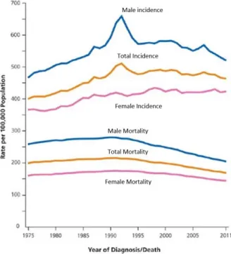
Cancer in general is a major health issue world-wide. According to the World Health Organization (WHO), 8.2 million people died of cancer world-wide in 2012 and 30 % of cancer cases can be prevented. With the increase of life expectancy, the likelihood of developing any type of tumour also increases (Figure 1.1). Combined with an improvement in detection technics, there was a rise of cancer incidence (2). In compensation, the advances in treatments have allowed to lower the mortality rate.
The tumorigenesis process can be influenced by many factors such as genetic susceptibility and lifestyle, leading to a heterogeneity of tumours amongst patients. Even
Figure 1.1.1 - Trends in cancer incidence and mortality rates. Trends for both sexes in the United States from 1975
to 2011. Adapted from Siegel et al.; Cancer Statistics, 2015; CA CANCER J CLIN 2015;65:5–29
2 the contribution of a person’s diet to the development of cancer is not yet fully understood. To add to the problem, there are many examples of tumour resistance to most treatments.
The first step in the standard treatment for a majority of cancers is surgery (if the tumour is located within an operable region). Assuming that the tumour can be removed, the patient will be subjected to adjuvant treatment that includes radiotherapy and/or chemotherapy. Radiotherapy consists of using radiation to damage the DNA and kill any actively dividing cells (including both normal and tumour cells). Chemotherapy uses drugs to achieve the same objective. Chemotherapy must take in consideration many factors such as type of cancer, its extent and localization. Both types of treatment are given in cycles to allow the patients to recover from the inherent toxicities of these therapeutic approaches.
The greatest problems in the treatment of cancer are late diagnosis, poor patient response to treatment and the re-emergence of resistant cancer cells following a variable remission period. Many patients are treated with a combination of chemotherapeutics and still have little to no objective response to therapy. Furthermore, the patients that do have a measurable response typically acquire resistance to treatments in a short period of time. This delay between remission and the emergence of resistant cancer cells is highly variable and is dependent on the type of cancer. Consequently there has been a great focus in developing new therapeutic agents and modalities. One of this new treatments is immunotherapy. This modality is based upon the interplay between the tumours and the microenvironment where they are located. Immunotherapy can be used to increase the patients’ immune system to recognise and subsequently eliminate the cancer cells (3). As cancers harbour many genetic mutations, a number of some tumours have proteins that can be recognize as foreign antigens (4). While promising, most tumours are identified as “self” by the host and thus are able to escape immune system detection. The use of targeted molecules capable of inducing an antitumour immune response can help gain a chemotherapeutic effect, by “boosting” the detection capabilities of the immune system (3).
Another approach to cancer treatment is targeted therapy. By investigating the aberrant molecular pathways in cancer, many therapeutic targets have been identified. These potential targets must be as exclusive to the tumour cells as possible to avoid indirectly affecting any normal cells exposed to the therapeutic. Many such chemotherapeutics have already been developed and have proven to be considerably more
3 effective than the previous non-specific drugs. A prime example is Gleevec© (Imatinib) that targets the mutant fusion protein Bcr-Abl and is used for the treatment of leukaemia. Another example is Vemurafenib that can inhibit the kinase of the mutation B-RAFV600E and is used to treat metastatic melanoma (5). The greatest advantage of targeted therapy is that allows to develop a personalized treatment for each patient according to the “characteristics” of their tumour. These (and others) are extensively reviewed in Shtivelman et al. (6) .
1.2- Melanoma
Melanoma is a type of skin cancer with its origin in mutated melanocytes. Although a minority amongst the other types of skin cancer (constituting less than 5% of all skin cancer cases), melanoma is responsible for over 80% of all skin cancer deaths (4). In the last decade, the incidence rate of melanoma has dramatically increased with an average increase of 3-8% per year that continues to rise annually (7). In the United States alone, there will be an estimated 73870 new cases and 9940 deaths for just 2015 alone (2). Men are more prone to develop malignant melanoma (1 in 34) than women (1 in 53). This is thought to be due to sex-specific behaviour and environmental exposure differences (2).
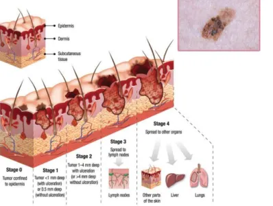
Melanoma is a malignant neoplasm of melanocytes, cells specialized in producing melanin (skin pigment) that are localized within the basal layer of the epidermis (8). The
transformation of
melanocytes can arise from multiple sources such as accumulation of genetic alterations, interaction between environmental factors and impaired DNA repair. Melanoma spreads along the epidermis initially and as the tumour develops it invades in depth through the skin layers (Figure 1.2).
Figure 1.2.1 – Progression of skin cancer. Squematic of
4 The primary method to recognize a suspicious cutaneous lesion as early melanoma is the ABCDE acronym (asymmetry, border irregularity, colour variegation, diameter and evolvinglesions) (8). This method is useful in early detection but it is limited since it cannot evaluate melanomas with vertical growth. This approach is complemented by dermoscopy, a non-invasive procedure that aids in the visualization of sub-surface structures (8).
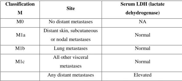
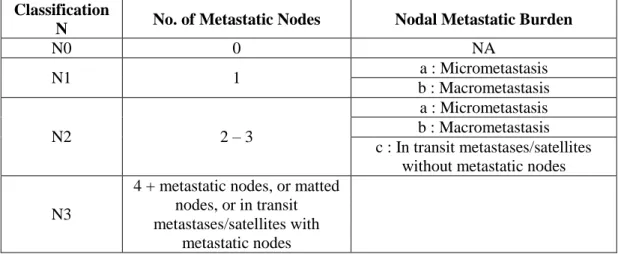
The staging of melanoma involves many parameters. Initially melanoma is classified based on the thickness (T), the number of lymph nodes affected (N) and the number of metastases (M) (9). Disease stage also takes in consideration ulceration, the rate of mitosis (expressed in number of mitosis per square millimetre of primary tumour) (8) and the levels of lactate dehydrogenase (LDH). The TNM categories are then combined to perform a stage grouping. These are summarized in Table 1.2.1, 1.2.2, 1.2.3 and 1.2.4.
Classification T Thickness (mm) Ulceration Status/Mitoses
Tis (in situ) NA (not applicable) NA
T1 ≤1,00 a : whiteout ulceration and mitoses < 1/mm
2
b : with ulceration or mitoses ≥ 1/mm2
T2 1,01 – 2,00 a : whiteout ulceration b : with ulceration T3 2,01 – 4,00 a : whiteout ulceration b : with ulceration T4 >4,00 a : whiteout ulceration b : with ulceration Classification M Site Serum LDH (lactate dehydrogenase) M0 No distant metastases NA
M1a Distant skin, subcutaneous
or nodal metastases Normal
M1b Lung metastases Normal
M1c All other visceral
metastases Normal
Any distant metastases Elevated
Table 1.2.1– Tumour thickness classification. According to the 7th edition of the Melanoma staging system by the American Joint Commission on Cancer (AJCC).
Table 1.2.2 – Lymph nodes classification. According to the 7th edition of the Melanoma staging system by the American Joint Commission on Cancer (AJCC).
5
Stage Characteristics Classification 5 year survival rate T N M
0 Carcinoma in situ (in epidermis
and has not spread to dermis) Tis N0 M0 99 – 100 %
I A/B Lesions up to 2 mm but no nodal or distant metastases
T1a N0 M0 A – 95% B –88 - 92 % T1b N0 M0 T2a N0 M0 II A/B/C
Lesions greater than 2 mm, no positive nodes or distant
metastases T2b N0 M0 A – 77 - 79 % B – 61 - 70% C – 43 - 45% T3a N0 M0 T3b N0 M0 T4a N0 M0 T4b N0 M0
III A/B/C Lesions of any size with positive lymph nodes Tx N1 M0 A – 57 – 73 % B – 41 – 57 % C – 20 – 34 % Tx N2 M0 Tx N3 M0 IV Lesions of any size with distant
metastases Tx Nx M1 5 – 22 %
Of all diagnosed melanomas, 90 % are superficial spreading melanoma, nodular melanoma and lentigno malignant melanoma (that are common in the elderly) (10). The
Classification
N No. of Metastatic Nodes Nodal Metastatic Burden
N0 0 NA N1 1 a : Micrometastasis b : Macrometastasis N2 2 – 3 a : Micrometastasis b : Macrometastasis c : In transit metastases/satellites
without metastatic nodes N3
4 + metastatic nodes, or matted nodes, or in transit metastases/satellites with
metastatic nodes
Table 1.2.3 - Metastases classification. According to the 7th edition of the Melanoma staging system by the American Joint Commission on Cancer (AJCC).
Table1.2.4 – Melanoma staging. According to the 7th edition of the Melanoma staging system by the American Joint Commission on Cancer (AJCC) and respective five-year survival rates.
6 remaining are acral lentiginous melanoma (palms of the hands and soles of the feet) or non-cutaneous melanomas (ocular, mucosal) (10).
The standard treatment for advance melanoma has for many years been Dacarbazine (DTIC) that was approved by the Food and Drug Administration (FDA) in 1975. Until 2011, DTIC used in combination with interleukin-2 (IL-2) was the only approved treatment for metastatic melanoma (4). Importantly, DTIC only has an average 12% response rate (RR). In Europe, some countries use fotemustine as a 1st line therapy because this nitrosoureas drug has shown a RR of 12-27% (4).
In 2011, Ipilimumab, a safer therapeutic has been approved. Ipilimumab is a recombinant monoclonal antibody raised against the cytotoxic T-lymphocyte associated antigen 4 (4) that can help the immune response to tumour antigens (11). CTLA-4 acts as an “immune checkpoint” that downregulates T-cell activation and prevents auto-immunity (3). By blocking CTLA-4, Ipilimumab allows the re-establishment of the proper interaction between T-cell and the antigen presenting cell (10). Many clinical trials with Ipilimumab showed an increase in overall survival (OS) of patients independent of age, sex, stage of tumour or previous treatment(s) (4).
Another group of successful chemotherapeutics is signal transduction small molecule inhibitors such as Vemurafenib, Dabrafenib and Trametinib. The first two are inhibitors of oncogenic BRAF-V600 protein kinase approved by the FDA in 2011 and 2013, respectively (10). Since 40 – 50% of melanomas harbour a BRAF mutation, these drugs can oppose its oncogenic activities. Trametinib acts in similar way but it inhibits MEK, a substrate of BRAF (10). Despite good initial responses to these treatments, many tumours develop resistance to these inhibitors (4). Consequently, the investigation to increase the treatment “arsenal” and its many combinations remains imperative.
1.3- The PI3K/AKT pathway
Overall, DTIC has a very poor response rate in the majority of melanoma patients. As such, researchers are trying to identify common conserved mutations in deregulated signalling pathways in cancer that could be therapeutically targeted. One such pathway that has demonstrated a very high number of potential “drugable” molecules is the PI3K/AKT pathway.
The PI3K/AKT pathway is a highly complex, critical cellular network involved in cell proliferation and survival (schematically represented in Figure 1.3.1) (12). This pathway is one of the most frequently mutated pathways (for example, the loss of PTEN)
7 in many human cancers where there is the constitutive activation of phosphatidylinositol-3 kinase (PIphosphatidylinositol-3K). In non-transformed cells, following growth or other proliferative signals, PI3K phosphorylates inositol phospholipids generating phosphatidylinositol 3, 4, 5-trisphosphate, (PIP3). PI3K is a heterodimer that is composed of a catalytic subunit (p110) and a regulatory subunit (p85) (13). Typically PI3K is activated by either a receptor protein tyrosine kinase (RPTK) or a G-protein coupled receptor (GPCR) (13). The conversion of the plasma membrane lipid PI(4,5)P2 (PIP2) into PIP3 by active PI3K is extremely rapid (normally within seconds) (12). Once generated, PIP3 specifically binds to at least two distinct protein-lipid binding domains, principally the FYVE (Fab-1, YGLO23, Vps27 and EEA1) domain and/or pleckstrin homology (PH) domains (14). These PH domains are present in many proteins including the protein kinase B (PKB, also known as AKT) and the serine/threonine kinase 3’-phosphoinositide-dependent kinase 1 (PDK1) (12).
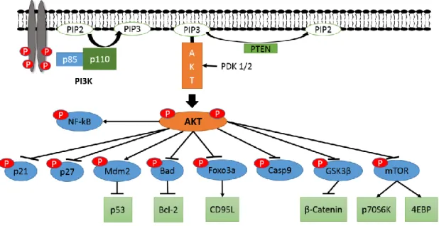
Figure 1.1.1 – Schematic of the PI3K/AKT pathway. Activation of RTK leads to recruitment of PI3K to
the membrane and activation of the catalytic subunit. This initiates production of PIP3, which in turn recruits PDK1/2 and AKT. Activated AKT uses phosphorylation to activate and inhibit many different targets involved in cell survival, growth and proliferation. PTEN is a negative regulator of the pathway by converting PIP3 back to PIP2.
AKT is a 57 kilo-Dalton serine/threonine kinase that acts by the phosphorylation of downstream protein targets. Mammalian species possess 3 AKT genes: AKT1, AKT2 and
AKT3 (15). However their protein expression varies in a tissue-specific manner. AKT1
expression is very high in brain, heart and lung. In contrast, AKT2 protein expression is highest in skeletal muscle and embryonic brown fat tissue (16). AKT3 is predominantly expressed in brain, kidney and embryonic heart(17).
8 Following AKT/PIP3 binding, AKT is activated by PDK1 and is phosphorylated at the threonine 308 residue (Thr308) (18). A secondary phosphorylation occurs at serine 473 (Ser473) by the action of different kinases such as mammalian target of rapamycin complex 2 (mTORC2) and integrin-linked kinase (ILK) (19). Although a dual phosphorylation is necessary for a full activation of AKT, a Thr308 p-AKT is still capable of phosphorylate some but not all of its substrates.
The active AKT is translocated (by a currently unknown mechanism) from the cell membrane into the cytoplasm and from here, into many others cellular compartments (including the nucleus, Golgi, endoplasmic reticulum and mitochondria), where its target substrates are located (19,20). Once activated, AKT inhibits the GSK3 (glycogen synthase kinase 3) protein by direct phosphorylation (20). With GSK3 inhibition, its targets such as β-catenin are not phosphorylated, which impedes their degradation. This allows β-catenin to translocate to the nucleus, where it interacts (both directly and indirectly) with transcription factors to regulate the expression of several genes such as
cyclin D1. Cyclins are proteins which are involved in cell cycle checkpoints, in
combination with cyclin-dependent kinases (CDKs) to regulate the cell cycle (21). The cyclin D1 protein is very important because not only is it a positive regulator for the G1/S checkpoint transition in the cell cycle, but it is also a direct target of GSK3β (22). Cyclin D1 binds to CDK4, forming a cyclin/CDK complex that phosphorylates the retinoblastoma protein (Rb). The phosphorylation of Rb leads to the activation of genes required for the cell cycle progression (23). As such, AKT can positively regulate G1/S cell cycle progression through inactivation of GSK3β and nuclear accumulation of cyclin D1.
AKT can also stop the inhibition of Cyclin/CDK complexes by the phosphorylation of p21Waf1/Cip1 (p21) and p27Kip2 (p27) (24,25). Once phosphorylated, the p21 protein is retained in the cytoplasm and cannot inhibit the cyclin D-CDK4 complex while p27 is relocated from the nucleus into the cytoplasm, preventing p27-mediated inhibition of cyclin E-Cdk2 and cyclin A-Cdk2 (19).
AKT can also affect protein synthesis through regulation of mTOR (mammalian target of rapamycin). The mTOR protein is a serine/threonine kinase that exists in two different protein complexes: mTORC1 and mTORC2 (26). Among its components, a relevant difference between them is the Raptor (regulatory-associated protein of mTOR) accessory protein in complex 1 and Rictor (rapamycin-insensitive companion of mTOR) accessory protein in complex 2 (26). mTORC1 can elevate mRNA translation by
9 activating the p70 ribosomal protein S6 kinase (p70S6K1) and inhibiting the repressor eIF4E-binding protein (4E-BP) (27). The regulation of mTORC1 is by the tuberous sclerosis complex (TSC), a heterodimer of hamartin (TSC1) and tuberin (TSC2). The TSC acts as a GTPase-activating protein (GAP) that negatively regulates Rheb (RAS homolog enriched in brain), a small GTPase that is required for mTORC1 activation (27). AKT can phosphorylate TSC2, impeding the formation of the TSC complex and leading to the activation of mTORC1.
Another interesting interaction between AKT and mTOR is through the mTORC2. This complex can regulate metabolism, cytoskeleton (28) and it can also phosphorylate AKT at Ser473 (26,27). Additionally, mTORC2 can phosphorylate the Thr450 residue upon the translation of nascent AKT, implying that mTORC2 activity is regulated by signals that promote AKT translation (28).
AKT can also promote cell survival by activating the cyclic AMP-response element binding protein (CREB). Phosphorylation of CREB induces the binding of accessory proteins that are needed for the transcription of anti-apoptotic genes, including Bcl-2 (B-cell lymphoma 2) and Mcl-1 (myeloid (B-cell leukaemia 1) (19). Related to this function, AKT can affect apoptosis by the phosphorylation of BAD (Bcl-2-associated death promoter). Bad controls the release of cytochrome c from mitochondria to initiate the caspase cascade, a process that is critical for the induction of apoptosis (12). BAD can also bind to Bcl-2 or Bcl-XL (B-cell lymphoma-extra-large), therefore blocking their anti-apoptotic activities (19). When phosphorylated by AKT, the BAD-Bcl-2/Bcl-XL complexes are disrupted and BAD is bound instead to the 14-3-3 chaperon protein, being sequestered into the cytosol. In addition, AKT will also phosphorylate BAX (Bcl-2-associated X protein), blocking its translocation to the mitochondria (19). Another way that AKT directly affects the apoptotic response is the inactivation of pro-caspase9, impeding the initiation of caspase cascade, and the phosphorylation of MDM2 (murine double minute 2), leading to the inhibition of p53-mediated apoptosis (19). A critical AKT protein target are the members of the FOXO (Forkhead box O) family of transcription factors including FOXO1a, FOXO3a and FOXO4 (19). These transcription factors have an essential role in promoting apoptosis and inducing cell cycle arrest. These (FOXOs and p53) are discussed in depth in section 1.4 and 1.5 respectively.
AKT can also activate the inhibitor of kappa B kinase-α (IKKα), leading to the phosphorylation of I-kappa-B (I-kB), marking it for degradation and stopping its inhibitory function over NF-kB (nuclear factor kappa-light-chain-enhancer of activated
10 B cells) (19). The free NF-kB can translocate to the nucleus and stimulate the transcription of pro-survival genes including the inhibitor of apoptosis protein 1 (IAP1) and IAP2 (19). The PI3K/AKT pathway is negatively regulated by PI-3, 4, 5-P3 phosphatases including the phosphatase and tensin homologue deleted on chromosome 10 (PTEN) (12,29). This phosphatase dephosphorylate PIP3 into PIP2, thus preventing the activation of AKT. Other phosphatases capable of regulating this pathway are protein phosphatase 2A (PP2A) and the pleckstrin homology domain leucine-rich repeat protein phosphatase (PHLPP) family (19). These phosphatases are able of dephosphorylating the thr308 and ser473 AKT residues respectively, thus inactivating the network.
1.4- FOXO proteins
The Forkhead box O (FOXO) family are transcription factors with a highly conserved DNA binding domain (30). In mammalian cells, four FOXOs have been identified: FOXO1, FOXO3a, FOXO4 and FOXO6. FOXOs can act either as transcriptional activators or as repressors, depending on additional co-factors recruited alongside them when they are bound to DNA (30). FOXOs are critical effectors of the PI3K pathway and when growth factors, cytokines or hormones activate the PI3K pathway, FOXOs are phosphorylated by AKT and are bound by the 14-3-3 chaperon protein (31,32). This FOXO/14-3-3 complex formation prevents FOXO-DNA binding and directs the cytoplasmic retention of FOXOs (30). Other kinases have been identified that are able to induce the cytoplasmic relocation of FOXO, such as serum- and glucocorticoid-inducible kinase (SGK), cyclin-dependent kinase- 2 (CDK2) and IκB kinase (33). By directing the cytoplasmic accumulation of FOXOs (and their subsequent ubiquitin-dependent proteomic degradation), activated AKT can prevent FOXO-dependent gene transcription preventing cell cycle arrest and apoptosis (summarized in figure 1.4.1).
11
FOXOs have a major role in proliferation, apoptosis, metabolism, inflammation, differentiation and stress resistance (32). Amongst the genes currently known that are regulated by FOXOs are p27, p21, p15 and p19. The p15 and p19 proteins are Cdk inhibitors that block the binding of cyclin D to CDK4 and CDK6 (30). As mentioned before, the p27 and p21 proteins are also CDK inhibitors necessary for cell cycle checkpoints regulation. Through the regulation of the expression of these genes/proteins, FOXOs are able to modulate the G1/S and G2/M transition of the cell cycle. In addition, it has been showed that activation of FOXO3 alone is sufficient for p27 upregulation and inhibition of proliferation (30).
During oxidative stress, FOXOs can coordinate the regulation of manganese superoxide dismutase (MnSOD) and catalase expression to decrease oxidative damage and increase cellular survival (30,34). Another way that FOXOs favours cell survival is by maintaining energy homeostasis through Sestrin3 (Sesn3) and Rictor expression. Inducing the expression of these genes results in inhibition of mTORC1 activity (major energy consumer) and an increase of mTORC2 activity, leading to the activation of AKT (which increase energy metabolism) (27).
In regard to regulating apoptosis, FOXOs can modulate the expression of BIM
(Bcl-2 interacting mediator of cell death, also known as BCL(Bcl-2L11), PUMA (p53 upregulated modulator of apoptosis) and Bcl-6 to induce the intrinsic apoptotic pathway.
Alternatively, FOXOs upregulate the death receptor ligands FasL (Fas-ligand, also
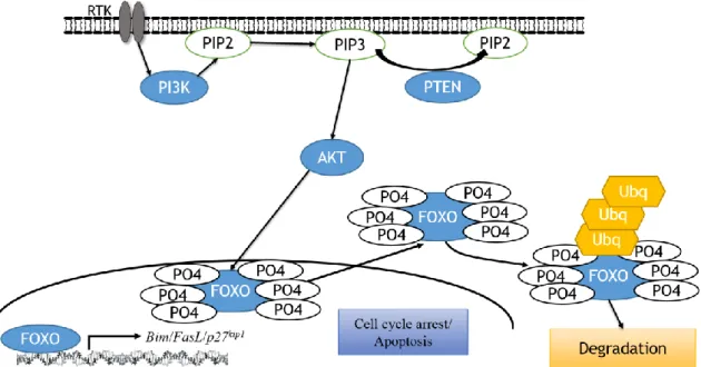
Figure 1.4.1 - Schematic of FOXO cytoplasmic translocation and sequential degradation.
Activated AKT translocate to the nucleus (unknown mechanism) and phosphorylates FOXO. FOXO is then translocated to the cytoplasm where it suffers polyubiquitination and subsequent degradation.
12 known as CD95L) and TRAIL (tumour necrosis factor-related apoptosis inducing ligand) to activate the extrinsic apoptotic pathway (30). FasL is the key death factor of receptor-triggered programmed cell death in immune cells (35,36). It has an important role in termination of immune responses, elimination of autoreactive cells and the establishment of immune privilege. Tumour cells that constitutively express FasL on their surface are able to generate a tumour-associated immune privilege, evading the immune surveillance and even kill tumour infiltrating lymphocytes (35). It has also been shown that FOXOs also play a role in preserving the self-renewal capacity of hematopoietic stem cells (33).
FOXO factors have been shown to be deregulated in many types of cancer such as breast, prostate, glioblastoma, rhabdomyosarcoma and leukaemia (33). Because of their role in regulating genes involved in apoptosis and cell cycle arrest, inactivation of FOXOs is an important step in tumorigenesis. As tumour suppressor genes, FOXOs are of extreme importance in the development of treatments against cancer. The study of how the many different signalling pathways affect the FOXO proteins have already shown that these factors are need for the current chemotherapeutics to have a cytostatic or cytotoxic effect on tumour cells (31,33). As such, FOXOs can be considered as biomarkers for treatment prognostics and risk assessment of patients. For example, cytoplasmic location of FOXO3a correlates with poor survival in breast cancer (33). New drugs that can influence the expression of FOXOs or that can affect their subcellular translocation will bring alternative approaches to chemotherapy. Combination treatments with drugs that activate FOXO can synergize with standard chemotherapeutics to sensitize tumour cells, opening new ways to counteract cancer resistance. However, the use of FOXO transcription factors must be well study because their ability to activate survival signalling and feedback mechanisms between FOXO family members may limit clinical utility.
The role of FOXOs on melanoma ranges from initial tumorigenesis all the way to treatment resistance. Melanoma is known to have the PI3K pathway constitutively active from either mutation of RTKs, PI3K or loss of PTEN. With AKT constantly active, the phosphorylation of FOXOs and its subsequent degradation will lead to uncontrolled proliferation. As for resistance, given that many current chemotherapeutics need FOXOs to exert their cytostatic/cytotoxic effect, tumour cells can counter treatments with cross-talk between signalling pathways that have FOXO as a common downstream target. For example, FOXO3a is a common target of three oncokinases: AKT, IKK and ERK (33). All three kinase-mediated phosphorylation induce FOXO3a ubiquitination and
13 subsequent degradation. Reverting their action over FOXO3a can re-sensitize tumour cells to chemotherapeutics.
1.5- The tumour suppressor p53
Similar to FOXO-family members, the tumour suppressor protein p53 is a transcription factor that regulates many genes involved in cell cycle arrest, apoptosis, senescence, autophagy and metabolism. Due to the importance of p53 in multiple cellular networks and preserving the cells genome, p53 is commonly referred to as “the guardian of the genome”(37).
Under normal cellular conditions, p53 exists in the cell bound to its negative regulator MDM2 (murine double minute 2) (schematically represented in Figure 1.5.1). In response to stress signals (for example DNA damage), p53 can be phosphorylated, abrogating the MDM2 interaction, allowing p53 levels to accumulate leading to the transcriptional induction of a vast array of target genes (37,38).
When MDM2 is phosphorylated, it will shift the balance p53/MDM2 in three ways (37). First, it abrogates the transcriptional activity by binding to the N-terminal of the transactivation domain of p53. Second, it induces nuclear export of p53 through mono-ubiquitination. Lastly, it destabilizes p53 through poly-ubiquitination and subsequent proteasomal degradation (Figure 1.5.1).
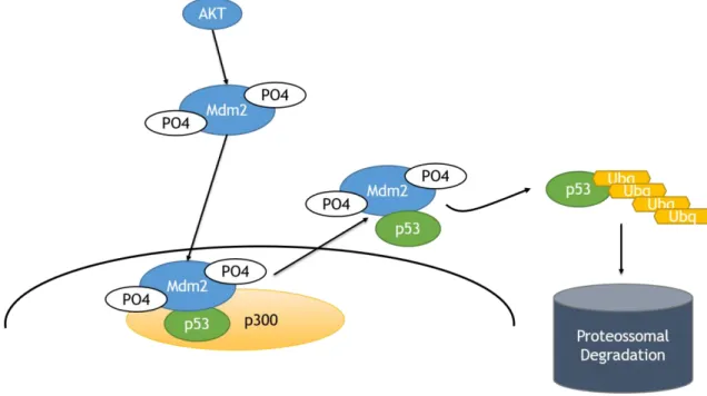
Figure 1.5.1 - Schematic of p53 degradation.
Activated AKT phosphorylates Mdm2, which in turn will bind to p53. P53 then suffers ubiquitination and is degraded by proteasomes.
14 As an E3 ubiquitin ligase, MDM2 has the ability to attach an ubiquitin chain of 76 amino acids to its target protein (38). This action is facilitated by MDM4, an E4 ligase (also known as ubiquitin chain elongating factors) that mediates the elongation of the ubiquitin chain previously established by MDM2 (38). Importantly, Mdm2 is a transcriptional target of p53, creating a feedback loop between them. This loop is the primary mode through which p53 autoregulates its levels.
The p53 transcription factor has always been of major interest in the fight against cancer. Because of its diverse biological functions that thwart tumorigenesis, the status of p53 has become a standard evaluation in cancer patients. In fact, p53 is the most mutated gene in human cancer, with a mutation rate that varies from 5% to 95% according to tumour type (39). Most mutations occur in the DNA binding domain. Even if the tumour cells retain the wildtype p53, it is often non-functional through its regulation mechanisms being altered (40).
In chemotherapy, p53 has an important role in chemosensitization and radiosensitization of tumour cells. As such, drug development is focusing p53 in four different approaches 1) activate wildtype p53, 2) reactivate mutant p53, 3) selectively kill cells with mutant p53 and 4) temporally inhibit wildtype p53 for normal cell protection (40).
In the particular case of melanoma, p53 has a mutation frequency of 10-20%. This is curious given that the background mutational load of melanoma is one of the highest in human cancer, comparable to lung and colon cancer (39). Although melanoma retains a wildtype p53, its functions are inactivated by other means. One way is through the deletion of CDKN2A (cyclin-dependent kinase inhibitor 2A), which leads to the loss of p14ARF, an inhibitor of MDM2 that prevents the targeting of p53 for degradation. Another mechanism is through the amplification of MDM2, leading to the destabilization of p53/MDM2 homeostasis (39,41).
Another possibility is the constitutive activation of the PI3K pathway. AKT phosphorylates MDM2 in both the nucleus and the cytoplasm, leading to the degradation of p53 through the methods described above. This link between p53 and AKT is of extreme importance in the development of new treatments for melanoma.
1.6- Novel Therapeutics in the Treatment of Melanoma
With modern drug development, many new drugs are being developed focusing in the cross-talk between different signalling pathways as a way to avoid resistance. As seen
15 with Vemurafenib, cancer cells can “counter-act” the inhibition of BRAF by, for example, re-activation of downstream targets such as ERK (4). A way to delay or circumvent the resistance to BRAF inhibitors is to combine them with MEK inhibitors such as Trametinib, which is being tested in clinical trials.
A key example of combined targeted therapies is the PI3K/AKT pathway. As this pathway interacts with and signals via RAS/RAF/MEK/ERK components at multiple points, resulting in cross-activation, cross-inhibition and pathway convergence, these targets could, in principle be individually targeted in combinational therapy to either restore drug sensitivity or to reduce the possibility of emerging resistant cancer cells(3). This is particularly noted in breast cancer resistance. Trastuzumab (marketed as Herceptin) is a recombinant monoclonal antibody that binds to the extracellular domain of HER2 (Human Epidermal growth factor Receptor 2) (42) and used to treat HER2+ breast cancers. Trastuzumab blocks the formation of HER2-HER3 heterodimers and also the binding of HER3 to PI3K (43). Many laboratory models of resistance have demonstrated that the loss of PTEN can reduce the anti-tumour effect of Trastuzumab as well as mutational activation of PI3K (42). As a consequence of this, many PI3K and AKT inhibitors are being tested in clinical trials as a means to reverse resistance to Trastuzumab in dual treatment regimens(43,44).
The PI3K, mTOR and AKT inhibitors can be used in many types of cancer, since they can target one of the most mutated pathways in human cancer. The mTOR inhibitors such as rapamycin and everolimus to test the PI3K signalling blockade in melanoma through inhibition of mTORC1 (45). Unfortunately, the efficacy of mTORC1 inhibitors is limited by dysregulation of negative feedback loops (45). PI3K inhibitors, such as wortmannin and LY294002 (46), and AKT inhibitors, such as afuresertib and MK2206 (44,47), showed positive results in pre-clinical models (47). Although positive, this results only confirmed that single-agent inhibition of the PI3K/AKT pathway is often cytostatic rather than cytotoxic, with activation of compensatory pathways (48).
The development of dual PI3K-mTOR inhibitors, such as BAG956, NVP-BBD130 and NVP-BEZ235, was an important step that showed impressive antiproliferative activity both in vitro and in vivo (45,46). In particular, BEZ235 is a potent pan-PI3K inhibitor that blocks mTOR by targeting the ATP-binding site of mTORC1/mTORC2 (46) and is currently in clinical trials (47,49). As mentioned before, cross-talking between signalling pathways has become a focus in the development of new therapies. The dual PI3K/mTOR inhibitors can synergize with BRAF and MEK
16 inhibitors, sensitizing back tumours who previously acquired resistance to this drugs (50– 52). A prime example is the combination of Vemurafenib with BEZ235, currently being tested in pre-clinical studies but already showing promising results. More combinatorial targeted therapies are extensively reviewed in Grazia et al. (47).
Another approach is the improvement of immunotherapies and their combination with cytotoxic agents and/or targeted therapies. Nivolumab (BMS-936558, MDX – 1106) is a fully human IgG4 antibody that blocks the 1 (programmed death 1) receptor. PD-1 interacts with PD-LPD-1 ( Programmed death ligand PD-1), inhibiting T-cell proliferation and cytotoxic functions (4). The interaction PD-1/PD-L1 plays an important role in tumour-induced immunosuppression in many advanced malignancies. Nivolumab has demonstrated clinical activity in patients with melanoma, renal-cell carcinoma and non-small-cell lung cancer (NSCLC) (10). Currently, a combination of Ipilimumab and Nivolumab is being studied and already shows early phase results of rapid and deep response of 80% (NCT01783938; NCT01024231) (53).
An alternative to complement current therapies is the use of phytochemicals. These are naturally occurring chemical compounds such as flavonoids and phytoalexins that can be found in common food (peanuts, red wine, grapes, strawberries, etc.).Phytochemicals are remarkably nontoxic and have proven their ability in preventing the development of cutaneous malignancies (45). They are currently being studied as potential adjuvant therapies for metastatic melanoma (45).
1.7- The Tribble2 homolog
Originally identified as tribbles in Drosophila, TRIB2 is pseudokinase which functions as a signalling modulator and mediator (54). Mammalian cells possess three Trib homologs: Trib1, Trib2 and Trib3 (55). All three Trib have been associated with human malignancies by interacting with pathways involved in cell survival and apoptosis. Trib proteins interact with many signalling molecules and transcription factors such as activating transcription factor 4 (ATF4), p65, mitogen-activated protein kinase kinase (MAPKK), AKT and constitutive morphogenesis 1 (COP1) (55).
Like all Trib proteins, TRIB2 has three main domains: an N-terminal, a C-terminal and a kinase domain. It belongs to the pseudokinase family because its central region (kinase domain) possess high homology with serine/threonine kinases but deviations in the catalytic loop eliminates its catalytic activity (Figure 1.7.1). Although it is a phosphoprotein, it is theorized that TRIB2 is unable of autophosphorylate itself (55).
17 TRIB2 has two important motifs on its C-terminal domain: MEK1 and COP1 binding sites. Through the MEK1 motif, TRIB2 is able to interact with the RAS/MEK/ERK signalling pathway and enhance ERK phosphorylation (54). TRIB2 can bind to COP1, an E3 ubiquitin ligase, through the COP1-binding site and promote proteasome-mediated degradation (54). One known target is the C/EBP
(CCAAT/enhancer-binding protein) family of transcription factors. By facilitating C/EBP-α degradation, TRIB2 aids in myeloid differentiation. This characteristic marked TRIB2 as an oncogene for acute myeloid leukaemia (AML). (54,56). Further studies brought to light that TRIB2 is commonly overexpress in many types of cancer such as lung adenocarcinoma, cervical carcinoma, liver cancer and malignant melanoma (55–59). In the case of melanoma, TRIB2 can be used as a biomarker since its expression correlates with the tumoral progression (60).
Another important fact is that TRIB2 was found to be a negative regulator of the FOXO family of transcription factors (59). TRIB2 can interact with AKT, leading to the inactivation and degradation of FOXO proteins. This implies that TRIB2 may be involved in mechanisms of cancer resistance.
Since TRIB2 is a pseudokinase, the mechanisms through which it exercises its functions is currently unknown. The main possibility is that TRIB2 acts as an adaptor that binds multiple proteins and brings them together. Given its proven role over FOXOs and in tumorigenesis, elucidation of how TRIB2 interacts and activates survival signalling can be an important step in cancer treatment.
1.8- Hypothesis
The previous work of our group showed that TRIB2 is a biomarker of diagnosis and progression of melanoma. Recent studies in our laboratory revealed that TRIB2 involved in mechanisms of drug resistance to many chemotherapeutics, including PI3K inhibitors currently in clinical trials. Based in our research, this work intends to characterize the TRIB2 pseudokinase following PI3K inhibition, its regulation, the domain responsible
Figure 1.7.1 - Representation of TRIB2. Kinase domain and C-terminal are highly conserved, with two
18 for the activation of survival signalling molecules such as AKT and how it relates to drug resistance in cancer.
19
2. Materials and Methods
2.1- Cell lines and reagents
The human cell lines HEK293T (embryonic kidney cells), U2OS [p53+/+], were maintained in DMEM supplemented with 10% FBS (Sigma, PT) and antibiotics (Gibco, US). Antibodies used in my studies are shown in Table 2.1.1 and were used for our immunoblots. Signal visualization was achieved using a ChemidocXRS+ system (BioRad, PT). Dacarbazine (Sigma, PT), gemcitabine hydrochloride (Eli Lilly #VL7502), AKT inhibitor VIII (Calbiochem, US), BEZ235 (Novartis, US), BAY806946, BAY 1082439, BAY1001931 (a gift from Bayer Healthcare, Germany), rapamycin (Sigma, PT), actinomycin D (Sigma, PT), and cycloheximide (Sigma, PT) and MG132 (Sigma, PT) were used at the following concentrations (Actinomycin D (ActD) 5 μg/ml, Cycloheximide (CHX) 50 μg/ml, MG132 at 1 μM).
Primary Antibodies Species Information Supplier
Actin goat I-19; sc-1616 Santa Cruz Biotechnology Total AKT goat C-20; sc-1618 Santa Cruz Biotechnology p-AKT rabbit Ser473; sc-7985 Santa Cruz Biotechnology BIM rabbit H-191; sc-11425 Santa Cruz Biotechnology P27 mouse F-8; sc-1641 Santa Cruz Biotechnology MDM2 rabbit C-18; sc-812 Santa Cruz Biotechnology p-MDM2 rabbit Ser 166; sc-293105 Santa Cruz Biotechnology
p-Mdm2 rabbit S166; 3521S Cell Signalling
Technology Lamin A/C mouse N-18; sc-6215 Santa Cruz Biotechnology
TRIB2 rabbit custom Home-made, generated at
CNIO (Madrid, Spain) FasL rabbit C-178; sc-6237 Santa Cruz Biotechnology Total Foxo3a
(FKHRL1) goat N-16; sc-9813 Santa Cruz Biotechnology p-Foxo3a
(p-FKHRL1) rabbit Ser 253; sc-101683 Santa Cruz Biotechnology
Table 2.1.1- Primary antibodies used. Datasheets of each antibody are on Appendix A1-A11.
2.2- Western blot analysis
For the preparation of whole cell lysate, cells were harvested and lysed using RIPA buffer (50 mM Tris-HCl pH 7.4, 1% NP-40, 0.5% Na-deoxychlorate, 150 mM NaCl, 1
20 mM EDTA, 2 mM NaF, 2 mM NaVO4 and 1x protease inhibitor cocktail (PIC) (Sigma). We used the Bradford assay, with the Quick Start TM Bradford 1x Dye Reagent (Bio-Rad). We prepare PCR tubes with 99 μl of Bradford. We dilute 1 μl of sample in 10 μl of H2O. We then add 1 μl of the dilution to the tubes. The blank tube was prepared with 100 μl of Bradford only. We analysed in the Nanodrop 2000/2000c Thermo Scientific by using 2 μl of each tube. For SDS–PAGE, protein samples were boiled for 5–10 min in protein sample buffer (50 mM Tris pH 6.8, 1% SDS, 10% glycerol, 0.01% Bromophenol Blue, β-mercaptoethanol [50 µL per 950 µL sample buffer]). Following electrophoresis, proteins were transferred onto nitrocellulose membrane (BioRad). The membrane was blocked for 1 hour at room temperature or overnight at 4°C 5% BSA 0.1% tween20 blocking buffer. Primary antibodies were added to the membrane (Supplemental Table 1) overnight at 4°C or for 2 hours at room temperature. Secondary antibody was added (Supplemental Table 2) at typically 1:5000 dilution for 1 hour at room temperature. Visualization of signal was achieved using a ChemidocXRS+ Imaging System (BioRad).
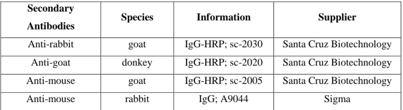
Secondary
Antibodies Species Information Supplier
Anti-rabbit goat IgG-HRP; sc-2030 Santa Cruz Biotechnology Anti-goat donkey IgG-HRP; sc-2020 Santa Cruz Biotechnology Anti-mouse goat IgG-HRP; sc-2005 Santa Cruz Biotechnology
Anti-mouse rabbit IgG; A9044 Sigma
Table 2.2.1 – Secondary antibodies used. Datasheets of each antibody are on Appendix A12-A15.
2.3- Constructs
A 3062 bp fragment encoding the entire human Trib2 (hTRIB2) cDNA was sub-cloned into pEGFP-N1 or pIREPuro2 plasmids, (one co-expressing GFP, the other containing a V5 tag.). Full length human TRIB2 (hTRIB2, 1-343 aa), dN (63-343aa hTRIB2, dC (1-304 aa) hTRIB2, KD (63-304 aa) hTRIB2, NT (ΔCT, 1-250 aa) hTRIB2, CT (ΔNT, 270-343 aa) hTRIB2 and ΔCOP1 hTRIB2 were kindly provided by W.S. Pear and shown in Table 3. Each was cloned into pMigR1-myc plasmid (55). All cDNAs were sequenced in their entirety to verify there were no mutations in any of our constructs. Each construct was transfected (Polyplus Transfection jetPRIME® transfection reagent) following the manufacturers guidelines (Appendix B). After transfection, the plates were incubated for 24 hours or selection and screening initiated.
21
Plasmids
constructs Mutant
#1 Full length TRIB2
#2 Δ COP1 #3 Δ N #4 Δ C #5 Kinase domain #6 C term #7 N term
Table 2.3.1 – TRIB2 plasmids constructs used
2.4- Flow cytometry cell cycle analysis
Cells were grown to 70% confluence. Cells were mock treated/exposed to each compound for the time points indicated. Samples were collected, washed (PBS) and fixed (70% ethanol). Ethanol was removed and samples were ressuspended in PBS. Propidium iodide (2.5 mg mL) was added to each sample. Samples were run on a Fluorescence Activated Cell Scanner (FACS) and the percentage populations (sub-G1, G1, S and G2 phases) determined. 10,000 total events were scored per study Data was analysed using Infinicyt (Cytognos).
2.5- FACS sorting
Cells were grown to 70% confluence. Cells were mock treated/exposed to each compound for the time points indicated. Samples were collected, washed (PBS) and fixed (70% ethanol). Ethanol was removed and samples were ressuspended in PBS. Samples were run on a Fluorescence Activated Cell Sorter (FACS) and the GFP positive and GFP negative populations separated. 50,000 total events were scored per study. Data was analysed using Infinicyt (Cytognos). The sorted samples were stored at 4⁰C until the following day prior to sample collection for immunoblotting.
2.6- Immunoprecipitation
The samples sorted by FACS were collected with 50 μl each of total protein lysis buffer (50 mM Tris pH 7.5, 1% NP-40, 0.1% Na-deoxychlorate, 130 mM NaCl, 5 mM EGTA, 10 mM NaF, 2,5 mM NaVO4 and 1x protease inhibitor cocktail (PIC)). Stored at -80⁰C for 24 hours. On the following day, added to each sample 5 μl of antibody and left them spinning on the “end-over-end” overnight at 4⁰C.
Figure 2.3.1 – Schematic of TRIB2 mutants. Different
22 Prepared beads with Protein G Sepharose Fast Flow (20 μl/sample) and PBS. Spin for 2 minutes at 1.300 rpm. Ressuspended with 1ml PBS and spin again. Repeated the last step but added PBS in an equal amount to the total volume of beads. Added 20 μl of the beads solution prepared and 300 μl of PBS to each sample. Left the samples spinning in the “end-over-end” for 1 hour at room temperature. Centrifuged for 2 minutes at 1.300 rpm. Removed supernatant and kept the beads. Added 300 μl of PBS and repeated the spin. Removed the supernatant and prepared the samples with 30 μl of 2x laemmli buffer. Boiled the samples for 5 minutes at 98⁰C. Proceeded by following the western blot protocol indicated above by loading 25 μl of each sample to the gel.
2.7- Co-Immunoprecipitation
Cells were harvested using total protein lysis buffer (50 mM Tris pH 7.5, 1% NP-40, 0.1% Na-deoxychlorate, 130 mM NaCl, 5 mM EGTA, 10 mM NaF, 2,5 mM NaVO4 and 1x protease inhibitor cocktail (PIC)). We used the Bradford assay and the Nanodrop 2000/2000c Thermo Scientific for protein quantification, as mentioned above.
Prepared α-GFP probes with Protein G Sepharose Fast Flow, antibody against GFP and PBS. Spin the probes for 1 h in the “end-over-end” at room temperature. Centrifuged for 1 minute at 1.300 rpm. Washed 2 times with PBS. Ressuspended with PBS.
Calculated amounts required for 250 μg of protein, probe and PBS for each sample. Add all to an Eppendorf and spin again in the “end-over-end” for 1h. Centrifuged for 1 minute at 1.300 rpm. Washed 2 times with 1 ml of PBS. Removed the supernatant and eluted with 50 μl of 2x Laemmli buffer. Boiled the samples for 5 minutes at 98⁰C and stored them at -80⁰C.
2.8- RNA extraction/First strand cDNA protocol
We used the TRI-reagent protocol from Sigma (Appendix C). The resulting RNA samples were then used to make first strand cDNA with NZY First-strand cDNA Synthesis Kit from Nzytech (Appendix E). Samples were stored at -18⁰C.
2.9- qRT-PCR /DNA electrophoresis agarose gel (1 %)/Primer validation
We used the LuminoCt® SYBR® Green qPCR Ready MixTM protocol from Sigma (Appendix F).
23 Primers Supplier GAPDH Nzytech TRIB2 Nzytech PTEN Nzytech MDM2 Nzytech FasL Nzytech P27 Nzytech TRAIL Nzytech Cyclin D1 Nzytech P19 Nzytech BIM Nzytech P21 Nzytech
Table 2.9.1 – List of primers used
We then ran a 1% agarose electrophoresis gel to analyse the amplified products.
2.10- Chromatin Immunoprecipitation assay (ChIP)
The plates were washed with ice-cold 1x PBS twice and added a 1% formaldehyde/PBS solution to each for cross-linking the proteins with the DNA. Plates were incubated at 37⁰C for 10 minutes and washed again with ice-cold 1x PBS twice. The plates were scraped with 1 ml Scrape buffer (1M Tris-HCL with 10 mM DTT), collected in eppendorfs and incubated in water bath at 30⁰C for 15 minutes. The tubes were centrifuged and washed with ice-cold 1x PBS, Buffer I (10% Triton X-100, 100 mM EDTA, 100 mM EGTA, 0,5 M HEPES) and Buffer II (100 mM EDTA, 100 mM EGTA, 0,5 M HEPES in 5M NaCl) . The pellets were ressuspended with Lysis buffer (10 % SDS, EDTA, Tris pH 8.0) prepared with PIC (protease inhibitor complex). The samples were sonicated 5-6 times, 10 seconds each, to break the DNA into approximately 250 base pairs fragments. The samples were then centrifuged at 15.000 rpm, 4⁰C for 10 minutes.
The supernatant was transferred to new eppendorfs and we added 300 μl of Buffer D (10% Triton X-100, 100 mM EDTA, Tris pH 8.0, NaCl) with PIC. We then removed 100 μl to the input eppendorfs. These input samples were left in the heat block at 65⁰C overnight (to reverse the cross-link). To the remaining samples we added antibody (5μl per sample) and left overnight at 4⁰C.
On the following day, we removed the input samples from the heat block and stored them at -20⁰C. Added protein G-fast flow beads (Sigma) to samples (10μl per sample)
24 and rotated for 1 hour at room temperature. Washed samples with TSE I (10% SDS, 1% Triton X-100, 100 mM EDTA, 3% NaCl (5M) in Tris pH 8.0), TSE II (10% SDS, 1% Triton X-100, 100 mM EDTA, 16% NaCl (5M) in Tris pH 8.0) and TSE III (1% NP-40, 100 mM EDTA, 10% Tris pH 8.0, 1% deoxycholate in LiCl (1M)) with rotation and centrifugation between washes. Washed 3 times with ice-cold TE buffer. Added a solution of SDS and NaCHO3 to each sample and rotated for 1 hour at room temperature. Transferred the supernatant to new eppendorfs, left them overnight on the heat block at 65⁰C and stored them at -20⁰C on the next day.
We used the NzyGelpure purification kit (Appendix D) to “clean” the previous ChIP and input samples. We used 1 ml of binding buffer on step 1 and 30 μl of dH2O on step 5 instead of the manufacturer indications. The purified DNA was stored at -20⁰C.
25
3. Results
3.1- TRIB2 protein levels increase in the presence of PI3K inhibitors
Our group has previously demonstrated that TRIB2 is a biomarker for malignant melanoma (60) and is a negative regulator of FOXO3a (59). As such, we know that TRIB2 is integrated within the PI3K pathway but where within this network and specifically how TRIB2 can regulate FOXO3a is
unknown. We have
previously generated a range of isogenic TRIB2 expressing cell lines and obtained a number of novel PI3K pathway inhibitors (summarized in Figure 3.1.1). We first questioned if there were any effects on the TRIB2 protein before and after PI3K inhibition within our isogenic cell lines. Using an immunoblotting approach, we found a discernible increase in TRIB2 protein in both the endogenous U2OS-Empty and the U2OS-TRIB2 cell lines (Figure 3.1.2).
Figure 3.1.1- PI3K pathway inhibitors.
Squematic of PI3K pathway and how the inhibitors target its components.
Figure 3.1.2- TRIB2 protein levels increase following PI3K inhibition.
Representative western blot of isogenic osteosarcoma cell lines with over expression of TRIB2 (U2OS-TRIB2) and endogenous TRIB2 protein expression (U2OS-Empty). Cell lines were treated with 100nM of each indicated compound and 50µg of total protein lysate was loaded per lane. β-actin is shown to indicate protein loading. .