Maria Rita Rebocho Lopes do Amaral
Licenciada em Biologia
Evaluation of a new vaccine based on
pDNA and recombinant protein against
Helicobacter pylori
Dissertação para obtenção do Grau de Mestre em Genética Molecular e Biomedicina
Orientador: Doutora Lídia Maria Diogo Gonçalves,
Co-orientador: António José Leitão Neves Almeida, Professor Catedrático,
Faculdade de Farmácia da Universidade de Lisboa
Juri:
Presidente: Prof. Doutora Ilda Maria Barros dos Santos Gomes Sanches Arguentes: Prof. Doutora Cecília Ribeiro da Cruz Calado
Prof. Doutora Rosario Mato Labajos Vogais: Doutora Lídia Maria Diogo Gonçalves
Prof. Doutor António José Leitão Neves Almeida
III Evaluation of a new vaccine based on pDNA and recombinant proteins against Helicobacter pylori
Copyright Rita Amaral, FCT/UNL, UNL
A Faculdade de Ciências e Tecnologia e a Universidade Nova de Lisboa têm o direito, perpétuo e sem
limites geográficos, de arquivar e publicar esta dissertação através de exemplares impressos
reproduzidos em papel ou de forma digital, ou por qualquer outro meio conhecido ou que venha a ser
inventado, e de a divulgar através de repositórios científicos e de admitir a sua cópia e distribuição
com objectivos educacionais ou de investigação, não comerciais, desde que seja dado crédito ao autor
V Em primeiro lugar, não podia deixar de agradecer à Doutora Lídia Gonçalves, pois sem a sua
valiosa orientação este trabalho não seria possível. Um obrigado especial por toda a sua simpatia,
alegria, companheirismo e muitas vezes pelo seu papel de amiga e mãe para me chamar à razão.
Obrigada pela paciência e por todos os ensinamentos. Aprendi bastante consigo, e sei que todos os
conhecimentos transmitidos farão de mim uma melhor pessoa e investigadora, e sei que contribuirão
tanto para a minha vida profissional futura como para a vida pessoal.
Em segundo, queria agradecer ao Professor Doutor António Almeida pela oportunidade de
ingressar neste trabalho, pelo voto de confiança e pela oportunidade de integrar este fantástico
laboratório. Obrigada também por todas as chamadas de atenção e conselhos, de forma a melhorar
sempre a o sentido critico e o espirito científico e por toda a disponibilidade e preocupação.
Ao Professor Doutor Jorge Vítor, por me ter aceitado tão prontamente no seu laboratório, por
todos ensinamentos transmitidos relativamente a essa complicada bactéria que é a Helicobater pylori,
e relativamente ao seu complicado cultivo, e ao Miguel por toda a disponibilidade e ajuda prestada em
todo o trabalho realizado. Contribuíram bastante para o progresso do meu conhecimento em
microbiologia, e especialmente da Helicobacter pylori.
Queria também agradecer a todos os investigadores do Lab112, por me terem acolhido tão
calorosamente e por me terem ajudado sempre que precisei. Um especial obrigado à Giuly por toda a
preocupação e ter sempre cuidado de mim. Obrigada pelos momentos passados, pela companhia, pelos
almoços e por todas as grandes reclamações que tinhas todos dias, tão característico teu e que fazem
de ti uma pessoa especial. Obrigada pela amizade.
A todas as minhas amigas de mestrado, à Carolina, à Inês, à Mónica e à Rita, por todos os
momentos vividos e partilhados. Não importa referir quais, vocês sabem. Por toda a amizade que se
mantem e que prevalecerá.
Não podia deixar de agradecer aos meus amigos de sempre, principalmente à Joana, ao Melão e à
Teresa toda a amizade ao longo destes anos, pelos momentos vividos e partilhados, por todas as
confidências e pelo ombro amigo, por estarem sempre presentes e por continuarem a fazer parte da
minha vida.
Um agradecimento muito especial à minha família, aos meus avós por todo o apoio e por
acreditarem sempre em mim, aos meus tios únicos e preferidos e às minhas primas mais importantes
VI Por fim, mas não menos importante, ao meu namorado, pois sem ele teria sido tudo bem mais
difícil. Obrigada por todo o amor, carinho, apoio e companhia ao longo deste ano. Por me chamares
sempre à razão, por me motivares, por todos os conselhos, todos os jantares, almoços e
pequenos-almoços e principalmente por todos os maravilhosos momentos partilhados.
Agradece-se o apoio financeiro da Fundação da Ciência e Tecnologia (FCT) e FEDER
(PTDC/BIO/69242/2006 e PEst-OE/SAU/UI4013/2011).
VII Helicobacter pylori is a bacterium capable of surviving and infecting a healthy human stomach
and it is estimated that infect more than a half of world population. Despite of being almost always
asymptomatic, in some cases, the infection can evolve to several gastric disease as chronic gastritis,
peptic ulcers, gastric cancer and MALT lymphoma. Vaccination against H. pylori is a promising
option due to emerging problems of antibiotics treatment. It is thought that oral immunization could be
a good approach for a more effective protection against infections by H. pylori, creating a first line of
defense in mucosal surfaces. Chitosan nanoparticles are a suitable vehicle for oral vaccines delivery
due to its immunogenic and mucoadhesive properties, protecting the DNA and allowing high levels of
transfected cells. Thus, this work aims to evaluate a new pDNA- and recombinant protein-based
vaccine, with multi epitopes of different H. pylori antigens. Following production and purification of
plasmid DNA and recombinant proteins, vaccines were formulated for oral and intramuscular
administration with the antigens encapsulated with chitosan nanoparticles.
The type of immune response induced and the effectiveness of protective immunity elicited
were assessed by ELISA, through analysis of specific IgGs, mucosal SIgA and cytokines levels
produced by immunized BALB/C mice. When give by the intramuscular route, the formulated pDNA
and recombinant protein-based vaccines efficiently stimulated the production of specific IgG2a and
IgG1, which is supported by cytokines levels, revealing a better and balanced systemic immune
response than oral immunizations. Nevertheless as expected, oral immunizations with either pDNA
vaccines or recombinant protein revealed high levels of SIgA, showing to be effective in gastric
mucosal immunization for a more protective immune response, contrasting with intramuscular
immunizations which did not induce SIgA.
The immunization results showed that both pDNA and recombinant proteins vaccines
encapsulated with chitosan nanoparticles are good candidates for the development of a future vaccine
to prophylactic and therapeutic use to improve the eradication of H. pylori infections.
IX
Resumo
Helicobacter pylori é uma bactéria capaz de sobreviver e infectar o estômago humano saudável e
estima-se que mais de metade da população mundial esteja infectada. Apesar de, na sua maioria, estas
infecções serem assintomáticas, em alguns casos poderão evoluir para várias doenças gástricas como
gastrite crónica, úlceras pépticas, carcinoma gástrico e linfoma MALT. A vacinação contra infecções
por H. pylori poderá ser uma alternativa promissora, devido aos problemas emergentes dos
tratamentos à base de antibióticos. Pensa-se que a vacinação oral poderá ser uma boa abordagem para
uma imunidade protetora mais eficaz contra infecções por H. pylori, criando uma primeira linha de
defesa ao nível das mucosas. Nanoparticulas de quitosano têm mostrado ser um vínculo adequado para
vacinação por via oral, devido às suas propriedades imunogénicas e mucoadesivas, conferindo
proteção do DNA, e permitindo níveis elevados de células transfectadas. Este trabalho visa avaliar
uma vacina baseada em DNA plasmídeo e proteína recombinante, constituída por múltiplos epítopos
de diferentes antigénios de H. pylori.
Após produção e purificação do pDNA e da proteína recombinante, foram formuladas vacinas
para administração oral e intramuscular com os antigénios encapsulados em nanoparticulas de
quitosano. O tipo de resposta imunitária gerada e a eficácia da imunidade protetora foi avaliada por
ELISA, através da análise dos níveis de IgGs especificos, de SIgA e de citoquinas produzidas,
presentes nos soros de ratos BALB/c imunizados. Quando administradas pela via intramuscular, as
vacinas formulada com pDNA e proteína recombinante estimularam eficientemente a produção tanto
de IgG1 como de IgG2a, resposta suportada pelos níveis de citocinas produzidas, revelando uma
resposta imunitária sistémica mais consistente e equilibrada do que imunizações por via oral. No
entanto, como esperado, imunizações por via oral revelaram níveis elevados de SIgA, mostrando-se
eficaz na imunização da mucosa gástrica conferindo uma maior imunidade protectora, em contraste
com imunizações por via intramusculares que não induziram secreção de IgAs.
Os resultados obtidos das imunizações mostraram que ambas as vacinas formuladas com pDNA e
proteína recombinante são boas candidatas para o desenvolvimento de uma vacina futura para uso
profilático e terapêutico de modo a erradicar infecções por H. pylori.
XI
Contents
Acknowledgments ... V Abstract ... VII Resumo ... IX Contents ... XI List of Figures ... XIII List of Tables ... XV Abbreviations and symbols ... XVII
1. Introduction ... 1
1.1. Historical perspective ... 3
1.2. Helicobacter pylori ... 3
1.3. Epidemiology of Helicobacter pylori infection ... 7
1.4. Helicobacter pylori Infection and Immune system ... 8
1.4.1. Mucosal immunity ... 10
1.5. Helicobacter pylori associated diseases ... 11
1.6. Diagnosis and Treatment ... 11
1.7. Vaccines ... 12
1.7.1. Types of vaccines ... 12
1.7.1.1. DNA based vaccines ... 14
1.7.2. Routes of vaccination ... 17
1.7.3. Adjuvants and Delivery systems ... 17
1.8. Aims ... 19
2. Materials and Methods ... 21
2.1. Antigen production ... 23
2.1.1. Cell transformations ... 23
2.1.2. Cell culture growth ... 24
2.1.3. Optimization of production conditions of 6T recombinant protein ... 24
2.1.4. 6T recombinant protein purification ... 24
2.1.5. SDS-PAGE ... 25
2.1.6. Western Blotting ... 25
XII
2.1.8. DNA Plasmid extraction and purification ... 27
2.1.9. Electrophoresis ... 27
2.1.10. Quantification of plasmid DNA ... 28
2.2. In vivo Assays ... 29
2.2.1. Nanoparticle preparation ... 29
2.2.2. Immunization studies / Vaccination ... 29
2.2.3. Sample collection and analysis ... 30
2.2.4. Quantification of immune response by ELISA ... 31
2.2.5. Cytokine production studies ... 32
2.2.6. Statistical analysis ... 33
2.3. Agglutination assays ... 33
3. Results and Discussion ... 35
3.1. Antigen production ... 37
3.1.1. Cell transformation ... 37
3.1.2. Cell culture growth ... 37
3.1.3. Optimization of protein expression conditions ... 38
3.1.4. Protein purification ... 40
3.1.5. Plasmid DNA purification ... 44
3.2. In vivo assays ... 46
3.2.1. Nanoparticle characterization ... 46
3.2.2. Immunization studies ... 47
4. Conclusions ... 59
XIII
List of Figures
Figure 1.2.1 –H. pylori infection: Colonization and infection of the most distal part of the stomach,
the antrum...4
Figure 1.2.1.1 - Mechanisms of action of VacA on host gastric epithelial cells and its effect on immune cells...6
Figure 1.3.1 - Prevalence of H. pylori infection around the world in asymptomatic adults (Crew and Neugut, 2006)...8
Figure 1.5.1 – Chronic H. pylori infection: Chronic H. pylori infection can lead to different gastric diseases such as chronic gastritis, duodenal ulcer and gastric cancer. Adapted from Amieva and El-Omar (2008)...11
Figure 1.7.1.1.1– Plasmid DNA backbone scheme for DNA vaccines with gene sequence of interest, polyA sequence, promoter and origin of replication, and antibiotic resistance gene for selection of competent bacteria (Ingolotti et al., 2010)...14
Figure 1.7.1.1.2 – Resume of the Mechanisms of action of different types of vaccination: attenuated vaccines, inactivated vaccines, subunits vaccines, toxoids vaccines and DNA vaccines; Advantages and disadvantages (Ingolotti et al., 2010)...16
Figure 2.1.1.1 - H. pylori DNA and protein vaccines.Schematic representation (not to scale) of the H. pylori vaccine construction ...23
Figure 2.1.8.1 - Adapted from “QIAfilter Plasmid Purification Handbook” of QIAGEN Inc, Germany...27
Figure 2.2.3.1 –Sample collection and immunoassays Scheme...31
Figure 3.1.2.1– Cell growth curve of E. coli XL1 Blue expressing 6T recombinant protei...38
XIV
Figure 3.1.3.2 - SDS-PAGE and Western Blot of 6T-protein extract processed with anti-HistTag
antibodies...40
Figure 3.1.4.1 –SDS-PAGE (10% polyacrylamide gel) and Western blot of samples from purification
process of recombinant 6T protein...42
Figure 3.1.6.1 – Confirmation of the purification of plasmid DNA by agarose gel electrophoresis...45
Figure 3.2.2.1 - Serum anti-H. pylori specific IgG, IgG1 and IgG2a titres induced after mice immunization ……….………...48
Figura 3.2.2.2 – Serum anti-H. pylori specific IgG, IgG1 and IgG2a titres induced by immunization with pDNA by both oral and i.m. route. Data represent a preliminary assessment concerning serum
pool of each group (n=5)……...…..49
Figure 3.2.2.3– Serum specific anti H-pylori IgG, IgG1 and IgG2a titres stimulated by immunizations with 6T recombinant protein by oral (G3 and G4) and i.m. (G7 and G8). Data represent a preliminary
assessment concerning serum pool of each group (n=5)...50
Figure 3.2.2.4– Serum specific anti-H. pylori IgG, IgG1 and IgG2a titres from immunized mice with pDNA and 6T protein by the oral route. Data represent a preliminary assessment concerning serum
pool of each group (n=5)...51
Figure 3.2.2.5– Serum anti-H. pylori specific IgG, IgG1 and IgG2a titres of mice immunized by i.m. route, with pDNA and recombinant proteins, both encapsulated and in solution. Data represent a
preliminary assessment concerning serum pool of each group (n=5)...51
Figure 3.2.2.1.1 - sIgA levels in intestine homogenates of mice immunised by i.m. and oral route....53
Figure 3.2.2.2.1 - Cytokine levels after splenocytes stimulation with H. pylori antigens (Note: G2 and
XV
List of Tables
Table 2.1.2.1 - Tryptic Soy Broth composition...24
Table 2.2.2.1 - Immunization groups with recombinant antigens 6T and 6T-plasmid by oral and i.m.
routes...30
Table 2.3.1–H. pylori selective medium composition per litre...33
Table 3.1.5.1 –
Amounts of purified protein obtained before and after buffer exchange and
desalting.
...43Table 3.1.7.1 –Total amount of purified 6T-plasmid...45
Table 3.2.1.1– Charecteristics of nanoparticle used in immunization studies. Values are expressed as
mean±S.D.(n=3)………...47
Table 3.3.1 – Results from agglutination tests with H. pylori bacterial suspension against sera of
XVII
Abbreviations and symbols
Abs Absorbance
AGS Human gastric adenocarcinoma epithelial cell line
APCs Antigen presentation cells
BB Brucella broth
BCA Bicinchoninic acid protein assay
BSA Bovine serum albumin
CagA Cytotoxin-associated gene A
Cag-PAI cag-pathogenicity island
CS/DS Chitosan/deoxycholate
CS/TPP/Alg Chitosan/Tripolyphosphate/alginate
CMV Cytomegalovirus
CT Cholera toxin
DC Dendritic cells
DNA Desoxirribonucleotic acid
dsDNA double strain Desoxirribonucleotic acid
DU Duodenal ulcer
E. coli LT E. coli heat-labile toxin
ELISA Enzyme-linked immunosorbent assay
FBS Fetal bovine serum
GC Gastric Cancer
GroEL Chaperonin
GU Gastric ulcers
Homb outer membrane protein
HpaA Neuraminil-lactose hemagglutinin connection
IFN-γ Interferon-gamma
i intermediate-region
i1 intermediate-region polymorphism 1
i2 intermediate-region polymorphism 2
IgA Immunoglobuline A
IgG Immunoglobuline G
IgG1 Immunoglobuline G1
IgG2a Immunoglobuline G2a
IL Interleucine
IMAC Imobilized metal ion affinity chromatography
IPTG Isopropyl b-D-1-thiogalactopyranoside
ISCOMs Immunostimulatory complexes
LPS Lipopolysaccharides
LT heat-labil toxin
m mid-region
m1 mid-region polymorphism 1
m2 mid-region polymorphism 2
MALT Mucosa-associated lymphoid tissue
MHC Major Histocompatibility Complex
XVIII
MHC II Major Histocompatibility Complex type II
mRNA mesenger Ribonucleotic acid
MW Molecular weight
NAP Neutrophil-activating protein
NBT Nitro-Blue Tetrazolium
PAMPs pathogen-associated molecular patterns
PBS Phosphate buffer saline
PCR Polymerase chain reaction
pDNA plasmid Desoxirribonucleic acid
poly[A] polyadenylation sequence
PPI proton pump inhibitor
PRR pattern recognition receptors
PUD peptide ulcer disease
RNA Ribonucleotic acid
SDS-PAGE Sodium dodecyl sulfate polyacrylamide gel electrophoresis
s s-region
s1 s-region polymorphism 1
s2 s-region polymorphism 2
T4SS Type IV secretion system
TcL T citotoxic lymphocites
Th T helper cells
TLRs Toll-Like Receptors
TNF-α Tumor necrosis factor-alpha
TPP/Alg Tripolyphosphate/alginate
Ty21a Typhoid vaccine
UBT Urea breath test
UreA Urease alpha subunit
UreB Urease beta subunit
1
3
1.1. Historical perspective
Since the early of 20th century several investigators have reported the presence of spiral
microorganisms in the stomach of several animals (Kusters et al., 2006; Dubois, 2007). In 1906,
Walter Krienitz was the first to report the observation of spiral bacteria in the human stomach,
suggesting that spiral-shaped bacteria were causing gastric diseases, but it was not possible to make a
consistent association between the presence of this bacteria and any specific disease. In 1983,
Helicobacter pylori were successful isolated and cultured for the first time by Warren and Marshall.
Self-ingestion experiments by Marshall demonstrated that these spiral bacteria can colonize the
healthy human stomach and inducing inflammatory response in the gastric mucosa, developing a
transient gastritis, as proved by gastric biopsy samples taken from B.J. Marshall (Marshall et al.,
1985). This discovery was worth the 2005 Nobel Prize in physiology or medicine to Robin Warren
and Barry Marshall for their “Discovery of the bacterium Helicobacter pylori and its role in gastritis and peptic ulcer disease.” (Kusters et al., 2006). This knowledge showed that gastric colonization with H. pylori can lead a variety of gastrointestinal disorders, such as chronic gastritis, duodenal ulcers
(DU) or gastric ulcers (GU), gastric mucosa-associated lymphoid tissue (MALT) lymphoma, and
gastric cancer (GC). This discovery prompted efforts by other scientists in the study of the physiology,
genetics, transmission and epidemiology of this bacterium (Kusters et al., 2006). The result was an
increasing number of scientific manuscripts up to about 30 thousand manuscripts published in the past
20 years.
1.2. Helicobacter pylori
H. pylori is a gram-negative bacterium, spiral-shaped, microaerophilic, with 3 to 7 unipolar
flagels, that allow the bacteria to move along the mucus layer to gastric epithelium and colonize the
antral region (the most distal region) of the human stomach after oral ingestion (Amieva and El-Omar,
2008).
Most H. pylori organisms are free living in the mucus layer, but some organisms attach to the
apical surface of gastric epithelial cells and after attachment always elicit an acute inflammatory
response (gastritis) in the stomach mucosa (Velin and Michetti, 2010).
The stomach has a too acid and hostile environment unfavorable to the presence of most
microorganisms, due to large amounts of gastric juice composed by digestive enzymes and
hydrochloric acid (Amieva and El-Omar, 2008; Zhou et al., 2009). H. pylori has a unique mechanism
that allows it to survive a transient exposure to this acid environment of the stomach for a few
minutes, by secreting a large amount of the enzyme urease that hydrolyses urea into ammonia and
4 known gastric Helicobacter species are urease positive but also highly mobile through flagella. This
motility permits rapid movement toward the more neutral pH of the gastric mucosa. Both factors are
prerequisites for colonization of the human stomach and will allow trigger an inflammatory response
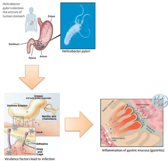
in epithelial cells (Figure 1.2.1) (Kusters et al., 2006). The enzyme urease along with lipases,
proteases and others virulence factors produced by the bacterium will disrupt the defenses of the
mucus layer and will lead to the success of infection.
Figure 1.2.1 –H. pylori infection: Colonization and infection of the most distal part of the stomach, the antrum. Adhesion, urease and other virulence factors are important to H. pylori surviving in a
transient exposure to low pH environments: to avoid the acid lumen of the stomach for long
exposures, H. pylori swims toward the mucosal cell surface using their polar flagella and chemotaxis
mechanisms. As near the epithelium, H. pylori actively adheres to the cell surface using a variety of
specific adhesions that recognize glycoproteins on the host cell. Attachment to the host cells allows H.
5
1.2.1. Virulence factors
H. pylori has a high genetic variability among strains, due to a high rate of mutation and
recombination, horizontal transfer of DNA and the lack of mismatch repair system of DNA. This leads
to a high diversity of genes and a high number of alleles, essential for adaptation and colonization of
the hostile environment of the stomach (Suerbaum and Michetti, 2002). Bacterial virulence factors
play an important role, since the virulent strains are more aggressive and increase the risk of
developing severe clinical manifestations. These virulence factors also play structural and critical roles
and functions such as adhesion, invasion, colonization and virulence. Some antigens have been
extensively studied and tested, known to be involved in the pathogenesis of the infection and shown to
be safe and promising, due to its pathogenic importance, immunogenicity and representativeness in a
large number of strains. These antigens include urease, the Neuraminil-lactose hemagglutinin
connection (HpaA), the vacuolating cytotoxin (VacA), the cytotoxin-associated antigen (CagA), the
neutrophil-activating protein (NAP) and others (Del Giudice et al., 2009; Velin and Michetti, 2010).
Urease is an enzyme that catalyzes urea into ammonia (NH3) and carbonic acid (H2CO3), which
in turn is converted into CO2 and H2O, and is crucial for the survival of the H. pylori in the human
stomach. This enzyme is present in all strains and because of that its activity is used for detected H.
pylori infections as in urea breath test (UBT) (discussed in section 1.7.) and gastric biopsy samples
(urease test). It is composed by 12 urease alpha (UreA) and 12 urease beta (UreB) subunits with
molecular mass of approximately 27 kDa and 60 kDa each, respectively, being the UreB subunit most
commonly used for immunization (Begue and Sadowska-Krowicka, 2010; Calvet et al., 2010).
The virulence factor HpaA or adhesion A is a protein that works as an adhesion present in
flagellar filament of flagella and on the bacterial surface, essential to colonization and infection.
Studies with hpaA mutants showed this protein is necessary for motility and for establishment of a
stable colonization. HpaA is highly immunogenic and leads to induction, maturation and antigen
presentation by dendritic cells (Lundstrom et al., 2001; Voland et al., 2003; Carlsohn et al., 2006).
HomB is a Helicobacter outer membrane protein (OMP) that permits the attachment of the
bacteria to the gastric epithelium to avoid the bacteria elimination by peristaltic movements. Bacterial
adherence to ephitelial cells leads to an induce of inflammatory response through activation of IL-8,
leading to PUD. This makes this protein a co-marker for strains associated with peptic ulcers, as well
as with the presence of other H. pylori disease-related genes like cagA, babA and vacA. (Oleastro et
al., 2008).
Vacuolization cytotoxin VacA, is an exotoxin that is involved in various mechanisms of virulence
such as induction of cytoplasm vacuolization in epithelial cells that can cause disruption of the
epithelial barrier due to formation of pores, membrane channels and cellular damage, targeted to the
6 Figure 1.2.1.1) and phagocytosis inhibition by macrophages (Suerbaum and Michetti, 2002; Wilson
and Crabtree, 2007).
Figure 1.2.1.1 - Mechanisms of action of VacA on host gastric epithelial cells and its effect on
immune cells. Surface bound VacA may function as an adhesion (1), while secreted VacA may bind to
a host receptor and induce pro-inflammatory response (2) or induce apoptosis mediated by cytochrome
c (in green) release (3), or induce vacuolation by alterations in endocytic vesicles (4), or form a
membrane channel leading in exit of nutrients and ions to the extracellular space (5), or achieve the
lamina propria where it binds to immune cells causing immune system modulation (6). Adapted from
Cover and Blanke (2005) and Jones et al. (2010).
The VacA gene is present in all known H. pylori strains but the activity of the protein depends on
this polymorphism of two regions of the gene, s-region (s) and mid-region (m). Each s and m regions
present two polymorphisms, s1 and s2, and m1 and m2. Strains having s1/m1 vacA gene have the
highest vacuolization activity, when s1/m2 strains present VacA activity but has a restricted number of
affected cells, and activity of the protein is absent in presence of s2/m2 strains. The S2/m1 genotype is
very rare (Letley and Atherton, 2000; McClain et al., 2001). VacA vacuolization activity is codified
by s-region while the toxin binding to epithelial cells is related to m-region. A third variable region,
located between the s- and m-region, was identified as intermediate (i) region, and i1 is associated to
the vacuolization form and i2 is associated to the non-vacuolization form. Was observed that while all
s1/m1 strains were of type i1 and s2/m2 were type i2, s1/m2 could be both i1 and i2, varying in their
7 VacA gene was identified as mostly common and encodes a highly antigenic protein due to
induce the production of pro-inflammatory cytokines, such as TNF-α, IL-6 and IL-8 in immune cells, and showed to have an immunosuppressive effect on them. It will inhibit T cell activation interfering
with calcium-signaling events inside the cell and prevented activation of the calcium-dependent
phosphatase calcineurin, inhibit the proliferation of T and B lymphocytes and will interfere with
phagocytosis and antigen presentation (Backert et al., 2010).
The cag-pathogenicity island (cag-PAI), a 40kb DNA segment of the H. pylori genome, encodes
both CagA protein and type IV secretion system (T4SS). This system translocates the CagA protein
into the epithelial cells through a pilus responsible for the delivery of bacterial proteins such as CagA,
inside the host cells, during infection (Covacci et al., 1993; Censini et al., 1996; Vitoriano et al.,
2011). The entry of CagA, will lead to increased inflammation and to activation of adaptative
immunity, disruption of tight junctions, cytoskeleton rearrangements, oxidative stress that leads to
apoptosis and DNA damage, and induce secretion of 8 cytokine activating Dendritic Cells and
IL-12 cytokine, that will stimulate Th1 cells too (Vitoriano et al., 2011). CagA protein is one of the most
important and well-characterized virulence factors of H. pylori. For that reason it is essential for
stimulating the immunity to protect the host against H. pylori infections.
When injected in the host cell, CagA is phosphorylated and interacts with several epithelial cells
effectors proteins of the host. Additional bacterial factors are injected through the T4SS, such as
peptidoglycan, that also interact with host proteins. These interactions with host protein effectors by
cag-PAI genes products, results in the deregulation of signalling pathways associated with host cell
motility and elongation, proliferation, inflammatory response, disruption of cell junctions and
phagocytosis inhibition. It was demonstrated that cag-PAI positive strains induces the development of
gastritis and GU, when compared to mutants for cag-PAI-encoding proteins (Tegtmeyer et al., 2011).
1.3. Epidemiology of Helicobacter pylori infection
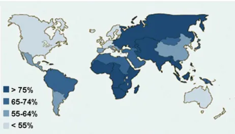
It is estimated that more than a half of global population is infected with H. pylori and its
prevalence differs according to geographic location. Within the same country, the prevalence of H.
pylori is influenced by patients’ socioeconomic status, age, gender and genetic predisposition. Among countries, the prevalence is higher in developing countries were achieves 80-90%, when compared to
developed countries with 10-60% of prevalence (Figure 1.3.1) (Del Giudice et al., 2009; Khalifa et al.,
2010). The prevalence of infection is especially higher in the rural developing areas in contrast to
urban developed ones because in rural environments people are probably exposed to an increased
number of infectious sources such as contaminated food and water, or intensive contact between
8 The route of transmission of H. pylori is not completely understood but is thought that new
infections occur as a consequence of direct human-to-human transmission or by environmental
contamination. There are evidences that infection by H. pylori can occur by gastro-oral, oral-oral and
fecal-oral routes, but these data are inconclusive (Vale and Vitor, 2010).
Infection occurs typically in early childhood, frequently transmitted within families through
mother-to-child transmission, but the role of these vehicles in H. pylori transmission remains to be
clarified (Kabir, 2007). Unless treated, colonization usually persists during lifelong (Kusters et al.,
2006).
Figure 1.3.1 - Prevalence of H. pylori infection around the world in asymptomatic adults (Crew and
Neugut, 2006).
Most infections remain asymptomatic, however, in some cases and if not treated the infected may
develop for a chronic gastritis, gastro-duodenal ulcer, gastric adenocarcinoma or mucosa-associated
lymphoid tissue lymphoma, depend on the inherent properties of the bacterial strain, virulence factors,
genetic predisposition and the immunological response of the host. Gastric cancer is the fourth most
common cancer and the second most common cause of cancer death in the world (Jemal et al., 2011).
Since 1994 the World Health Organization classifies the bacterium as a type I carcinogen (Kabir,
2007; Lima and Rabenhorst, 2009).
1.4. Helicobacter pylori Infection and Immune system
The innate immune system represents the first line of defence against pathogens and provides an
unspecific response. After colonization by H. pylori, the pattern recognition receptors (PRR), like
Toll-Like Receptors (TLRs), are the first involved in the recognition of conserved microbial
9 (LPS), peptidoglycan (PG), flagellins, RNA and DNA molecules. TLRs are expressed by epithelial
cells, including gastric epithelial cells and by specialized antigen presenting cells (APCs) such as
macrophages and dendritic cells (DC). Activation of TLRs triggers several signaling pathways,
including activation of cytokines production, stimulate the migration of neutrophils, and induce the
activation of macrophages and maturation of DCs in the gastric mucosa, leading to an inflammatory
response (Kindt et al., 2006).
APCs will trigger and stimulate the adaptive immune response through the ability to capture,
process and present antigens to other cells, presenting the peptides fragments of antigens connected to
surface molecules of Major Histocompatibility Complex (MHC) class I and II. Intracellular antigens
(like viral proteins) are presenting by molecules of MHC I which activate T CD8+ lymphocytes
(CTLs) and extracellular antigens are endocyted and presenting to molecules of MHC II which
activate T helper (Th) CD4+ lymphocytes (Kindt et al., 2006).
Macrophages are one of those APCs and are also involved in the amplification of the
inflammatory immune response producing more cytokines, such as IL-1, TNF-α and IL-6, and along with DCs are activators of adaptive immunity producing cytokines such as IL-12, that stimulate
differentiation of Th lymphocytes (Kawai and Akira, 2011). Other APCs involved in activation of
adaptive immunity are DCs. These cells, after capture and process antigens, which are transported
linked to molecules of MHC class I and II, migrate to secondary lymphoid organs, leading to
activation of T lymphocytes and differentiation of B lymphocytes thereby initiating a specific immune
response against the antigens of the pathogen (Wilson and Crabtree, 2007).
The adaptive immune system is the second line of defence and is able to provide highly specific
responses against pathogens. After presentation of antigens to the specific surface receptors linked to
MHC class II, both humoral and cellular immune responses in the infected host are triggered.
T lymphocytes are divided into two lineages: T helper (Th) lymphocytes that express CD4 +,
stimulated by MHC II, and cytotoxic T lymphocytes (CTL) expressing CD8+ to the surface,
stimulated by MHC I (Kindt et al., 2006).
The cytokines produced by APCs such as IL-12, will stimulate activation and recruitment of
lymphocytes and the development of T helper (Th) response. Two types of T CD4+ lymphocytes will
be stimulated: Th1 lymphocytes and Th2 lymphocytes. Th1 cells are induced in the presence of
intracellular pathogens and mediate the cellular immune response through the production of a set of
cytokines that include interferon (IFN)-γ, tumor necrosis factor (TNF)-α and interleukin (IL)-2. These cytokines will activate the CTLs and enhance phagocytosis. Thus CTLs will eliminate the infected
cells by a mechanism mediated by antibodies. In the other hand, Th2 are stimulated in the presence of
extracellular pathogens, mediating the humoral immune response, characterized by production of
cytokines such as IL-4, IL-5, IL-6, IL-10 and IL-13 (Wilson and Crabtree, 2007).
After activation, Th cells will stimulate B lymphocytes to produce immunoglobulins. Associated
10 The CD4+ T cells activated by antigens linked to MHC II molecules are crucial to protection
against H. pylori infections. It is known that H. pylori induces a polarized Th1 response and being a
non invasive microorganism was expected that induce a mediate Th2 response, but this is not observed
(Suerbaum and Michetti, 2002). Studies have shown that H. pylori-infected patients have an increased
of IFN-γ producing T-cells, consistent with Th1 cytokine response and mucosal T-cells produce high levels of Th1 cytokines such as IFN-γ and IL-2, unlike Th2 cytokines such as IL-4 and IL-5 that has not been detected in the gastric mucosa of infected individuals (Peek et al., 2010; Velin and Michetti,
2010). The persistent H. pylori infections typically result in Th1-polarized responses, whereas
successful H. pylori immunization could result in Th1/Th2 balanced response (Algood and Cover,
2006). It could then be concluded that the immune system is unable to clear off H. pylori infection.
Studies have shown that H.pylori is capable of compromise the host immune response through
virulence factors that interfere with T-cell proliferation (Velin and Michetti, 2010).
Thus, the immune response production in presence of H. pylori is inefficient to eliminate the
infection and will contribute to the tissue damage like apoptosis and DNA damage, duo to the
inefficient immune response by Th2 cells which leads to continuous neutrophil and macrophag
activation, consequent inflammation persistence and constant production of cytokines such as IFN-γ (Wilson and Crabtree, 2007).
1.4.1. Mucosal immunity
M cells are the major effector cells in the mucosal lymphoid tissues, taking up the antigens or
microorganisms by phago-, endo-, or pinocytosis and delivery to the APCs. The mucosal lymphoid
tissues can be divided into effectors sites and inductor sites. Antigens are presented the inductor sites
leading to an activation of immune system cells. The effector sites are the locals where the antibodies
and the immune cells act. The main sites of induction of mucosal immune system are the MALT
components, which include the gut associated lymphoid tissue (GALT), the bronchus-associated
lymphoid tissue (BALT), the nasopharynx associated lymphoid tissue (NALT) and the genital
mucosa-associated lymphoid tissue (GENALT). At the MALT, some PPRs interact with PAMPs of
the microorganisms for the translocation the bacteria across the epithelial barrer (Azizi et al., 2010).
The main effector cells at mucosal surfaces are constituted by CD8+ and CD4+ T. The APCs like macrophages, DCs and B lymphocytes process and present antigens to activate CD4+ T cells and CD8+ T cells. Therefore, the proximity of APCs to the T and B lymphocytes at the induction sites of mucosa
11
1.5. Helicobacter pylori associated diseases
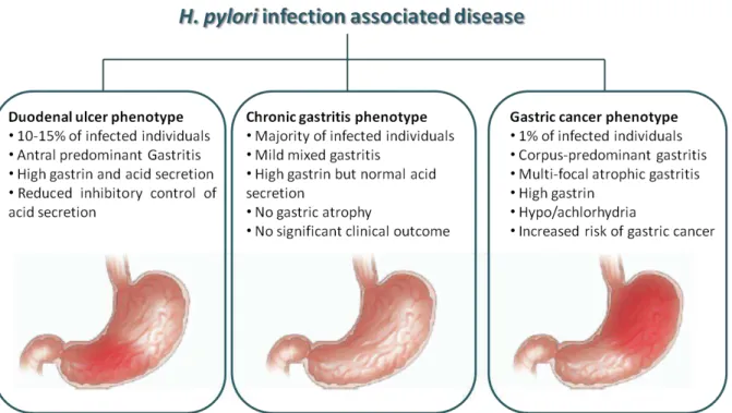
H. pylori is therefore considered the main etiologic factor in chronic gastritis, PUD (peptide ulcer
disease) and GC. The infection, despite being asymptomatic in most individuals, can evolve and cause
different gastric disorders, such as those shown in Figure 1.5.1. In most of symptomatic infected
individuals the colonization by H. pylori can cause acute gastritis which if not treated will progress to
a chronic gastritis that will affect both antrum and corpus. In 10-15% of infected patients, chronic
gastritis occurs only in gastric antrum and these cases will result in DU outcomes, characterized by
excessive acid and gastrin secretion in antral zone of the stomach. Another gastric phenotype is
characterized by gastritis in corpus zone of the stomach, which occurs in 2-5% of the infected
individuals that leads to GU with hypochlorhydria and acid reduction and may evolve into GC
(Amieva and El-Omar, 2008; Schubert and Peura, 2008; Konturek et al., 2009).
Figure 1.5.1 – Chronic H. pylori infection: Chronic H. pylori infection can lead to different gastric diseases such as chronic gastritis, duodenal ulcer and gastric cancer. Adapted from Amieva and
El-Omar (2008).
1.6. Diagnosis and Treatment
Infection with H. pylori can be diagnosed by both non-invasive and invasive methods. Invasive
tests are based on endoscopic biopsy of the gastric mucosa, collecting samples for diagnosis of
12 presence of H. pylori tests are performed such as urease test, bacterial culture and histological analysis
of the biopsy samples. In addition, H. pylori can also be detected using molecular methods, such as
PCR and real-time PCR applied to specific H. pylori genes (Suerbaum and Michetti, 2002).
Non-invasive tests include the urea breath test (UBT), serologic tests, search of stool antigens and
urine tests. The commonly used is the urea breath test (UBT) and is based on the abundant H. pylori
-derived urease activity in the stomach (detection of 13CO2 and 14CO2). For routine diagnostic the most
used test is UBT with an accuracy of >90%, while for large number of samples, such as
epidemiological studies, faecal antigen test is most appropriate, although less accurate (Suerbaum and
Michetti, 2002).
Current triple therapies for H. pylori eradication include two antibiotics in combination with a
proton pump inhibitor (PPI). Commonly used antibiotics for H. pylori eradication therapy include
macrolides (usually clarithromycin), tetracycline, amoxicillin and imidazoles (predominantly
metronidazole) as the first-line therapies. Antimicrobial resistance is the primary cause of treatment
failure, but antibiotic treatment has others emerging problems such as antibiotic side effects,
re-infections, and recurrence and has high-costs (Zhou et al., 2009).
For these reasons is essential to develop new therapies and other therapeutics approaches such as
an effective preventive and therapeutic vaccine against H. pylori, that stimulates an effective humoral
response as well as cellular response, capable of prevent and effectively eliminate the infection.
1.7. Vaccines
Thought to be therapeutic vaccination the solution for clearance and eradicate infection by H.
pylori, efforts are being made in toward to develop a vaccine that has a prophylactic use to prevent
infection and a therapeutic use to eradicate an ongoing infection (Kabir, 2007). Vaccination presents a
major benefit/cost ration and has major advantages such as financial, while avoiding re-infections and
decrease antimicrobial resistances.
Some prophylactic and therapeutic vaccine strategies using a wide variety of H. pylori antigens
have been reported (Del Giudice et al., 2009) and will be referred to in the following sections.
1.7.1. Types of vaccines
Several clinical studies have been made in order to develop an efficient vaccine capable of induce
a protective immune response for eradicate ongoing infections and in order to test the
13 Vaccines with attenuate H. pylori are extendedly study, due to have the advantage to having all
the H. pylori antigens, but these vaccines can lead to safety problems due to mutations that lead a
reversion on virulence or to an insufficient attenuation, which may ultimately induce the infection
(1.7.1.1.2). Alternatively there are inactivated whole killed vaccines that such as attenuated vaccines
have the advantage of containing all bacteria antigens, but without the danger of virulence reverting.
Studies with H. pylori inactivated whole-cells combined with LT adjuvant showed a significant rise in
specific IgA antibodies, but did not show success in eradicating the pre-existing infection (Wilson and
Crabtree, 2007). Despite inactivated whole-cell vaccines have the advantage of elicit immune response
against all antigens known and unknown of the bacteria, this can be also a disadvantage since it
contains other dangerous components of the bacterium that could induce a cross-reacted immunity
responses (Kabir, 2007; Wilson and Crabtree, 2007; Ingolotti et al., 2010).
The protection induced by several H. pylori antigens was described, including recombinant
antigens and strain-specific virulence factors of the bacterium. In addition to urease, protection with
CagA, VacA, NAP and GroEL has been demonstrated (Wilson and Crabtree, 2007), but it has been
concluded that given the genomic variability and variation of H. pylori antigens, an optimal vaccine
must be contain multiple antigens. As an example, recombinant H. pylori urease with E. coli LT (E.
coli heat-labile toxin) orally administrated to H. pylori infected volunteers, demonstrated the
immunogenicity of the vaccine through the observation of a reduction in bacterial load in vaccinated
individuals compared with controls, but the eradication of the infection was not accomplished and the
degree of gastritis remained unchanged (Kabir, 2007).
Other approach for immunization against H. pylori has been delivering antigens in live vectors
such as attenuated Salmonella typhimurium and modified poliovirus. Salmonella-based recombinant
vaccine expressing urease was orally administrated but most of the volunteers presented specific IgG
to the Salmonella vector and none of them showed any detectable mucosal or humoral immune
response against urease. Vaccines vectors using poliovirus genomes (replicons) also have been
described as a protective way to immunized mice: capsid genes were replaced with urease B subunit
and administered by systemic route and showed both prophylactic and therapeutic efficacy against H.
pylori, clearing an established infection in mice (Wilson and Crabtree, 2007).
Although these types of vaccines have proven their efficacy in inducing some type of immune
response, they do not retain the security requisites needed and the strong humoral immune response
induced alone do not provide protective immunity against subsequent contacts with the pathogen
14
1.7.1.1. DNA based vaccines
On other hand, there are the DNA vaccines, whereby naked plasmid DNA, introduced into the
host, is taken up by eukaryotic host cells and the encoded antigen is expressed (Figure 1.7.1.1.1). The
gene sequence of the antigens of interest is inserted in a bacterial plasmid backbone. The
transcriptional unit containing the nucleotide sequence should be under the control of an eukaryotic
housekeeping gene promoter and should be followed by a polyadenylation sequence (poly[A]) to
ensure the stability of the mRNA molecule and its translation. One of the most commonly used is the
cytomegalovirus (CMV) promoter. The plasmid backbone should also contain an origin of replication
for amplification the plasmid in bacteria and an antibiotic resistance gene to enable the selective
growth of transformed bacteria. Thereby, there is a commercially available plasmid approved by the Food and Drug Administration, “pVAX1” (Invitrogen) specially designed for DNA vaccines (Ingolotti et al., 2010).
Figure 1.7.1.1.1 – Plasmid DNA backbone scheme for DNA vaccines with gene sequence of interest,
polyA sequence, promoter and origin of replication,
and antibiotic resistance gene for selection of
competent bacteria (Ingolotti et al., 2010).
The advantage of DNA vaccines, compared to protein-based vaccines (figure 1.7.1.1.2), is the
possibility to triggering both humoral and cellular immune response, so that the infected host can
quickly and effectively respond to the infection and efficiently eliminate the pathogen. DNA vaccines
stimulate the antigen presentation both by MHC I to the CD8+ T cells (TCL) and by MHC II to the CD4+ T (Th) (Wilson and Crabtree, 2007).
In a simplistic view of the DNA vaccine mechanism, the DNA plasmid upon transfection is
translocated to the cell nucleus and uses the host cell machinery to transcribe and translate the genes.
The resulting proteins are recognized by the host cells as non-self and undergo cleavage resulting in
fragments that will be presented on the cell surface by MHC I. Moreover, the antigenic protein
secreted by the transfected host cells can be taken by APCs, which present it on the cell surface MHC
II. Also, apoptotic transfected cells can be engulfed by APCs which will present the antigen through
15 CD4+ Th cells and a cascade of cytokines will activate B cells which induce antibody production. The CD8+ T cells will lyse transfected cells which presenting antigens through MHC I causing the release of more antigens and CD4+ Th cells activated by DCs, which will repeat this cycle of activation. So both T and B cells migrate to the site of immunization and are re-stimulated (Kutzler and Weiner,
2008; Ingolotti et al., 2010).
To improve the efficacy of DNA vaccines could be encoded only some epitopes, instead the
whole antigen in order to avoid potential dangerous components of the bacterium such as those
sharing homologies with self-antigens and thus avoid cross-reactive immune responses against host
epitopes. These vaccines have the advantage of including only the potential epitopes selected,
considering only the PAMP highly conserved among strains. (Del Giudice et al., 2009; Zhou et al.,
2009; Moss et al., 2011).
Studies suggest that a DNA-prime and peptide-boost vaccine designed with multiple H. pylori
epitopes has a therapeutic efficacy in mice previously infected with H. pylori. Has showed that the
multi-epitope vaccine induced a broad immune response that lead to a significant reduction of H.
pylori colonization. So these studies suggest that further development of an epitope-based mucosal
vaccine against H. pylori can potentially lead to a novel approach to prevent H. pylori associated
16
Figure 1.7.1.1.2 – Resume of the Mechanisms of action of different types of vaccination:
attenuated vaccines, inactivated vaccines, subunits vaccines, toxoids vaccines and DNA vaccines;
Advantages and disadvantages (Ingolotti et al., 2010).
A variety of factors could affect the frequency of integration of plasmid DNA vaccines into host
cells, including DNA sequences within the plasmid, the expressed gene product (antigen), the
formulation, delivery system and route of administration. A proper route of administration and a
suitable delivery system could provide a better and highly immune response against infections H.
pylori. So, in this and others studies in our research group, effords were made for chose a proper route
of administration and develop an appropriated delivery system for enhance the potential of immune
17
1.7.2. Routes of vaccination
Most vaccines are administered through parenteral administration, such as intramuscular or
subcutaneous. However, mucosal vaccination presents several advantages since most pathogens infect
their host through mucosal membranes, as the respiratory tract, gastrointestinal, vaginal and urinary
tract, suggesting mucosal vaccination is essential to create a first line of immunization at mucosal
level. Thus, mucosal vaccines have the benefit of not only stimulate the mucosal immunity as well as
trigger systemic immune response, resulting in a stronger protective immunity. Other advantages
include non-invasiveness, which improve patient compliance, avoiding the use of needles, ease
self-administration, low costs and ease production and application, and low risks of needle infections
(Vajdy et al., 2004; Sijun and Yong, 2009; Azizi et al., 2010; Chadwick et al., 2010; Cadete et al.,
2012).
Recent studies indicate that oral route is the ideal mucosal route for induction predominantly
secretory IgA antibody in gastric mucosa to confer effective for protection against H. pylori
infections, preventing direct contact of the bacteria with epithelial cells (Strugnell and Wijburg, 2010;
Velin and Michetti, 2010).
Some studies have been made but with reduced success (Moss et al., 2011), since mucosal
inoculation require a successful delivery system for the vaccines efficiency, which constitutes a
challenge because mucosal membranes are constituted mainly of epithelial tissue highly vulnerable
with viscous mucus secretion that acts as a barrier against pathogens and against vaccine antigens.
These disadvantages may decrease the lifetime of antigens and difficult the determination the amount
that effectively penetrates the mucosa (Neutra and Kozlowski, 2006).
Thus, these vaccines have to be capable of effectively penetrate the mucus epithelial cells without
any damage, penetrate through the epithelium and interact with APCs or directly with MALT. For the
success of mucosal DNA-based vaccines it is essential to use suitable vaccine formulations working
both as adjuvants and delivery systems (Del Giudice et al., 2009).
It is known that vaccination through mucosal membranes requires potent adjuvants or delivery
systems to enhance the immunogenicity of the antigens as well as to protect the antigens form
degradation and to prolong the contact with the epithelium.
1.7.3. Adjuvants and Delivery systems
Adjuvants are molecules, compounds or macromulecules that do not have any specific effect of
antigen but can be co-administered with pathogen-derived antigens improving the efficacy of vaccines
with less reactive antigens and enhance a longer and stronger specific immune response, without
18 immunogenicity, through a proper transport and delivery system for presenting antigens to specific
cells of the immune system and to confer ability to prolong drug release. On the other hand, have to be
cost-effective, easily produced and present high stability (Vajdy et al., 2004).
Adjuvants can be divided into antigen-binding systems such as emulsions, micro and
nanoparticles, immunostimulatory complexes (ISCOMs) and liposomes, and immunostimulatory
adjuvants such as lipopolysaccharides (LPS), monophosphoryl lipid A, etc (Shahiwala et al., 2007).
The chemical composition of each vaccine is critical to defining the type of immune response that
will develop, stimulating a specific type of antibodies of interest secreted by B cells, and stimulating
specific cytokines secreted by T cells. This cascade of events can be controlled through the
combination of antigens associated with adjuvants (Vajdy et al., 2004; Shahiwala et al., 2007).
It is well known that some antigens in their soluble form are not recognised by APCs and
therefore do not induce a protective immune response. The therapeutic use of particulate carriers for
the development of protective immune responses is currently one of the most promising strategies to
fight infectious diseases (Shedlock and Weiner, 2000; Florindo et al., 2009).
There are a few adjuvants approved for human use. These usually raise issues of potential
toxicity, intolerable reactogenicity and side effects. A number of experiments with H. pylori vaccines
with different adjuvants, such as E.coli heat labile enterotoxin (LT), mutant LT, cholera toxin (CT),
typhoid vaccine Ty21a and aluminum hydroxide have been described, but none of these adjuvants
showed protective immunity and some were unable to eliminate ongoing infection (Kabir, 2007).
Aluminum hydroxide is the widely used adjuvant, with reduced side effects but has the disadvantage
of triggering an unbalanced response essentially the prevalence of Th2 type with humoral component,
having reduced stimulation of the cellular component. Other approach to delivering H. pylori vaccines
is the use of attenuated Salmonella typhimurium and modified poliovirus, but none of them succeeded
in eradication of H. pylori without triggering a successfully immune response in the host (Kabir, 2007;
Velin and Michetti, 2010; Moss et al., 2011).
Accordingly, therapeutic use of particulate carries for the development of protective immune
responses is currently one of the most promising strategies against infectious diseases. It is believed
that particulate systems for delivery the antigens could be the most advantageous because they confer
antigen protection against mucosal enzymes, promote the sustained release of antigens increasing the
time of contact between antigens and APCs, and possibility of increased time of retention in the
administration site through adhesion. Since particulate systems have dimensions similar to pathogens,
they are easily recognized and phagocytosed by APCs (Florindo et al., 2010).
Chitosan nanoparticles are thought to be a suitable vehicle for oral DNA vaccine delivery because
it is one of the most promising polymers for drug delivery through mucosal routes. Chitosan is
biocompatible and biodegradable, as well as mucoadhesive and permeation-enhancing. Its
mucoadhesive characteristics allow prolonged exposures of the antigens with the mucosal epithelium.
19 Studies reported no effects on cell viability, high levels of gene expression in the epithelial cells of
both stomach and intestine. Nanoparticles made of chitosan, showed stability under physiological
conditions of the intestine and also for short periods of time in the stomach. For these reasons chitosan
particles are good candidates for the development of novel gastrointestinal drug and DNA delivery
systems. Nanoencapsulation of DNA into chitosan nanoparticles showed efficient nanoparticles
binding to cellular membranes leading to transfection and effective immune system stimulation
triggering both humoral and cellular responses (Boyoglu et al., 2009). Other studies show that
intranasal administration with tetanus toxoid-loaded chitosan nanoparicles increases humoral response
as compared to the soluble antigens.
1.8. Aims
The ultimate aim of this research project, where the work of this thesis is included, is to develop a
prophylactic and therapeutic efficient multigenic DNA-based vaccine against H. pylori. In this
direction, this work aims to evaluate a pDNA- and recombinant protein-based vaccine encoding multi
epitopes from different H. pylori antigens capable to induce an effective humoral as well as cellular
immune response against H. pylori infections and capable of eradicating the pathogen. For that
propose, a plasmid DNA was produced, encoding the specific epitopes of selected antigens, and
expressed in an appropriated expression vector. To evaluate the immune response triggered after
vaccination, the recombinant proteins and plasmid DNA were purified and encapsulated in a specific
nanoparticulate delivery system and the immune response was assessed by ELISA. The results could
contribute to the positive development of an efficient prophylactic vaccine against H. pylori infections.
Positive results will lead to a new approach in the development of novel vaccines for other infectious
21
1.9.
23
2.1. Antigen production
2.1.1. Cell transformations
Escherichia coli XL1-Blue cells (Stratagene,USA) (200 μL), made competent by CaCl2
treatment, were transformed by heat shock method with two plasmids vectors pQE30 (6T) (Qiagen,
Germany) and pVAX1 (6T-plamid) (Invitrogen, UK), containing the recombinant gene for the
following fragment antigens of H. pylori CagA, UreB, HpaA, VacA, GroEL, Homb (Figure 2.1.1.1).
The positive clones were selected in agar with ampicillin for clone 6T and kanamycin for
6T-plasmid. The cells were expanded and a cell bank was produced and preserved with 20% glycerol at
-80ºC. Each production either of protein or plasmid was obtained from the same cell bank.
Figure 2.1.1.1 - H. pylori DNA and protein vaccines. Schematic representation (not to scale) of the H.
24
2.1.2. Cell culture growth
Cell culture growth of E. coli XL1-Blue cells with 6T recombinant vector, was followed by
measuring the absorbance at 610nm for 30 hour after inoculation of 1L of tryptic soy broth medium
(Table 2.1.2.1) (Biokar, France) containing 100mM ampicillin at 37°C, under agitation (250 rpm).
Table 2.1.2.1 - Tryptic soy broth composition (Sterilized at 121ºC, 20min)
For 1 L of medium:
- Tryptone ... 17.0 g
- Papaic digest of soybean meal ... 3.0 g
- Glucose ... 2.5 g
- Dipotassium phosphate ... 2.5 g
- Sodium chloride... 5.0 g
pH of the ready-to-use medium at 25°C: 7.3±0.2.
2.1.3. Optimization of production conditions of 6T recombinant protein
E. coli XL-1 Blue expressing 6T recombinant protein was cultivated in agitated flasks with
tryptic soy broth culture medium containing 100 mM of ampicillin and cultivated at 37 ºC after
induction with isopropyl b-D-1-thiogalactopyranoside (IPTG) collecting samples for 1h, 2h and 3h.
The final concentration of IPTG added in culture medium was 1 mM.
The expression of 6T protein was evaluated by SDS-PAGE and western blot using anti-His
antibody, after cell homogenization in denaturing buffer (8 M urea at pH 8.00).
2.1.4. 6T recombinant protein purification
The expression of recombinant protein was carried out as follows. E. coli XL-1 Blue transformed
using pQE30-6T plasmid was grown at 37ºC, after 3 hours of induction with IPTG, in 2 L of culture
media, overnight, until mid-log phase (Abs610nm=0.600). Cells were harvested, washed with ice-cold
10 mM phosphate buffer, pH 7.4 and frozen for further use. For each assay, 1 g (wet weight) of cell
pellet was resuspended in 10 mL lysing buffer (8M urea, 50 mM sodium phosphate, 500 mM NaCl,
1% (v/v) Triton X-100, 30 mM Imidazole, pH 8.0) and homogenized with an ultrasound probe for 3
cycles of sonication for 5 min. each, for cells lysis. Insoluble material was separated by centrifugation
at 30,000g for 30 min at 4 ºC (Beckman 64R, USA). The supernatant was purified in a HisTrap FF 1
25 The protein was loaded onto the column twice times and washed with 10 bed column volumes of
a solution containing 8M urea, 50 mM sodium phosphate, 500 mM NaCl, 1% (v/v) Triton X-100, 30
mM Imidazole, pH 8.0. Bound proteins were eluted with 8M urea, 20 mM sodium phosphate, 500 mM
NaCl, 1% (v/ v) Triton X-100, 250 mM Imidazole, pH 8.0 solution. The eluted proteins were analyzed
by 10% (w/v) SDS–PAGE under reduced conditions, by western blot using anti-His antibody following the NuPAGE Novex Bis-Tris Mini Gels Technical Guide (Invitrogen, UK) and further
quantified by the BCA Protein Assay kit (Pierce, USA) using BSA as standard.
The purified protein from the 2.5 mL nickel column was desalted on Sephadex G-25 medium
pre-filled PD-10 column (GE Healthcare) using 100 mM Heppes buffer pH 7.4.
Samples taken along the purification steps were analyzed in 10% SDS-PAGE gel followed by
Western Blot to confirm the presence of recombinant protein in different solutions filtered.
After this process, the obtained proteins are frozen with 10% of trealose and then lyophilized.
This was the protein used in in vivo assays and ELISA analysis.
2.1.5. SDS-PAGE
To confirm the presence and the structural integrity of proteins extracted and purified, the
samples were analyzed by electrophoresis in pre-casted 10% (w/v) polyacrylamide gel (NuPAGE®,
Invitrogen, UK) and run at a constant voltage of 200 V for 35 min using a Electrophoresis Power
supply EPS 3501XL system (GE Healthcare Life Science, USA) under denaturing conditions, through
comparison with BenchMarckTM pre-stained molecular markers in the range of 6-180 kDa (Invitrogen, UK) and purified proteins. After migration, proteins were visualized by SimplyBlueTM SafeStain solution (Invitrogen, UK).
2.1.6. Western Blotting
Samples were transferred from polyacrylamide gel onto the PVDF membrane using a semi-dry
transfer system (Hoefer Semiphor Amersham, GE Healthcare Life Science, USA) for 1 h at 0.8
mA/cm2 membrane. The membrane was then washed and blocked by its incubation with 10% (w/v) skimmed milk powder (Merck KGaA, Germany) dissolved in 10 mM PBS at pH 7.4 containing 0.05%
(v/v) of Tween 20 (PBST; Sigma Aldrich, Co., Germany), for 1 h under constant agitation in an orbital
shaker (100 rpm). The membrane was further incubated for 2 h at room temperature with mouse
anti-6XHis tag protein diluted in the blocking buffer (1:1700), under constant agitation. After washing, the
membrane was incubated with a goat anti-mouse IgG conjugated to phosphatase alkaline (Sigma