Lídia Alexandra Santos Cavaca
Licenciatura em Bioquímica na Faculdade de Ciências e Tecnologia da Universidade Nova de Lisboa
New Synthetic Routes for the Valorization
of Easily Accessible Bio-renewable
Resources
Dissertação para obtenção do Grau de Mestre em Química Bioorgânica
Orientador: Carlos Alberto Mateus Afonso, Professor
Catedrático, Faculdade de Farmácia da Universidade de
Lisboa
Co-orientador: Catarina Alexandra Baptista Rodrigues,
Investigadora, Faculdade de Farmácia da Universidade de
Lisboa
Júri:
Presidente: Prof. Doutora Paula Cristina de Sério Branco, FCT-UNL Arguente: Prof. Doutora Luísa Maria da Silva Pinto Ferreira, FCT-UNL
Vogal: Prof. Doutor Carlos Alberto Mateus Afonso, FF-UL
Lídia Alexandra Santos Cavaca
Licenciatura em Bioquímica na Faculdade de Ciências e Tecnologia da Universidade Nova de Lisboa
New Synthetic Routes for the Valorization
of Easily Accessible Bio-renewable
Resources
Dissertação para obtenção do Grau de Mestre em Química Bioorgânica
Orientador: Carlos Alberto Mateus Afonso, Professor
Catedrático, Faculdade de Farmácia da Universidade de
Lisboa
Co-orientador: Catarina Alexandra Baptista Rodrigues,
Investigadora, Faculdade de Farmácia da Universidade de
Lisboa
Júri:
Presidente: Prof. Doutora Paula Cristina de Sério Branco, FCT-UNL Arguente: Prof. Doutora Luísa Maria da Silva Pinto Ferreira, FCT-UNL
Vogal(ais): Prof. Doutor Carlos Alberto Mateus Afonso, FF-UL
iii
Indicação sobre os direitos de cópia
v
Agradecimentos
Aqui deixo o meu agradecimento a todos os que contribuíram para a realização desta tese e para a minha aprendizagem e evolução como cientista e química.
Ao Professor Carlos Afonso, por me ter recebido e acolhido tão bem no seu laboratório, pelo apoio durante este ano e por toda a orientação científica.
À Professora Paula Branco, por me ter aceite no Mestrado em Química Bioorgânica e assim, através das aulas, permitir alargar os meus conhecimentos e me incentivar a seguir química orgânica.
À Doutora Catarina Rodrigues, por toda a paciência e disponibilidade, e por todo o conhecimento em química orgânica que me transmitiu. Obrigada pela confiança e por todos os bons momentos passados dentro e fora do laboratório.
Ao Doutor Svilen Simeonov, pela orientação e pelos constantes trabalhos de casa e perguntas, que me ajudaram muito a aprender mais sobre química orgânica e, ao Rafael Gomes, pela ajuda na fase final do trabalho.
Ao Dr. Carlos Monteiro, Dr. Hélio Faustino, Fábio Santos e Roberto Russo por toda a ajuda no laboratório, e a todos os meus colegas de laboratório, pela amizade, profissionalismo, constante disponibilidade e partilha de conhecimentos, que me fizeram crescer muito durante este ano e também a ser mais responsável.
Ao Dr. Jaime Coelho, pela ajuda com os cálculos computacionais.
À minha família pelo apoio incondicional, por todos os conselhos e pela confiança que depositam em mim e nas minhas decisões.
Aos meus amigos, pelo apoio e por estarem ao meu lado em todos os momentos.
vii
Resumo
A oliveira (Olea europaea) é fonte natural de uma variedade de compostos fenólicos, incluindo os
secoiridóides. A oleuropeína é o secoiridóide que existe em maior quantidade nas oliveiras e noutras plantas da família Oleaceae. É encontrada nas azeitonas e pequenos ramos, mas são as folhas que
possuem um teor mais elevado deste composto. As folhas de oliveira são produtos secundários provenientes da indústria de produção de azeite, e não têm aplicações significativas, sendo por isso consideradas uma fonte acessível e bio renovável de oleuropeína.
A estrutura molecular da oleuropeína é constituída por três unidades: hidroxitirosol, glucose e monoterpeno. A glucose confere-lhe solubilidade em água, enquanto o hidroxitirosol é responsável pela maioria das suas propriedades biológicas. A unidade monoterpénica é, no entanto, mais interessante do ponto de vista químico, possuindo dois centros assimétricos e sete posições reativas disponíveis para transformações químicas.
Este trabalho consistiu na extração e isolamento de oleuropeína de folhas de oliveira, e no estudo de uma série de transformações semissintéticas ao seu esqueleto, visando a obtenção de novos derivados pela remoção das unidades de glucose e hidroxitirosol, e transformações do core da unidade
monoterpénica, focando-se num estudo mais completo e aprofundado da reação de metanólise da oleuropeína em condições acídicas. Este baseou-se num screening de ácidos próticos e resinas acídicas
e um estudo do efeito da temperatura, com análise quantitativa por HPLC-UV, permitindo estudar o mecanismo da reação. Métodos de fluxo contínuo também foram explorados, com resultados positivos.
Numa única reação, a metanólise permitiu várias transformações à estrutura da oleuropeína, com obtenção de um composto com atividade antiviral reportada e utilizado como precursor na síntese de um anti-hipertensivo, para além de compostos com potencial para serem utilizados na síntese de derivados com novas propriedades farmacêuticas ou com outros interesses adicionais.
ix
Abstract
The olive tree (Olea europaea) is a natural source of a diversity of phenolic compounds, including
secoiridoids. Oleuropein is the major secoiridoid found in olive tree and other plants from the Oleaceae
family. It is found in olive fruit and small branches, although exists in higher amounts in olive leaves. Olive leaves are by-products from olive oil industries, and have no practical applications, being considered an easily accessible bio-renewable resource of oleuropein.
The molecular structure of oleuropein is divided in three subunits: hydroxytyrosol, glucose and monoterpene moieties. The glucose unit confers solubility in water, while hydroxytyrosol is responsible for most of its biological properties. However, the monoterpene moiety is more interesting in chemical reactivity point of view, having two asymmetric centers and seven potential reactive positions, available for synthetic modifications.
This work consisted on the extraction and isolation of oleuropein from olive leaves and the study of semi-synthetic transformations in order to obtain new derivatives, by removal of the glucose and hydroxytyrosol moieties, and modifications at monoterpene core, focusing on a complete study of the methanolysis reaction of oleuropein in acidic conditions. It was based on a screening of protic acids and acidic resins and a study of the temperature effect, with quantitative analysis by HPLC-UV, complemented by a mechanistic study of the reaction. A continuous flow approach was also performed, with positive results.
In one pot, the methanolysis reaction allowed several transformations on the molecular structure of oleuropein, with formation of a compound with reported antiviral activity and used as precursor on an anti-hypertensive drug synthesis, as well as compounds with potential to be used in the synthesis of derivatives with novel pharmaceutical properties and other additional interest.
xi
Subject Index
1. Introduction ... 1
1.1. Olive tree as a natural source of bioactive compounds ... 1
1.2. Phenolic compounds in olive leaves ... 3
1.3. Biosynthesis of oleuropein ... 5
1.4. Biodegradation of oleuropein ... 7
1.5. Quantification of polyphenols in olive leaves ... 9
1.5.1 Influence of biotic and abiotic factors ... 9
1.5.2 Influence of treatment conditions ... 9
1.5.3 Extraction of polyphenols and analytical methods ... 11
1.6. Synthetic transformations of oleuropein ... 19
1.7. Biological activity of oleuropein and its derivatives ... 25
2. Objectives... 29
3. Results and Discussion ... 31
3.1. Extraction and isolation of oleuropein from olive leaves ... 31
3.2. Semi-synthetic transformations of oleuropein ... 35
3.2.1. Krapcho decarbomethoxylation of oleuropein 1……...………35
3.2.2. Hydrolysis of the glucose moiety by β-glucosidase………..36
3.2.3. Reduction of hydroxytyrosol and methyl esters……….………..38
3.2.4. Hydrolysis of hydroxytyrosol ester………..……….39
3.2.5. Methanolysis of oleuropein 1in acidic conditions………...40
Continuous flow approach of the methanolysis of oleuropein 1……….51
Acid methanolysis of the olive leaves extract……….53
4. Conclusions ... 55
5. Materials and methods ... 57
5.1. General Remarks ... 57
5.2. Extraction and isolation of oleuropein 1 ... 59
xii
Oleacein (2)……….61
Compound (3)……….62
Jaspolyside (4)……….62
Compound (5)……….63
Compound (6)……….64
Compound (7)……….65
Screening of acids and temperature effect studies in methanolysis reaction……….66
Continuous flow experiments under heterogeneous conditions……….66
Methanolysis reaction of crude mixture……….66
6. References ... 67
7. Appendices ... 75
7.1. Appendix I – NMR spectra and LC-MS-ESI of oleuropein 1 ... 75
7.2. Appendix II – NMR spectra and ESI-MS of jaspolyside 4 ... 79
7.3. Appendix III– NMR spectra and ESI-MS of compounds 5 ... 83
7.4. Appendix IV – NMR spectra and ESI-MS of compounds 6 ... 87
7.5. Appendix V – NMR spectra and ESI-MS of compounds 7 ... 91
7.6. Appendix VI – Screening of acids and acidic resins experiment ... 95
7.7. Appendix VII – Continuous flow experiments ... 101
xiii
Figure Index
Figure 1.1. Examples of polyphenols in olive leaves. Glc - Glucose; Rut - Rutinose. Adapted from
Talhaoui et al., Food Research International, 77 (2015).[7] ... 3
Figure 1.2. Molecular structure of oleuropein. Representation of hydroxytyrosol, monoterpene and glucose moieties. ... 4
Figure 1.3. Representation of the hydrogen bond network between hydroxytyrosol and glucose moieties of oleuropein. Hydrogen bonds are represented by dashed lines. Adapted from Gikas et al., J. Molecular Structure: THEOCHEM, 821 (2007).[32] ... 4
Figure 1.4. General representation of iridoids (A) and secoiridoids (B) molecular structures... 5
Figure 1.5. Monoterpene unit chemical features. Reactive positions represented by arrows; stereocenters represented by (*). ... 19
Figure 1.6. Molecular structures of peracetylated forms of oleuropein, hydroxytyrosol and aglycones. ... 22
Figure 3.1. 1H NMR spectrum of oleuropein 1, in CD 3OD. ... 33
Figure 3.2. 1H NMR spectrum of oleacein 2, in CDCl 3. ... 36
Figure 3.3. 1H NMR spectrum of compounds 3, in CDCl 3. ... 37
Figure 3.4. 1H NMR spectrum of jaspolyside 4, in D 2O. ... 39
Figure 3.5. Structures of the compounds 5-7 resulted from the methanolysis of oleuropein 1. ... 41
Figure 3.6. 1H NMR spectrum of compounds 6, in CDCl 3. ... 42
Figure 3.7. 1H NMR spectrum of compounds 5, in CDCl 3. ... 43
Figure 3.8. Methanolysis reaction profile using pTsOH.H2O, at 70 ºC. ... 45
Figure 3.9. Structures and relative free energies (Kcal/mol) of truncated diastereoisomers 5a and 5b computed at a B3LYP/6-31G(d) level of theory. ... 46
Figure 3.10. Methanolysis reaction profile for compound 5a, at different temperatures, using pTsOH.H2O; Graphic ampliation until 30 minutes. ... 47
Figure 3.11. Methanolysis reaction profile for compound 5b, at different temperatures, using pTsOH.H2O. ... 47
Figure 3.12. Methanolysis reaction profile for compounds 6, at different temperatures, using pTsOH.H2O. ... 48
Figure 3.13. 1H NMR spectrum of compounds 7, in CDCl 3. ... 49
Figure 3.14. Correlations at distance of the major diastereoisomer of 7. ... 50
Figure 3.15. NOESY spectrum of compound 7, in CDCl3. ... 50
Figure 3.16. Methanolysis reaction profile of crude mixture using pTsOH.H2O, at 70 ºC. ... 53
Figure 4.1. Molecular structures of compounds 5a, 6 and the major diastereoisomer of 7. ... 55
Figure 7.1. 13C NMR spectrum of oleuropein 1, in CD 3OD. ... 75
xiv
Figure 7.3. HMBC spectrum of oleuropein 1, in CD3OD. ... 76
Figure 7.4. COSY spectrum of oleuropein 1, in CD3OD. ... 76
Figure 7.5. LC-ESI-MS of oleuropein 1, in positive mode. ... 77
Figure 7.6. LC-ESI-MS spectrum of oleuropein 1, in negative mode. ... 77
Figure 7.7. 13C NMR spectrum of jaspolyside 4, in D 2O. ... 79
Figure 7.8. HSQC spectrum of jaspolyside 4, in D2O. ... 79
Figure 7.9. HMBC spectrum of jaspolyside 4, in D2O. ... 80
Figure 7.10. ESI-MS spectrum of jaspolyside 4, in positive mode. ... 80
Figure 7.11. ESI-MS spectrum of jaspolyside 4, in negative mode. ... 81
Figure 7.12. 13C NMR spectrum of compounds 5, in CDCl 3. ... 83
Figure 7.13. HSQC spectrum of compounds 5, in CDCl3. ... 83
Figure 7.14. HMBC spectrum of compounds 5, in CDCl3. ... 84
Figure 7.15. COSY spectrum of compounds 5, in CDCl3. ... 84
Figure 7.16. ESI-MS spectrum of compounds 5, in positive mode. ... 85
Figure 7.17. 13C NMR spectrum of compounds 5, in CDCl 3. ... 87
Figure 7.18. HSQC spectrum of compounds 6, in CDCl3. ... 87
Figure 7.19. HMBC spectrum of compounds 6, in CDCl3. ... 88
Figure 7.20. COSY spectrum of compounds 6, in CDCl3. ... 88
Figure 7.21. ESI-MS spectrum of compounds 6, in positive mode. ... 89
Figure 7.22. 13C NMR spectrum of compounds 7, in CDCl 3. ... 91
Figure 7.23. HSQC spectrum of compounds 7, in CDCl3. ... 91
Figure 7.24. HMBC spectrum of compounds 7, in CDCl3. ... 92
Figure 7.25.COSY spectrum of compounds 7, in CDCl3. ... 92
Figure 7.26. ESI-MS spectrum of compounds 7, in positive mode. ... 93
Figure 7.27. Calibration curves of oleuropein 1, compounds 5 and 6, at 230 nm. ... 95
Figure 7.28. Chromatograms of oleuropein 1, compounds 5 and 6. ... 96
Figure7.29. Examples of methanolysis reaction profiles for different acids and acidic resins, at 70 ºC. ... 97
Figure 7.30. Chromatograms of the methanolysis reaction of oleuropein 1, using pTsOH.H2O; 2 represents compound 5 and 3 represents compound 6. ... 98
Figure 7.31. Chromatograms of the methanolysis reaction with crude, using pTsOH.H2O, at 70 ºC; 2 represents compound 5 and 3 represents compound 6. ... 99
Figure 7.32. Reactor for continuous flow experiments. ... 101
Figure 7.33. Continuous flow system. ... 101
Figure 7.34. Chromatograms from continuous flow experiences, of 1 min, 5 min and 10 min of residence time. ... 102
xv
Scheme Index
Scheme 1.1. Proposed biosynthetic pathway for the formation of oleuropein. Adapted from Obied et
al.,Natural Product Reports, 25 (2008).[33] ... 5
Scheme 1.2. Molecular structures of elenolic acid glucose (A) and demethyloleuropein (B), formed by esterase action, and aglycone forms (C and D) and oleuropein aglycone (E) by β-glucosidase action. ... 7
Scheme 1.3. Proposed transformations of oleuropein aglycone by solvents as methanol, chloroform and acidic solutions. Solvents are identified by dashed boxes. Adapted from Paiva-Martins et al., J. Agric. Food Chem., 56 (2008). [84] ... 20
Scheme 1.4. Acid hydrolysis of oleuropein with sulfuric acid to produce elenolic acid. ... 21
Scheme 1.5. Molecular structures of the aglycone forms of oleuropein after treatment with β-glucosidase or hydrolysis by lanthanides. ... 21
Scheme 1.6. Hydrolysis of hydroxytyrosol ester and acetylation of glucose. Adapted from Hanessian et al. Organic Letters, 8, 2006, 4047-9 . [88] ... 22
Scheme 1.7. Synthesis of pyridine alkaloids from oleuropein; Adapted from Ranarivelo et al., Nat. Prod. Lett., 2001, 15(2), 131-7. ... 23
Scheme 1.8. Reduction of oleuropein to oleuropeinol; hydrolysis of oleuropeinol to compounds a and b; acetylation and reduction of the mixture of compounds a and b... 23
Scheme 1.9. Acid rearrangement of oleuropeinol in methanol. ... 24
Scheme 1.10. Krapcho decarbomethoxylation of oleuropein to produce oleacein. ... 24
Scheme 3.1. Two routes for Krapcho decarbomethoxylation of oleuropein 1. ... 35
Scheme 3.2. Proposed transformations of oleuropein 1 by β-glucosidase. ... 37
Scheme 3.3. Reduction of oleuropein 1 with NaBH4 to produce oleuropeinol. ... 38
Scheme 3.4. Basic hydrolysis of hydroxytyrosol ester of oleuropein 1 to produce jaspolyside 4. ... 40
Scheme 3.5. Acid methanolysis of hydroxyornoside. ... 41
Scheme 3.6. Methanolysis reaction under optimum conditions, to obtain compound 7. ... 48
xvii
Table Index
Table 1.1. Published values of oleuropein concentration according to extraction method. ... 14
Table 1.2. Health benefits of olive leaf extracts, oleuropein and its derivatives. ... 25
Table 3.1. Extraction and purification conditions of oleuropein from dried olive leaves. ... 31
Table 3.2. Conversion and best yields obtained with different acid promoters; time is shown in parenthesis. ... 44
Table 3.3. Results for compound 5a from continuous flow experiments. ... 52
Table 7.1. Data for calibration curves of oleuropein 1, compounds 5 and 6. ... 95
Table 7.2. Main results obtained from the screening of acids experiments, after 6 hours. ... 98
Table 7.3. Results from temperature effect experiments, after 2 hours. ... 99
Table 7.4.Yields from methanolysis reaction of crude mixture. ... 99
Table 7.5. Weights for flow calculation. ... 101
xix
Abbreviations
ACN – Acetonitrile
CAL-B –Candida antarctica lipase B
COSY – Correlation Spectroscopy
ESI – Electron Spray Ionization
EtOAc – Ethyl Acetate
FD – Freeze Drying
HAD – Hot Air Drying
HCl – hydrochloric acid
HMBC – Heteronuclear Multiple Bond Correlation Spectroscopy
HPLC – High Performance Liquid Chromatography
HSQC – Heteronuclear Single Quantum Coherence Spectroscopy
LC – Liquid Chromatography
MAE – Microwave Assisted Extraction
MS – Mass Spectrometry
NMR – Nuclear Magnetic Resonance
NOESY – Nuclear Overhauser Effect Spectroscopy
PPO – Polyphenol oxidase
pTsOH.H2O – para-toluene sulfonic acid monohydrated
TFA – Trifluoracetic Acid
TLC – Thin Layer Chromatography
TMS – Tetramethylsilane
1
1.
Introduction
Natural products include a large and diverse group of substances obtained from natural sources, as marine organisms, bacteria, fungi and plants.
Nature offers a great variety of chemical scaffolds with several stereocenters and functionalities that by synthetic chemistry are difficult or impossible to obtain. Therefore, unlike human synthesized molecules, natural products are able to interact with a broad range of biological macromolecules, showing their relevance for pharmaceutical research.(1) Because of this, the isolation, structural
elucidation and formulation of key active compounds from natural sources has gained increasing importance, which was only possible due to the scientific progress of the last century.
Primarily, synthetic chemists were just focused on improving natural compounds drug-like features. However, natural products can serve as powerful starting materials to generate drug substances with novel therapeutic utility, through synthetic modifications.(2)
1.1.
Olive tree as a natural source of bioactive compounds
Plants are one of the most important sources of natural bioactive products since they have developed an effective defense system against predators, involving the production of a large number of different chemical compounds.(3)
Phytochemicals are high-added value natural substances used as ingredients in foods, dietary supplements, cosmetics and pharmaceuticals. The consumers preference for products containing natural additives, generally perceived as safer and healthier, has increased the demand for phytochemical ingredients.(4)
Olea europaea, commonly known as olive tree, is native to Mediterranean countries and has provided
huge economic and dietetic benefits since ancient years. Olive trees are cultivated for production of olive oil and table olives, which are two of the most representative ingredients of the traditional Mediterranean diet.(5-7)
Epidemiological studies revealed that lower incidence of coronary heart diseases and some forms of cancer in Mediterranean countries, such as Greece, Italy and Spain, are generally associated to their diet, and is especially due to the high consumption of olive oil. It is also known that the majority of health benefits credited to olive oil are due to the presence of a variety of phenolic compounds.(8) Although
they are found in olive oil, polyphenols are also present in other parts of the tree, including leaves and small branches. In fact, leaves are richer in polyphenols than olive oil itself.(9)
Industries generate a large quantity of by-products as waste, which contain high-added value chemical compounds. A high number of by-products and residues are collected from olive tree production and olive processing industry, but most of them have no practical applications.
2
the trees. Usually, leaves are directly thrown away, burned, or grinded and scattered on the field, potentially causing environmental damage, but also with increasing cost for producers due to their removal, storage and elimination. Given this, and the fact that olive leaves are very rich in phenolic substances, they might be considered as a cheap and easily available natural source of these bioactive compounds, with advantages at many levels.(4, 7, 10-15)
Olive leaf extract gained popularity in the global nutraceutical market due to a range of claimed health attributes; they are traditionally used in the treatment of a wide spectrum of diseases. Therefore, olive leaves can be an interesting raw material for research on food and pharmaceutical sciences, and its valorization must be encouraged.(14, 16)
So far, published reviews, focus on olive leaves valorization due to high content in phenolic compounds and their biological properties, especially oleuropein, on the extraction and analytical separation methods of oleuropein and other phenolic compounds(14, 17-24), and also on the influence of
different environmental and treatment factors in phenolic compounds content.(7) From 1994 to 2010,
physical and spectral data, and information about biological and pharmacological properties of natural secoiridoids, including oleuropein, have been collected and reported in four article reviews. (25-28)
3
1.2.
Phenolic compounds in olive leaves
Polyphenols are the largest group of phytochemicals. Olive biophenols include a major group of secondary plant metabolites, distinguished by their water solubility and high molecular weights. They are of considerable physiological and morphological importance in plants, displaying high structural variety and performing several key activities. Actually, their excellent properties are a consequence of their function in the tree, namely, in the reactivity against pathogens attack and response to insect injury.(5, 14, 15)
Olive leaves contain a large variety of phenolic derivatives, consisting of simple phenols (phenylethanoids, hydroxybenzoic acids, hydroxycinnamic acids), flavonoids (flavones, flavanones, flavonols, 9-flavanols) and secoiridoids, as shown in Figure 1.1.(17)
Flavonoids and secoiridoids are the major active components in olive leaves. Secoiridoid byphenols are monomers of seco-conjugate structure, between phenolic, terpenic and glucoside moieties. They are
also characterized by an exocyclic 8,9-olefinic functionality at the terpene ring, and are present only in plants belonging to the Oleaceae family (Figure 1.1).(13, 17, 18)
Oleuropein is the major secoiridoid compound in olive leaves. Leaves are richer in oleuropein than other parts of the tree. Oleuropein was detected in olives for the first time in 1908 by Bourquelot and Vintilesco, but its chemical structure was assigned by Panizzi et al. in 1960. It is a heterosidic ester of
β-glucosylated elenolic acid and 3,4-dihydroxy-phenylethanol (hydroxytyrosol), being responsible for the bitter taste in olive oil.
4
It has a stereocenter at C-5 with (S) configuration and an exocyclic 8,9-double bond with E
configuration, which are characteristic of secoiridoids. The molecular structure of oleuropein can be divided into three subunits: hydroxytyrosol, monoterpene and glucose moieties (Figure 1.2.).(18, 19, 29-31)
Figure 1.2. Molecular structure of oleuropein. Representation of hydroxytyrosol, monoterpene and glucose moieties.
Gikas et al. found the lower energy conformation of oleuropein through semi-empirical and ab initio
calculations, which was confirmed by molecular dynamics simulations. Their results indicated that oleuropein’s minimum energy conformation has a closed geometry where the glucose moiety is in close proximity with hydroxytyrosol, and the structure of the molecule is stabilized by the formation of hydrogen bonds between the phenolic hydrogens and the hydroxyl groups of the sugar moiety. (Figure 1.3.). (32)
Figure 1.3. Representation of the hydrogen bond network between hydroxytyrosol and glucose moieties of oleuropein. Hydrogen bonds are represented by dashed lines. Adapted
5
1.3.
Biosynthesis of oleuropein
Iridoids (A) are monoterpenes characterized by a bicyclic fused ring system comprising a six-membered heterocyclic ring and a cyclopentane ring. Secoiridoids (B) are derived from iridoids by opening of the cyclopentane ring at the 7,8 bond (Figure 1.4.).(33)
Figure 1.4. General representation of iridoids (A) and secoiridoids (B) molecular structures.
Secoiridoids characterized by an exocyclic 8,9-olefinic functionality, which includes oleuropein, are termed oleosidic secoiridoids or oleosides, and are unique to oleaceous plants.
Secoiridoid conjugates that contain an esterified phenolic moiety, such as oleuropein, result from a branching in the mevalonic acid pathway in which oleoside moiety (monoterpene) synthesis and phenylpropanoid metabolism (phenolic part) merge.(33)
Mevalonic acid comes from the initial condensation of three acetyl-SCoA molecules, originating the ester β-hydroxy-β-methylglutaryl-CoA (HMG-CoA), which produces mevalonic acid, through an hydrolysis and enzymatic reduction.(34)
The proposed biosynthetic pathway for the formation of oleuropein in Oleaceae is shown in Scheme
1.1., via 7-epi-loganic acid, with ligstroside as direct precursor.(33)
Scheme 1.1. Proposed biosynthetic pathway for the formation of oleuropein. Adapted from Obied et al.,Natural Product Reports, 25 (2008).[33]
7
1.4.
Biodegradation of oleuropein
Oleuropein levels are affected by several factors, including harvesting season. It has been shown that oleuropein levels decrease during the growth period of fruit, with the lowest content of the year being measured in July, when the olive fruit is fully developed. However, in July, the total phenolic content increases, which suggests that oleuropein suffers degradation and other substances are formed.(35)
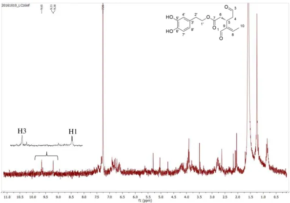
In olive fruit, elenolic acid glucoside (jaspolyside) and demethyloleuropein accumulate during ripening of olives, while the oleuropein levels decrease. Oleuropein degradation to these glucosylated derivatives involves cleavage by specific endogenous esterases. Activation of endogenous β-glucosidase during crushing or malaxation might produce the aglycone forms (C, D and E) by cleavage of glucose (Scheme 1.2).(36-38)
Ryan et al. have reported that different tissues of the olive tree have different phenolic composition,
and as such, the metabolism of each plant tissue is characteristic.(39)
However, according to Briante et al., the degradation of oleuropein is similar in olive leaves, since
many molecules isolated from leaves are thought to have originated from oleuropein, with endogenous enzymes playing an important role.(29)
Recently, De Leonardis et al. found that the enzyme polyphenol oxidase (PPO) is also involved in
oleuropein degradation in olive leaves. Fresh leaves and dried leaves showed different enzymatic activity on oleuropein. In the extracts from fresh leaves both the β-glucosidase and PPO enzymes were found active. On the other hand, the decrease of oleuropein concentration in dried leaves was mainly attributable to the PPO activity, leading exclusively to the formation of oxidation products.(40)
Scheme 1.2. Molecular structures of elenolic acid glucose (A) and demethyloleuropein (B), formed by esterase action, and aglycone forms (C and D) and oleuropein aglycone
9
1.5.
Quantification of polyphenols in olive leaves
1.5.1 Influence of biotic and abiotic factors
In olive leaves, oleuropein and total phenolic levels are affected by abiotic (non-living chemical and physical parts of the environment that affect the tree, such as soil, water, air, temperature, moisture, etc.) and biotic (other plants, fungi and bacteria, animals and human influences) factors.(7) Thus, oleuropein
and other phenolic content depend, essentially, of cultivar, harvesting season, maturity of leaves, geographical origin and moisture content.(15)
These differences have been widely reported in the literature, and different extraction and analytical methods have been employed for the determination of polyphenols content.(7, 8, 30, 39, 41-57)
Ortega-García et al. reported that oleuropein concentration in leaves was higher after exposure to
cold stress, which has been related to oleuropein antioxidant capacity, offering protection against oxidative damage induced by freezing.(44) Petridis et al. studied the effect of water deficit on olive leaf
phenolic composition, concluding that water stress induces the accumulation of phenolic compounds, especially oleuropein, suggesting their role as antioxidants.(49) Oleuropein is also involved in the olive
tree protection against salinity stress in leaves, serving as a glucose-reservoir for osmoregulation and high energy-consuming processes required for plant adaptation to salinity, increasing oleuropein content.(58)
Total phenolic levels in leaves decline as the altitude decreases, which can be related to changes in the climatic conditions. For instance, leaves from trees cultivated in windy and humid air have lower levels of phenolic compounds.(59)
An important biotic factor affecting the phenolic content is the age of the leaves. Oleuropein amount is higher in younger leaves than in mature ones, which suggests a gradual oleuropein degradation with the ageing of the leaves.(39, 52)
1.5.2 Influence of treatment conditions
The conditions in which olive leaves are treated influence their phenolic content. Currently, there are no widely accepted guidelines for the drying of olive leaves and data from literature are insufficient or contradictory. However, in order to stabilize the leaves and to avoid quality losses and undesirable degradation during storage, the dehydration of olive leaves has to be carried out immediately after harvesting.
Traditional methods of drying, as shade or sun drying, are still practiced, but this operation is not well controlled, influencing the final quality of the product. Thus, for industrial purposes, hot air drying is the most used method, allowing an accurate control of the process variables.(20)
Several studies have focused on the investigation and modelling of the drying behaviour of olive leaves. For example, Boudhrioua et al. studied the effect of blanching and infrared drying on the colour,
10
50 ˚C, 60 ˚C and 70 ˚C. Infrared drying seemed promising, inducing a considerable moisture removal from the fresh leaves (>85%), short drying time (15 min at 70 ˚C), and increased polyphenol content for leaves dried at this temperature.(60)
Erbay et al. used hot air for drying olive leaves. Optimum operating conditions by process
intensification were found to be at 53.43 ˚C, air velocity of 0.64 m/s, and process time of 288.32 min. At these conditions, moisture loss was of 69.55%, with minimum total phenolic content and antioxidant activity losses.(61)
Ahmad-Qasem et al. studied the freezing-drying of olive leaves. Hot- and freeze- air drying showed
a significant effect on the concentration of polyphenols. Hot air drying at 120 ˚C provided a higher phenolic content than freeze-drying, especially in oleuropein, which can be explained by the influence of cold in olive leaves, previously explained in section 1.5.1. The sharp decrease in oleuropein levels observed after brief thawing is probably due to the mixing of oleuropein with endogenous enzymes that are compartmentalized in fresh leaf cells.(62)
Using a laboratory convective solar dryer, Bahloul et al. investigated the effect of solar drying
conditions on the drying time and on some quality parameters of olive leaves, such as colour, total phenol content and radical scavenging activity. They tested different temperatures (40 ˚C, 50 ˚C and 60 ˚C) and air velocities (1,6 and 3,3 m3/min) to show that the total phenolics content is influenced by
drying air conditions and that it decreases as the drying time increases.(63)
Aouidid et al. used a microwave oven, drying leaves twice for two minutes at maximum power of
800 W and lyophilisation were employed prior to oleuropein determination in olive leaves.(64)
The application of an ultrasound energy system was also investigated for drying olive leaves. Kamran
et al. studied the recovery of phenolic compounds from fresh, air-dried, freeze-dried and oven-dried (60
˚C and 105 ˚C) olive leaves. Extracts of oven-dried leaves at 105 ˚C showed the highest phenol recoveries. Olive leaves oven-dried at 105 ˚C for three hours increased oleuropein recovery as compared with fresh olive leaves.(16) Cárcel et al. studied the influence of the ultrasound power application during
drying of olive leaves, at 40 ˚C. Ultrasound influenced the external resistance of the leaves, reducing it and increasing the drying rate.(65)
Erbay et al. determined and tested the most appropriate thin-layer drying model with air temperatures
of 50 ˚C, 60 ˚C and 70 ˚C, and air velocities of 0.5, 1.0 and 1.5 m/s, to understand the drying behaviour of olive leaves. It was concluded that leaves could be dried successfully by this drying method, and the drying rate depends on velocity and temperature of the air. However, temperature has more influence, and drying rate increase as temperature increases.(66)
11
Blanching processes are another factor studied. Olive leaves dried using steam blanching showed increased total phenolic content compared to the fresh ones. However, losses of oleuropein were higher using hot water blanching in leaves than using steam blanching.(6)
1.5.3 Extraction of polyphenols and analytical methods
The most important parameters that affect the recovery of phenols are the method employed and the extraction temperature, solvent and time. The methods and results reported in the literature are related to samples obtained from different species, different sources or different regions and, thus there is a large heterogeneity of results regarding phenol recovery. Besides, sample handling, processing, clean-up and storage conditions, extract stability, analytical technique sensitivity and the purity of standards used for preparation of calibration curves are commonly factors that can account for the wide variation of values in literature. Furthermore, it is difficult to develop a single method for optimal extraction of all phenolic compounds contained in olive leaves or other natural sources, because their polarities vary significantly.
Extraction is one of the most important steps in sample pre-treatment for polyphenols analysis. The most common has been solid-liquid extraction, such as Soxhlet extraction, by maceration of the olive leaves in a solvent, such as methanol, ethanol, acetone, ethyl acetate, and diethyl ether, as well as aqueous alcohol mixtures. However, these methods require large amounts of toxic solvents and long extraction times, with high costs, time waste and environmental pollution. Besides, being time consuming and having poor efficiency, some degree of heating is required, which can easily lead to thermo-sensitive ingredients losing their biological activities or degrading into other substances.(67)
In order to minimize these disadvantages, the extraction techniques have been continuously improved. In this way, techniques like microwave extraction, supercritical fluid extraction, superheated liquid extraction, liquid-liquid extraction, pressurized liquid extraction, derivatized polar extraction, fractionation by solid-phase extraction, dynamic ultrasound assisted extraction and microwave assisted extraction have been studied.(14)
High pressure extraction is a novel technique that utilizes high pressure conditions to extract active ingredients from plants. It increases the mass transfer rate by changing the concentration gradient and diffusivity, causing damage to the plant cell membrane and increasing its permeability. This increases the extracting power, albeit at the expense of process safety.(67) On the other hand, Xie et al. applied
reduced-pressure boiling extraction to extract oleuropein from olive leaves, and obtained higher yields of oleuropein.(68)
Japón-Luján et al. performed the ultrasound-assisted extraction of oleuropein and related biophenols
12
Luque designed and constructed an ultrasound-assisted Soxhlet extractor combined with supercritical CO2 extraction with improved rates and yields.(69) Supercritical fluid extraction has received increasing
interest, because supercritical fluids provide high solubility, improved mass-transfer rates and the process can be optimized by changing the temperature or pressure. Carbon dioxide (CO2) is the most
used solvent because it is safe, non-toxic, non-flammable, high selective and has moderate critical conditions (31.3 °C and 72.9 atm). However, this technique is limited to compounds of low or medium polarity.(20)
The microwave-assisted extraction technique has the advantages of low consumption of organic solvents and low costs, providing shorter extraction times compared to conventional procedures. In addition, it provides higher extraction efficiency and selectivity, which makes it a desirable method for the extraction of phenolic compounds from olive leaves.(14) Dang et al. investigated the optimal
operating conditions for enhancing the yield of oleuropein extracted from olive leaves by testing supercritical CO2 fluid extraction, microwave-assisted extraction and Soxhlet extraction techniques.
They have shown that the microwave-assisted technique can provide reduced extraction times.(70) In this
case, heating occurs in a targeted and selective manner with practical no heat being lost, since it is a closed system. The moisture when heated up inside the plant cells, evaporates and generates tremendous pressure on the cells wall, culminating with cell wall rupture, which facilitates leaching out of the phytoconstituents. Thus, this unique heating mechanism can significantly reduce the extraction time.(71)
This extraction technique has also gain much attention because of minimum degradation of target components.(7)
Recently, biopolymers and polymeric absorbents have been used to separate and purify target compounds from natural plant sources, with low cost and harmless to operator’s health. The polymer silk fibroin, used since the 1940s due to its hydrophobic and bonding characteristics, was used by Altiok
et al. and Bayçin et al., to increase the purity of oleuropein from olive leaves.(72, 73) Besides biopolymers,
macroporous resins are important as polymeric absorbent, usually applied in separation and purification of bioactive compounds from natural sources.(13)
Comparing all the different extraction techniques, microwave-assisted extraction and conventional extraction showed to be the best choice for extracting more polar compounds, such as oleuropein and its derivatives.(20)
Green approaches have also been tried with a continuous adsorption/nanofiltration hybrid process and use of imprinted polymer with in situ recovery of solvent for the isolation of oleuropein.(74)
13
matrix is always entirely immersed and the heating efficiency of the solvent under microwave should be considered, depending on solvent evaporation.(71)
A polar solvent is necessary to be able to extract oleuropein. Laboratory-scale methods use extractants such as methanol-water mixtures or hexane for the extraction of oleuropein and derivatives from olive leaves. But the increased human use of these compounds makes mandatory the development of methods based on nontoxic extractants, like water.(5)
Recently, Apostolakis et al. studied the efficiency of heated water/glycerol mixtures in extracting
polyphenols from dried olive leaves. Glycerol is a bio-solvent, with no toxicity and it is a natural constituent of foods. Authors concluded that the use of a heated aqueous glycerol solution was more efficient as compared with a hydro alcoholic solution in extracting olive leaf polyphenols, providing the same differentiated selectivity.(4)
Regarding to the analytical methods, the colorimetric Folin-Ciocalteau assay is the most used and rapid quantitative technique for the determination of total polar phenolics in olive leaves. High performance liquid chromatography (HPLC), usually in reversed-phase mode and coupled with several detectors (UV-Vis, MS, NMR, DAD, etc.), is used for the determination of individual compounds.(20)
Nuclear magnetic resonance spectroscopy (NMR) is used in the analysis of complex mixtures without previous separation of the individual components. Gas chromatography (GC) has lower detection limits and better separation, but needs sample pre-treatments, using derivatization reagents. Nevertheless, high/ultra-performance liquid chromatography (HPLC/UPLC) coupled to diode-array detection (DAD) and/or coupled to mass spectrometry (MS) is the most used to quantify and characterize phenolic compounds.(7)
Columns most used in the HPLC analytical technique are a reversed-phase C18 with 5 µm particle size. Gradient elution mode is commonly utilized, because the complexity of the phenolic profile makes it not able to be well-separated by the isocratic elution mode. UV-Vis detection system continued to be one of the most used detection systems for phenolic compounds, generally using as wavelength 280 nm. However, it should be considered that no universal absorbance maximum exists for olive leaf phenolics.(20)
The development of existing methods of separation and introduction of new techniques of high resolution are needed to give rise to the discovery of new effective compounds from phyto-pharmaceutical sources and improve its separation.(71)
14
Table 1.1 shows a compilation of values of oleuropein content regarding leaves conditions, extraction method and olive tree variety.
Table 1.1. Published values of oleuropein concentration according to extraction method.
Variety Leaves
Conditions
Extraction
Method Oleuropein Quantification
Oleuropein Concentration
(mg/g)1
Observations References
Leccino, Moraiolo
and Frantoio Fresh leaves
Solid-liquid extraction
CH3OH, 1 week, rt
Then, (CH3)2CO/H2O (1:1)
Extraction with n-pentane,
CH3Cl and ethyl acetate
Purification
Column chromatography with silica gel
CH3Cl/CH3OH (9:1) then
CH3Cl/CH3OH (4:1)
2,95 Oleuropein isolated from ethyl acetate extract (Gariboldi et
al., 1985) (75)
Moraiolo
Dried leaves (freeze-dried with
liquid N2)
Solid-liquid extraction
Different volume ratios of H2O/Ethanol
Analytical
HPLC
14,4
Best extraction solvent mixture: H2O/Ethanol (1:1)
Moraiolo variety possesses the
highest oleuropein content
(Briante et al.,
2002) (29)
N3 (Don Carlo) 8,39
N2 7,06
Coratina 6,10
Nociara 3,70
Frantoio 3,19
I-77 3,03
Leccino 1,05
Unknown Dried leaves
Microwave-assisted extraction (MAE)
100-200 W, 5-15 min Ethanol 80%-100%
Analytical
HPLC-DAD (280, 330, 340, 350 nm)
Lichrospher 100 RP18 column
(250×4 mm, 5 µm); Kromasil 5 C18 (15×4,6 mm, 5 µm)
6% acetic acid, 2 mM sodium acetate in H2O, and ACN
Flow: 0,8 mL/min Gradient
23,2
MAE – faster extraction method
Optimal extractant – Ethanol/H2O (8:2)
(Japón-Luján et al., 2006) (76)
15
Unknown Dried leaves (40 ˚C, 8 h)
Superheated liquid extraction (SHLE)
Variables: temperature, static and dynamic extraction time,
extractant flow-rate and extractant composition
Analytical
HPLC-DAD (280, 330, 340 and 350 nm)
Lichrospher 100 RP18 (250×4
mm, 5 µm)
Kromasil 5 C18 column (15×4,6
mm, 5 µm) 6% acetic acid, 2 mM sodium
acetate in H2O, and ACN
Gradient
Flow: 0,8 mL/min
23,0
Pressure: 6 bar
Solvent – EtOH: H2O (7:3), 140
˚C, 6 min
Dynamic mode, extractant: 7 min at 1 mL/min
Extraction time: 13 min
(Japón-Luján et al., 2006) (77)
Unknown Dried leaves
Ultrasound-assisted extraction
20KHz, 450 W Variables: irradiation time,
extraction flow-rate, Ethanol%, probe position,
temperature, radiation amplitude and duty cycle
Analytical
HPLC-DAD (280, 330, 340 and 350 nm)
Lichrospher 100 RP18 (250×4
mm, 5 µm)
Kromasil 5 C18 column (15×4,6
mm, 5 µm)
6% acetic acid, 2 mM sodium acetate in H2O, and ACN
Gradient Flow: 0,8 mL/min
22,6
Optimal conditions: irradiation time – 25 min; extraction flow-rate – 5 mL/min; 59 % EtOH;
Probe position – 4 cm; temperature – 40 ˚C;
Faster and more efficient than traditional methods.
(Japón-Luján et al., 2006) (5)
Unknown (December 2004)
Dried leaves (37 ˚C, 3 days)
Solid-liquid extraction
Ethanol, CH3OH, acetone and
their aqueous forms (10%/90%, v/v)
Analytical
HPLC-UV (280 nm)
C18 Lichrospher 100 analytical
column (250×4 mm, 5 µm), 30 ˚C
Flow: 1 mL/min
Acetic acid/H2O (2,5:97,5) and
ACN
Gradient, 60 min
74,5
70% EtOH: best extractant solvent
Silk fibroin as promising adsorbent for oleuropein
purification.
(Baiçın et al.,
16
Chemlali
Dried leaves
Solid-liquid extraction
CH3OH/H2O (4:1)
24 hours
Analytical
HPLC
Shim-pack VP-ODS (250×4,6 mm), 40 ˚C
0,1% phosphoric acid in H2O,
and 70% ACN in H2O
Flow: 0,5 mL/min Gradient, 40 min
43,2 - (Jemai 2008) et al.(78) ,
Dried leaves
Solid-liquid extraction
CH3OH/H2O (4:1)
24 hours
Analytical
HPLC
Shim-pack VP-ODS (250×4,6 mm), 40 ˚C
0,1% phosphoric acid in H2O,
and 70% acetonitrile in H2O
Flow: 0,5 mL/min Gradient, 40 min
24,4 - (Jemai 2009) et al.(79) ,
Coralina Dried leaves
Microwave-assisted extraction
H2O, 800 W, 10 min
Purification
LC Supelco Versa Flash
CH2Cl2/MeOH (8:2)
8,8 –21,7
Optimal extraction time: 10 min giving high oleuropein yield
(21,7×10⁻³ mg/g)
(Procopio et al.,
2009) (80)
Koroneiki Air dried leaves
Supercritical fluid extraction (SFE)
30 MPa Extraction: 50 ˚C Separation: 55 ˚C Solvent-to-feed ratio: 120 or
290
Co-solvent: 5% or 20 %
Analytical
HPLC-DAD (248 nm) SupelcoAnalytical Discovery
HS C18 (250×4,6 mm; 5 µm)
25 ˚C
H2O+1% acetic acid, and
CH3OH
Gradient Flow: 1 mL/min
51
Best solvent-to-feed ratio: 290
Co-solvent: 20% EtOH
Extraction: 50 ˚C
Separation: 55 ˚C
Pressure: 30 MPa
(Xynos et al.,
17
Unknown
(November 2010)
Fresh leaves Steam and hot water blanching; air-dried at 60 ˚C
Solid-liquid extraction
Ethanol/H2O (7:3)
Analytical
HPLC-DAD (280 nm)
Pinnacle II RP C18 (150×4,6
mm, 3 µm), 40 ˚C Kromasil 100-5 C18 (3,0/4,6
mm)
0,02 M sodium acetate pH 3,2 + acetic acid, and ACN
Flow: 1 mL/min
Hot water blanched
4,63-5,06
Oleuropein content was higher when steam or hot water
blanching were used
(Stamatopoulou s et al., 2012)
(6)
Non-blanched
0,21-2,14
Chemlali, Chetoui, Meski, Sayali and
Zarrazi
Dried leaves (MW-assisted,
800 W, 2 × 2 min)
Solid-liquid extraction
CH3OH/H2O (4:1)
24 hours
Analytical
Mid-infrared spectroscopy (4000 cm-1 and 700 cm-1)
109,2 to 171,7
Mid-infrared spectroscopy as rapid tool to predict oleuropein
content;
(Aouidi et al.,
2012) (64)
Serrana
Fresh leaves Hot air (HAD) or
freeze (FD) drying
Solid-liquid extraction
Ethanol/H2O (4:1)
24 hours
Analytical
HPLC-DAD/MS-MS (240, 280 and 330 nm) Lichrospher 100 RP18 (250×4
mm, 5 µm)
2,5% acetic acid in H2O, and
acetonitrile
Linear gradient Flow: 1 mL/min ESI – negative mode Desolvation temperature: 365
˚C
Vaporizer temperature: 400 ˚C Dry gas: N2 12 L/min
Nebulizer: 4,83 bar
Fresh HAD 70 ˚C 69
Hot air drying of leaves gives high oleuropein recovery
(Ahmad-Qasem
et al., 2013) (62)
Fresh HAD 120 ˚C 108,6
FD conventional
3
FD N2 16,44
HAD 70 ˚C+FD N2 30,1
HAD 120 ˚C+FD
18
Koroneiki Air dried leaves
Pressurized liquid extraction (PLE)
Variables: temperature, static time, extraction cycles and
Ethanol %
Pressure: 1500 psi
Analytical
HPLC-DAD (248 nm)
Supelco Analytical Discovery HS C18 (250×4,6 mm; 5 µm)
25 ˚C
H2O+1% acetic acid, and
CH3OH
Gradient
Flow: 1 mL/min
261,0
Extraction yield affected by temperature, static time and
extraction cycles
Optimal conditions for oleuropein extraction: 190 ˚C,
EtOH: H2O (56:44)
19
1.6.
Synthetic transformations of oleuropein
As reported in chapter 1.2, the molecular structure of oleuropein can be divided into three subunits: hydroxytyrosol, monoterpene and glucose moieties (Figure 1.2.). The glucose moiety is responsible for the water solubility of oleuropein, but to fully take advantage of all oleuropein chemical features it is essential to remove this problematic moiety to produce more handlable molecules.
Secoiridoids easily react with solvents and are transformed into other compounds during extraction processes. This is caused by the easy opening of the secoiridoid ring after hydrolysis of the glucose moiety. Possible transformation pathways of oleuropein aglycone in olive leaves when methanol, chloroform and acidic solvents are used, are shown in Scheme 1.3. (83, 84)
The monoterpene moiety is interesting from chemical reactivity point of view, since it has seven potential reactive positions (→) available for synthetic chemical modifications. In addition, it has two stereocenters (*), a feature that by synthetic chemistry is difficult to obtain (Figure 1.5.).
20
Scheme 1.3. Proposed transformations of oleuropein aglycone by solvents as methanol, chloroform and acidic solutions. Solvents are identified by dashed boxes.
Adapted from Paiva-Martins et al., J. Agric. Food Chem., 56 (2008). [84]
Several procedures for the cleavage of the glucose moiety were reported in the literature, using enzymes or by chemical hydrolysis.
Gariboldi et al. proposed an acid hydrolysis of oleuropein using sulfuric acid (H2SO4) in
21
Scheme 1.4. Acid hydrolysis of oleuropein with sulfuric acid to produce elenolic acid.
Guiso et al. made the hydrolysis of oleuropein using commercial endogenous β-glucosidase
from almonds, concluding that oleuropein aglycone by the enzymatic hydrolysis is actually a mixture of compounds with different structures. The relative amounts of these compounds in the mixture depend on the employed enzyme and the hydrolysis protocol. (85) Kikuchi et al. also
studied the enzymatic hydrolysis of oleuropein using this enzyme. (86) Briante et al. used
hyperthermophilic β-glucosidase for the hydrolysis of commercially available oleuropein. (87)
Procopio et al. reported the use of lanthanides, namely erbium (Er), to hydrolyse the glucose
moiety.
The molecular structures assigned revealed the presence of two elenolic acid forms, hemiacetal and dialdehyde, as well as a product which results from the rearrangement of the oleuropein aglycone, possibly through a 1,4 - addition of the enolic form of the aldehyde closer to methyl ester to the double bond (Scheme 1.5).
22
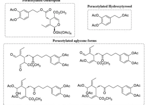
The authors also synthesized the peracetylated forms of oleuropein, hydroxytyrosol and aglycone compounds in 65 % yield using Er(OTf)3 as catalyst for the peracetylation (Scheme
1.6.). (80)
Figure 1.6. Molecular structures of peracetylated forms of oleuropein, hydroxytyrosol and aglycones.
Hanessian et al. reported the hydrolysis of hydroxytyrosol ester of oleuropein to the respective
carboxylic acid, using sodium hydroxide, followed by acetylation to afford the oleoside monomethyl ester peracetate (Scheme 1.6) (88)
Scheme 1.6. Hydrolysis of hydroxytyrosol ester and acetylation of glucose. Adapted from Hanessian et al. Organic Letters, 8, 2006, 4047-9 . [88]
Other transformations of oleuropein were also reported, to produce different scaffolds. Ranarivelo et al. reported the treatment of oleuropein with β-glucosidase in the presence of
23
Scheme 1.7. Synthesis of pyridine alkaloids from oleuropein; Adapted from Ranarivelo et al., Nat. Prod Lett., 2001, 15(2), 131-7.[89]
Bianco et al. examined the acid rearrangement of secoiridoids. The reduction of oleuropein
with sodium borohydride (NaBH4) in water produced oleuropeinol, with reduction of both
hydroxytyrosol and methyl esters moieties to the respective alcohols. The further acidic rearrangement of oleuropeinol using aqueous HCl removed the glucose moiety. By thin layer chromatography (TLC) only one compound was observed, but proton spectra in nuclear magnetic resonance (1H NMR) showed an equilibrium between the open dialdehyde form b (20 %) and the
closed acetal form a (80 %). A longer rearrangement of oleuropeinol does not give a major
compound, leading to degradation instead. The equilibrium mixture of a and b was acetylated,
giving only one main product c (98 %), found to be the monoacetyl derivative of the open form
b. This means that the equilibrium shifts towards the open form, that is further acetylated. The
mixture of a and b was also reduced with sodium borohydride, in methanol, producing only
compound d (98 %) (Scheme 1.8). (90)
Scheme 1.8. Reduction of oleuropein to oleuropeinol; hydrolysis of oleuropeinol to compounds a and b; acetylation and reduction of the mixture of compounds a and
24
The only reported modification of oleuropein in methanol was the transesterification of hydroxytyrosol ester to a methyl ester. In contrast, the acid rearrangement of oleuropeinol in methanol gave two major products. Compound e results from the formation of a cyclic hemiacetal
moiety between the primary alcohol function and the aldehyde function, which affords the cyclic acetal (40 %). Compound f probably arises from the open aldehyde form. 1H NMR showed four
signals characteristic of the methoxy groups present. This reaction was left for several hours, giving compound e as a major product, and suggesting the existence of an equilibrium between
compounds e and f, which slowly converts into the more stable e (Scheme 1.9). (90)
Scheme 1.9. Acid rearrangement of oleuropeinol in methanol.
Vougogiannopoulou et al. developed an efficient procedure for a one step semi synthesis of
oleacein from oleuropein, under Krapcho decarbomethoxylation. The possible conversion of oleuropein to oleacein involved goes through a three-step procedure: cleavage of the glucose moiety, secoiridoid ring-opening followed by the formation of the two aldehyde groups and decarbomethoxylation. Krapcho reaction was successfully applied in order to perform these three steps in one pot (Scheme 1.10). (91)
25
1.7.
Biological activity of oleuropein and its derivatives
The study of biological and pharmacological activity has revealed that secoiridoids exhibit a wide range of bioactivity.(28)
Oleuropein has potent biological and pharmacological properties. Its main pharmacological activities are anticancer, cardioprotective, neuroprotective, gastroprotective, hepato-protective, anti-diabetes, anti-obesity and radioprotective. These properties are in large part attributed to its antioxidant and anti-inflammatory effects.(92) Oleuropein derivatives were also found to have
biological and pharmacological properties.
The accumulation of free radicals on cellular membrane lipids play a major role in various pathological disorders, including atherosclerosis, cancer, aging, rheumatoid arthritis and inflammation. (93) It is known that compounds sharing an orthodiphenolic (catecholic) structure
possess antioxidant activity and the main reason of high antioxidant capacity of oleuropein is the existence of catechol structure, having the ability to scavenge free radicals that were possibly formed.(19)
The health benefits associated to olive leaf extracts, oleuropein and its derivatives are presented in Table 1.2. For this, several studies were performed in vivo and in vitro in animal
models, cell lines and human volunteers.
Table 1.2. Health benefits of olive leaf extracts, oleuropein and its derivatives.
Studies Compounds Subjects/Cell models Effects
In vivo
Animal studies
Olive leaf extract
Adult male Swiss mice; whole-body irradiated with a single dose of 48
cGy
Antioxidant and radioprotective effects; hydroxyl radical
scavenging capacity (94)
Oleuropein
Adult male Spargue-Dawley rats; oleuropein
plus ethanol (12 mg/Kg body weight; 10 days)
Antioxidant effect by scavenging of ROS, produced by
ethanol that initiate lipid peroxidation (95)
Oleuropein and Hydroxytyrosol
Adult male Wistar rats; diabetes induced with
alloxan
Anti-diabetic and antioxidant effects (79)
Hydroxytyrosol and its triacetylated
derivative
Wistar rats fed a standard laboratory diet or cholesterol-rich diet
Lipid-lowering and antioxidant effects (10)
Olive leaf extract
Wistar rats exposed to cold restraint stress; olive
leaf extract (80 mg/Kg daily) dissolved in
distilled water
Modulation of cold restraint stress oxidative changes in rat
liver by inhibition of lipid peroxidation (96)
Olive leaf extract
Sprague-Dawley rats receiving gentamycin (25 mg/Kg, 50 mg/Kg or 100 mg/Kg daily, 12 days)
Amelioration of gentamycin nephrotoxicity by inhibition of
26 Oleuropein
Female C57BL/6 mice; azoxymethane (AOM)/dextran sulphate
sodium (DSS)-induced colorectal cancer; oleuropein (50 mg/Kg or 100 mg/Kg) dissolved in
distilled water
Protection from AOM/DSS-induced colorectal cancer associated with acute colitis; suppression of the growth and multiplicity of colonic tumours
(98)
Hydroxytyrosol
Adult male Wistar rats; C6 glioma cell
implantation; subcutaneous injections
of 100 µg oleuropein, 100 µg hydroxytyrosol or both daily, for 5 days
Only hydroxytyrosol inhibited tumour growth (99)
Oleuropein
Visceral leishmaniasis model L. donovani
-infected BALB/c mice; intraperitoneal injection of oleuropein 14 times
Oleuropein and hydroxytyrosol selectivity for L. donovani;
Oleuropein gave parasite depletion of >95% in liver and
spleen after 6 weeks (100)
In vivo
Human studies Oleuropein
10 healthy female 20-30 years having skin Fitzpatrick types II and
III
Soothing effect in the treatment of UVB-induced erythema (101)
In vitro
Animal studies
Oleuropein
Normal mouse hepatocyte FL83B cells; HepG2 and FL83B cells
Decrease of the number size of lipid droplets in free fatty
acid-treated cells and reduced intracellular triglyceride
accumulation (102)
Olive leaf extract
Bacillus cereus CECT
148; B. subtilis CECT
498; Staphylococus aureus ESA 7; Escherichia coli CECT
101; Pseudomonas aeruginosa CECT 108;
Klebsiella pneumoniae
ESA 8.
Candida albicans CECT
1394; Cryptococcus neoformans ESA 3.
Antimicrobial effects observed with contribution of oleuropein
and hydroxytyrosol (48)
In vitro
Human studies
Oleuropein healthy male volunteers Whole blood of 11
Inhibition of platelet activation by scavenging of H2O2 produced
in arachidonic acid metabolism cascade that leads to platelet
aggregation (103)
Oleuropein and Hydroxytyrosol
Human breast cancer cell
27 Oleuropein and its
semi-synthetic peracetylated
derivatives
Human breast cancer cell lines (MCF-7 and
T-47D)
Anti-proliferative and antioxidant effects; the peracetylated compounds exerted higher antiproliferative
effects than oleuropein (105)
Hydroxytyrosol rich extract
MCF-7 human breast
cancer cells inhibition of MCF-7 cells A dose-dependent growth (106)
Olive leaf extract
Peripheral blood leukocytes from six
healthy volunteers
Protective effect on the peripheral blood leukocytes against adrenaline induced DNA
damage (107)
Olive leaf extract
Human endothelial cells from bovine brain, MCF-7 cells and T-24 cells (human urinary bladder
carcinoma)
Antiproliferative effect against cancer and endothelial cells (108)
Olive leaf extract (oleuropein and
apigenin-7-glucoside)
Human HL-60 cells
Apigenin-7-glucoside of the olive leaf extract was mainly responsible for the HL-60 differentiation and oleuropein
showed to exert an influence
over this differentiation (109)
Oleacein Human recombinant 5-lipooxygenase (5-LO) Inhibition of 5-LO (ICµM), acting as anti-50 = 2 inflammatory agent (91)
29
2.
Objectives
The lack of research on the synthetic transformations of oleuropein, lead to the purpose of this work to explore some of the reactions already reported, and a complete study of the methanolysis of oleuropein in acidic conditions.
The work started by the extraction and isolation of oleuropein from olive leaves, followed by the study of several reactions in order to remove the problematic glucose and hydroxytyrosol moieties, including methanolysis. For the optimization of methanolysis reaction, several conditions were screened including protic acids, acidic resins and the effect of temperature, which also gave us some mechanism insight of the reaction. A continuous flow approach of the methanolysis of oleuropein was also studied.