BJRS
RADIATION SCIENCES
07-02A (2019) 01-12ISSN: 2319-0612
Accept Submission: 2018-10-31
Semiquantitative evaluation of [
99mTc]TRODAT-1
binding potential by two methods of SPECT image
reconstruction.
Leite
1,3M. F. L, Reis
1,2,3M. A., Oliveira
3C. M., Castiglioni
3M. L. V., Bressan
1,2R. A.
1. Laboratório Interdisciplinar de Neurociências Clínicas – LiNC Universidade Federal de São Paulo – UNIFESP
Rua Pedro de Toledo, 669 3o andar, Edifício de Pesquisas II – Vl. Clementino 04039-032 São Paulo, SP, Brazil.
2. Departamento de Psiquiatria da Universidade Federal de São Paulo Universidade Federal de São Paulo – UNIFESP
Rua Borges Lagoa, 570 – Vl. Clementino 04038-000 São Paulo, SP, Brazil. 3. Departamento de Diagnóstico por Imagem – DDI
Universidade Federal de São Paulo – UNIFESP Rua Napoleão de Barros, 800 – Vl. Clementino
04024-002 São Paulo, Brazil.
Corresponding author: mariliaalvesdosreis@gmail.com
ABSTRACT
TRODAT-1 is a radiopharmaceutical derived from tropane and linked to tecnecium-99m ([99mTc] TRODAT-1)
has been used in studies of dopamine transporter (DAT) in central nervous system. Associated with the SPECT technique of acquisition, is able to detect changes in neurological disorders like Parkinson´s disease, evaluating the binding potential (BP) of DAT. The aim of this study was to evaluate the influence of the image reconstruc-tion methods, Filtered Back Projecreconstruc-tion (FBP) and iterative reconstrucreconstruc-tion (OSEM), in BP values at the striatal region in 30 healthy volunteers. Images were analyzed by visual inspection and semi-quantitative analysis. Re-gions of interest (ROI) were made over striatal areas on both sides. Nonparametric Wilcoxon statistical analysis was performed between the BP values from the FBP and OSEM methods. Our results showed that the
recon-struction methods have a statistical significant BP values difference in the total striatum (Z = -2,2787 p = 0.005), right striatum (Z = -2,602 p = 0.009) and left striatum (Z= 2,746 p = 0.006). The effect size was calculated to see if there influence in this test: the "large effect size" for all measurements was observed (total striatum r= -0.51; right striatum r= -0.48; left striatum r= -0.50). FBP is the usual method of reconstruction for brain SPECT im-ages, and our results showed influence of the OSEM method in BP. It is concluded that the method of image reconstruction adopted should be standardized to avoid incorrect evaluations of BP values using [99mTc]TRODAT-1.
1. INTRODUCTION
Dopamine (DA) is a catecholamine that participates in motor, emotional, and cognitive func-tions. Is the most important neurotransmitter of the central nervous system (CNS) [1,2]. There are different DA receptors at synaptic cleft that interact with this neurotransmitter. The DA transporter (DAT) is a molecule in the presynaptic neuron that modulates the uptake of DA [3]. Seventy per-cent of the DA removed from striatal synaptic clefts is mediated by DAT, regulating the local levels of available DA, and its concentration reflects the homeostatic tone of the dopaminergic system [4].
Alterations in dopaminergic system can lead to neurologic and neuropsychiatry disorders. Parkinson's disease (PD) is a common neurodegenerative disorder characterized by progressive de-generation of dopaminergic neurons in the substantia nigra, with loss of their nerve terminals in the basal ganglia structures, especially in the striatum [5]. The overall prevalence of PD is estimated at 0.2% but rises with increasing age, affecting about 0.5-1% of individuals aged 65-69 years and about 1-3% of individuals older than 80 years [6]. PD diagnosis is based on clinical symptoms and is characterized by resting tremor, rigidity, bradykinesia, and postural instability [5,7].
Researchers have been interested in developing sensitive diagnostic techniques for early PD diagnosis assessing DAT concentrations in the striatum. Radioligands are used to assess DAT den-sity and have been proving to be useful for investigating dopaminergic neurotransmission in hu-mans [8]. ]. These tracers are labeled with [123I] and [99mTc] for Single Photon Emission Computed Tomography (SPECT), or [11C], [18F], [15O] and [13N] for Positron Emission Tomography (PET) [8]. These radioligands allow in vivo evaluation of receptor density and affinity, measured as bind-ing potential (BP) [9].
TRODAT-1 is a radiopharmaceutical derived from tropane and linked to 99mTc ([99mTc]TRODAT-1) has a high affinity for DAT [10]. The resulting binding/labeling is demon-strated by SPECT images, and site-specific DAT density in striatum could be analyzed [11]. It is still necessary to standardize methods of acquisition, processing, reconstruction and analysis of SPECT images with [99mTc]TRODAT-1. These steps procedures may interfere in the results of the DAT density measured through BP values. There are two methods of brain SPECT reconstruction,
Filtered Back Projection (FBP) and iterative method (OSEM). FBP is easy to use, robust and repro-ducible and is a well established method for brain SPECT images, widely used in the literature. The OSEM method of reconstruction, introduced more than a decade allow better image quality for qualitative analysis, but there is no data in the literature to support its use for brain structures quan-tification.
The aim of this study was to verify if it is possible to find differences in two image recon-struction methods in the measurements of the DAT-BP in healthy individuals.
2. MATERIALS AND METHODS
Images were selected from the TRODAT-1 image database of Laboratório Interdisciplinar
de Neurociências Clínicas da Universidade Federal de São Paulo (LiNC-UNIFESP). This database
complains with 140 normal volunteers images, acquired from 2006 to 2014, and this study chose 30 healthy subjects (17 men and 13 women; 18-80 years old), without a previous diagnosis of one of the following the exclusion criteria: 1) Axis 1 psychiatric disorders (CID 10/DSM-IV), 2) severe clinical disease, 3) history of traumatic brain injury with loss of consciousness, and 4) functional illiteracy. This study was approved by the Research Ethics Committee of UNIFESP.
The images this database were acquired on a GE Hawkeye Infinia System with ultra-high resolution fan beam collimators, 4 hours after the intravenous injection of 814-888 MBq/2mL [99mTc]TRODAT-1 (INER; Taiwan, R.O.C.), in a matrix of 128 x 128 x 16, on a circular orbit of 128 steps and 3600 rotation, thirty seconds by projection with a zoom factor of 1.45. A sinogram was used to control the quality of exam, revealing the presence of possible patient movements dur-ing acquisition.
Qualitative and semi-quantitative evaluation image analysis was realized on Xeleris GE software. A single examiner applied two different reconstruction methods at the same sample: 1- Filtered Back Projection (FBP) and 2- iterative method (OSEM). Chang attenuation correction and a Butterworth filter with a constant parameter of 0.45 cut off order 10 were applied on both meth-ods. Images were reconstructed in transaxial, coronal and sagittal slices with a slice thickness of 8 mm. Semi-quantitative evaluation was performed in transaxial slices making manually regions of
interest at the striatal region (region with high DAT concentration) as region specific binding of [99mTc]TRODAT-1 with 150-155 pixel, and the occipital lobe area (region with low DAT concen-tration) with 400 pixel as region of non-specific binding of [99mTc]TRODAT-1 (Figure 1). The BP was calculated using the formula:
BP= Binding Potential
STR= Striatal Region, specific binding region of [99mTc]TRODAT-1 to DAT OCC= Occipital Lobe, non-specific binding region of [99mTc]TRODAT-1 to DAT
3. RESULTS
The sample include [99mTc]TRODAT-1 SPECT brain images from 30 healthy subjects:
56.7% men and 43.3% women, with a mean age of 47.93 + 13.23 (table 1). Kolmogorov-Smirnov test was used to calculate the distribution of data. The average striatal BP were compared between FBP and OSEM reconstructions methods. with nonparametric Wilcoxon statistical analysis and a p-value of <0.05 was considered significant. Version 22 of the SPSS program was used to make the statistical analyzes.
Table 1. Subjects Descriptive Statistics. Frequency Percent Valid Percent
Sex Male 17 56.7 56.7
Female 13 43.3 43.3
Total 30 100 100
Median 38.00
Mode 45.00
S.D. 13.23
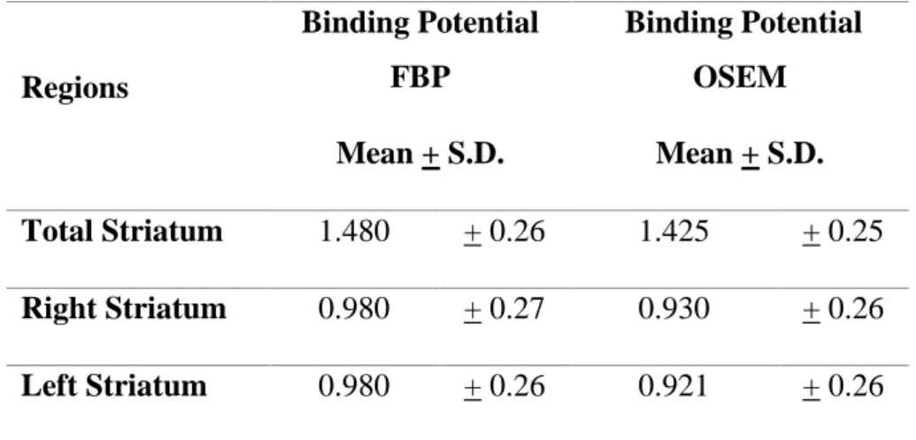
Table 2. DAT Binding Potential difference between the FBP and OSEM image reconstruction methods in striatum regions in the subjects.
Regions Binding Potential FBP Mean + S.D. Binding Potential OSEM Mean + S.D. Total Striatum 1.480 + 0.26 1.425 + 0.25 Right Striatum 0.980 + 0.27 0.930 + 0.26 Left Striatum 0.980 + 0.26 0.921 + 0.26
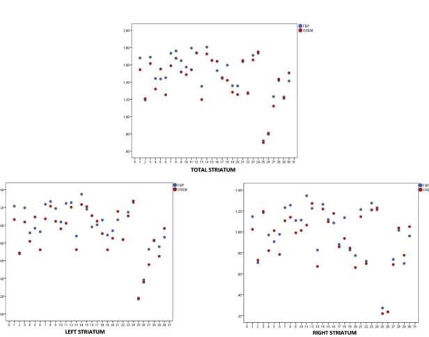
Graph 1. Scatterdot graphs DAT Binding Potential difference between the FBP and OSEM image reconstruction methods in striatum regions.
The mean values of the BP showed a difference, which can be observed in graph 1. Then, nonparametric Wilcoxon statistical analysis showed that the OSEM method have a statistical signif-icant BP difference when compared FBP method, with minor values for OSEM, in all regions: total striatum (Z= -2.2787 p= 0.005), right striatum (Z = -2.602 p = 0.009) and left striatum (Z= 2.746 p= 0.006). Subsequently, we observed the large effect size for all measurements (table 3). The ef-fect size was calculated by the formula:
r= effect size
Z= statistical Wilcoxon score N= Total number of observations
Table 3. Wilcoxon statistical analysis between the FBP and OSEM image reconstruction methods in striatum regions in the subjects.
Statistics Total Striatum OSEM – FBP Right Striatum OSEM - FBP Left Striatum OSEM – FBP Z -2.787 -2.602 -2.746 p Value 0.005 0.009 0.006 r* -0.51 -0.48 -0.50 *effect size
Figure 1. [99mTc]TRODAT-1 SPECT image of healthy volunteer, woman, 47 years old, to illustrate regions of interest drawing. Transaxial cut thickness 8mm. Manual ROI in the right and left striatal regions and elliptical ROI in the occipital area, to calculate the DAT Binding Potential. Acquisition date 03/17/2007.
Figure 2. [99mTc]TRODAT-1 SPECT image of healthy volunteer, man, 44 years old. Transaxial cut thickness 8mm. Two image reconstruction methods: A – Filtered Back Projection (FBP) and B –
Iterative Method (OSEM). Acquisition date 03/22/2012. Note that less noisy aspect of OSEM could enhance the contrast of structures, but has different quantitative data when compared with FBP.
4. DISCUSSION AND CONCLUSION
Several nuclear medicine imaging techniques were developed to diagnose and evaluate DAT and the progression of Parkinson’s disease. DAT ligands, such as [99mTc]TRODAT-1, are
estab-lished useful markers in evaluating changes in presynaptic DAT in vivo [8]. Semi-quantitative ROI techniques could be used to evaluate the specific DAT binding potential in the striatum and corre-sponds to the product of free receptor density, calculated as the ratio of striatal specific binding to nonspecific radiotracer concentration in the occipital region [8].
FBP remains the most commonly used reconstruction method for brain SPECT images, since it is a simple, fast and robust approach to image reconstruction. OSEM offers an alternative with a tendency to be less noisy (Figure 2), leading to a more accurate result in the process of re-construction [12].
Our results showed minor numeric value of OSEM when compared with FBP, with statisti-cal significance and this has to be validated in new researches. Sometimes less noisy and smoothed images, despite of a having good visual quality, were obtained with a loss of data information,
cau-sing biases in quantitative evaluation, and this phenomenon can explain in part our results. Clinical validation of this data is also necessary, in order to choose the best method of image reconstruction to evaluate a true decline of BP in several neurological diseases [13]. One limitation of our study was that the small sample of subjects tends to increase data dispersion, imposing some statistical biases in the analysis, that could be attenuated with the use of a larger sample in next works.
In Brazil [99mTc]TRODAT-1 is already been used to investigate dopaminergic neurotrans-mission in Parkinson’s disease [14-19].
We concluded that FBP should be the image reconstruction method of choice to evaluate [99mTc]TRO1- BP. No sufficient data in the literature support OSEM method in DAT-BP analysis. The differences encountered in our work could be important considering the evaluation of clinical borderline cases. We suggest that further studies should be carried out to evaluate the influence of image reconstructions methods in semi-quantitative analysis in patients and normal subjects.
5. ACKNOWLEDGMENT
This work was performed in the Nuclear Medicine of the Sao Paulo Hospital – University Hospital (HSP-HU) of the Imaging Diagnostic Department (DDI) in collaboration with the Psychia-try Department and Laboratório Interdisciplinar de Neurocências Clínicas (LiNC) of the Federal university of Sao Paulo (UNIFESP).
REFERENCES
[1]. MOZLEY LH, GUR RC, MOZLEY PD, GUR RE. Striatal dopamine transporters and cogni-tive functioning in healthy men and women. Am J Psychiatry. v. 158, p. 1492–9, 2001.
[2]. NIEOULLON A, COQUEREL A. Dopamine: a key regulator to adapt action, emotion, motiva-tion and cognimotiva-tion. Curr Opin Neurol. v. 16 (Suppl 2), p. S3–9, 2003.
[3]. REITH E, XU C, CHEN NH. Pharmacology and regulation of the neuronal dopamina trans-porter. Eur J Pharmacol. v. 324, p. 1–10, 1997.
[4]. JABER M, JONES S, GIROS B, CARON MG. The dopamine transporter: a crucial component regulating dopamine transmission. Mov Disord. v. 12, p. 629–33, 1997.
[5]. GELB DJ, OLIVER E, GILMAN S. Diagnostic criteria for Parkinson disease. Arch Neurol. v. 56(1), p. 33-9, 1999.
[6]. TANNER CM, GOLDMAN SM. Epidemiology of Parkinson’s disease. Neurol Clin. v. 14(2), p. 317-35, 1996.
[7]. HUGHES AJ, DANIEL SE, KILFORD L, LEES AJ. Accuracy of clinical diagnosis of idio-pathic Parkinson’s disease: a clinicopathological study of 100 cases. J Neurol Neurosurg Psychia-try. v. 55(3), p. 181-4, 1992.
[8]. SHIH MC, HOEXTER MQ, ANDRADE LAF, BRESSAN RA. Parkinson’s disease and dopa-mine transporter neuroimaging – a critical review. Sao Paulo Med J. v. 124(3), p.168-75, 2006. [9]. BRESSAN RA, ERLANDSSON K, JONES HM, MULLIGAN RS, ELL PJ, PILOWSKY LS. Optimizing limbic selective D2/D3 receptor occupancy by risperidone: a [123I]-epidepride SPET study. J Clin Psychopharmacol. v. 23(1), p. 5-14, 2003.
[10]. ACTON PD, KUSHNER SA, KUNG MP, MOZLEY PD, PLOSSL K, KUNG HF. Simplified reference region model for the kinetic analysis of [99mTc]TRODAT-1 binding to dopamine trans-porters in nonhuman primates using single- photon emission tomography. Eur J Nucl Med. v. 26(5), p. 518-26, 1999.
[11]. MEEGALLA SK, PLOSSL K, KUNG MP, ET AL. Synthesis and characterization of techne-tium-99m-labeled tropanes as dopamine transporter—imaging agents. J Med Chem. v.40, p. 9–17, 1997.
[12]. HARVEY A. ZIESSMAN & JAMES H. THRALL. Medicina Nuclear. Elsevier & Brasil, 2014.
[13]. KOCH W, SUESSMAIR C, TATSCH K, PÖPPERL G. Iterative reconstruction or filtered backprojection for semi-quantitative assessment of dopamine D2 receptor SPECT studies?. Euro Jour Nucl Med Mol Imag. v. 38(6), p.1095-1103, 2011.
[14]. SHIH MC, AMARO JR E, FERRAZ HB, ET AL. Neuroimaging of the Dopamine Transporter in Parkinson´s Disease – First study using [99mTc]-TRODAT-1 and SPECT in Brazil. Arq Neuro-psiquiatr. v. 64(3-A), p. 628-634, 2006.
[15]. BARSOTTINI OGP, FELÍCIO AC, AGUIAR PC, GODEIRO-JUNIOR C, SHIH MC, HO-EXTER MQ, BRESSAN RA, FERRAZ HB, ANDRADE LAF. Clinical and molecular neuroimag-ing characteristics of Brazilian patients with Parkinson’s disease and mutations in PARK2 or PARK8 genes. Arq Neuropsiquiatr. v. 67(1), p. 7-11, 2009.
[16]. SHIH MC, ANDRADE LAF, AMARO-JR E, FELÍCIO AC, FERRAZ HB, ET AL. Higher nigroestriatal dopamine neuron loss in early than late onset Parkinson´s disease? A [99mTc]-TRODAT-1 SPECT study. Mov. Disord. v. 22, p. 863-866, 2007.
[17]. FELÍCIO AC, GODEIRO-JUNIOR C, MORIYAMA TS, SHIH MC, ET. AL. Degenerative parkinsonism in patients whit psychogenic parkinsonism: A dopamine transporter imaging study. Clin. Neurol. & Neurosurg. v.112(4), p. 282-285, 2010.
[18]. FELÍCIO AC, MORIYAMA TS, GODEIRO-JUNIOR C, SHIH MC, HOEXTER MQ, BOR-GES V, SILVA SM, AMARO-JUNIOR E, ANDRADE LA, FERRAZ HB, BRESSAN RA. Higher dopamine transporter density in, Parkinson's disease patients with depression. Psychopharmaco-logy (Berl). v. 211(1), p. 27-31, 2010.
[19]. FELÍCIO AC, GODEIRO-JUNIOR C, SHIH MC, BORGES V, SILVA SM, AGUIAR PDE C, HOEXTER MQ, BARSOTTINI OG, ANDRADE LA, BRESSAN RA, FERRAZ HB. Evalua-tion of patients with Clinically Unclear Parkinsonian Syndromes submitted to brain SPECT imag-ing usimag-ing the technetium-99m labeled tracer TRODAT-1. J Neurol Sci. v. 291(1-2), p. 64-8, 2010.

![Figure 2. [ 99m Tc]TRODAT-1 SPECT image of healthy volunteer, man, 44 years old. Transaxial cut thickness 8mm](https://thumb-eu.123doks.com/thumbv2/123dok_br/18283115.881901/8.892.166.725.201.429/figure-trodat-spect-image-healthy-volunteer-transaxial-thickness.webp)
