* Corresponding author: Henry Hee-Seung Bom, Department of Nuclear Medicine, Chonnam National University Hwasun Hospital, 322 Seoyang-ro, Hwasun-Gun, Jeollanamdo, 519-809, Republic of Korea. Tel: 82-61-379-7270; Fax: 82-61-379-7281; E-mail: [email protected]
© 2014 mums.ac.ir All rights reserved.
This is an Open Access article distributed under the terms of the Creative Commons Attribution License (http://creativecommons.org/licenses/by/3.0), which permits unrestricted use, distribution, and reproduction in any medium, provided the original work is properly cited.
Effect
of
Post
‐
Reconstruction
Gaussian
Filtering
on
Image
Quality
and
Myocardial
Blood
Flow
Measurement
with
N
‐
13
Ammonia
PET
(yeon Sik Kim , Sang‐Geon Cho , Ju (an Kim , Seong Young Kwon , Byeong‐
il Lee , (ee‐Seung Bom *
Department of Nuclear Medicine, Chonnam National University (wasun (ospital, (wasun‐Gun, Jeollanamdo, South Korea
Department of Nuclear Medicine, Chonnam National University (ospital, (wasun‐Gun, Jeollanamdo, South Korea Department of Cardiology, Chonnam National University (ospital, (wasun‐Gun, Jeollanamdo, South Korea Korea Photonics Technology )nstitute, Gwangju City, South Korea
ARTICLE INFO ABSTRACT
Articletype:
Original article Objective(s):on image quality and myocardial blood flow MBF measurement by dynamic N‐ )n order to evaluate the effect of post‐reconstruction Gaussian filtering ammonia positron emission tomography PET , we compared various reconstruction and filtering methods with image characteristics.
Methods: Dynamic PET images of three patients with coronary artery disease male‐
female ratio of : ; age: , , and years were reconstructed, using filtered back projection FBP and ordered subset expectation maximization OSEM methods. OSEM reconstruction consisted of OSEM_ ), OSEM_ ), and OSEM_ ) with , , and iterations, respectively. The images, reconstructed and filtered by Gaussian filters of , , and mm, were obtained, as well as non‐filtered images. Visual analysis of image quality )Q was performed using a ‐grade scoring system by independent readers, blinded to the reconstruction and filtering methods of stress images. Then, signal‐to‐ noise ratio SNR was calculated by noise and contrast recovery CR . Stress and rest MBF and coronary flow reserve CFR were obtained for each method. )Q scores, stress and rest MBF, and CFR were compared between the methods, using Chi‐square and Kruskal‐Wallis tests.
Results: )n the visual analysis, )Q was significantly higher by mm Gaussian filtering,
compared to other sizes of filter P< . for both readers . (owever, no significant
difference of )Q was found between FBP and various numbers of iteration in OSEM
P= . and . for readers and , respectively . SNR was significantly higher in
mm Gaussian filter. There was a significant difference in stress and rest MBF between several vascular territories. (owever CFR was not significantly different according to various filtering methods.
Conclusion: Post‐reconstruction Gaussian filtering with a filter size of mm
significantly enhances the )Q of N‐ ammonia PET‐CT, without changing the results of CFR calculation.
Articlehistory:
Received: Feb Revised: Mar Accepted: Apr
Keywords:
Gaussian filtering Myocardial blood flow PET image reconstruction
►Pleasecitethispaperas:
Kim (S, Cho SG, Kim J(, Kwon SY, Lee B, Bom (S. Effect of Post‐Reconstruction Gaussian Filtering on )mage Quality and Myocardial Blood Flow Measurement with N‐ Ammonia PET. Asia Oceania J Nucl Med Biol. ; : ‐ .
Introduction
Reconstruction and filtering of acquired images in myocardial perfusion imaging MP) are of extreme importance, since they are
Filtering of N-13 ammonia PET image Kim HS et al
Asia Oceania J Nucl Med Biol. 2014; 2(2):104-110. 105
wrong decision making regarding the treatment choice or prognostic stratification.
)mage reconstruction and filtering harbor even more significant importance in cardiac positron emission tomography PET because it provides quantitative myocardial blood flow MBF, ml/g/min of tissue as well as coronary flow reserve CFR in addition to traditional relative tomographic images. Different recon‐ struction methods of PET images are directly correlated with different MBF results.
Generally, reconstruction methods are divided to analytic and iterative methods. Analytic methods include filtered back projection FBP , Fourier rebinning FORE , and three‐dimensional reprojection DRP algori‐ thms. )terative approaches consist of ordered subsets expectation maximization OSEM and maximum likelihood expectation maximization
ML‐EM .
The quality of OSEM images is superior to
that of FBP images ‐ . FBP is a back‐
projected image after filtering of the sinogram. The noise of FBP image is less than that of back‐ projected BP image. Although the FORE method is developed for reducing the calculation time, it is not used considering its distorting effects on images.
ML‐EM is an iterative image estimation method among the iterative approaches. Starting with an initial image guess, this method iteratively selects a new estimated image, based on the measured projections. )f ML‐EM method uses a total of subsets, OSEM method uses a subset of a total of subsets; therefore, OSEM method can reduce the calculation time.
OSEM method is developed for improving
the disadvantages of ML‐EM method . A
disadvantage of this method is the longer processing time of iterative approaches, compared to the processing time of FBP method. (owever, iterative approaches can potentially increase the accuracy of images, compared to analytic approaches. The OSEM algorithm has been usually used for PET studies, due to noise reduction properties in regions of low uptake . (owever, it is established that noise increase is associated with an increasing number of iterations , .
)mage filtering methods, including the popular Gaussian filtering, are used to reduce background noise and improve signal‐to‐noise ratio SNR of the image with better contrast . (owever, Gaussian filtering also generates image distortion, considering the size of filter, which makes the quantitative analysis of MBF
difficult. The purpose of this study was to compare various combinations of FBP and OSEM with Gaussian filtering in the measurement of MBF, using N‐N( dynamic PET, and to find an appropriate method.
Methods
Materials
N‐N( dynamic PET images were obtained
from three patients with coronary artery disease male‐female ratio= : ; age: , , and
years .
Imageacquisition
After CT transmission scan for an attenuation correction, MBq/kg of N‐N( was injected as a bolus < s , followed by min
of dynamic image acquisition × s, × s,
× s, × s for rest MBF measurement.
Thereafter, a ‐min gated image acquisition
was performed. After an additional min for the decay of N activity, pharmacologic stress
was given by infusing adenosine .
mg/kg/min for min. Stress imaging was performed immediately after the injection of
MBq/kg of N‐N( , which was done at peak
stress min after the start of adenosine
infusion . The stress image acquisition was done in the same method with that of the rest imaging.
Imagereconstructionandfiltering
Acquired PET data were reconstructed by FBP and OSEM methods. )n the FBP reconstruction method, the transaxial filter was set on enhanced (anning, and the cutoff was set to . mm. )n the OSEM reconstruction method, the subset was fixed to and z axial filter was set to standard; Full Width at (alf Maximum
FW(M of post filter was fixed at . mm. OSEM reconstruction method was divided into OSEM with two iterations OSEM_ ) , four
iterations OSEM_ ) , and six iterations
OSEM_ ) . Other factors of FBP and OSEM were set to default values. The reconstructed images were filtered by Gaussian filter, with sizes of ,
, and mm.
MBFmeasurement
The MBF of reconstructed images was measured by the cardiac pixel‐wise modeling
software PMOD . ; University (ospital
106 Asia Oceania J Nucl Med Biol. 2014; 2(2):104-110.
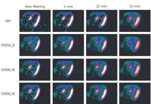
Figure1. N‐N( cardiac PET image was reconstructed by FBP and OSEM. OSEM reconstruction method was divided into OSEM_ ),
OSEM_ ), and OSEM_ ), since the number of iterations was set to , , and . The reconstructed images were filtered by , , and mm Gaussian filters. The noise increased with an increasing number of iterations. The noise was decreased and blurring was intensified as the Gaussian filter size increases
"C_PET t V_lv V_rv C_ t C_ t V_lv C_lv t V_rv C_rv t"
Where Vlv and Vrv are spill‐over fractions of
blood activity in the left ventricle Clvt and right
ventricle Crv t .
We used the polar map of segments; other factors were set to default values. The rest and stress PET images were automatically reoriented by the PMOD software. Finally, we could obtain the MBF for stress and rest and CFR of each segment, each territory: left anterior descending coronary artery LAD , right coronary artery RCA , left circumflex LCX , and total myocardium.
Imageanalysis
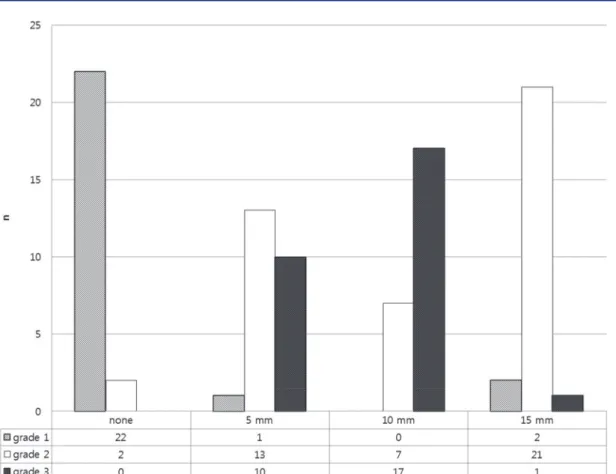
The visual image quality )Q was assessed by two experienced nuclear medicine phy‐ sicians, who were blinded to the reconstruction methods and filter size. Sixteen stress images, created by different reconstruction methods and filter sizes, were randomly rearranged and given to the two readers. Visual )Q was graded using a ‐grade scoring system, with , , and representing poor, acceptable, and good quality, respectively. Total score was the sum of counted scores from each reader.
Signal‐to‐noise ratio SNR was calculated by noise and contrast recovery CR . Noise was
measured by standard deviation σB and mean
μB in the right ventricle and CR was measured
in myocardium. SNR was calculated by the
equation of SNR=CR/ σB/μB . We also
calculated the perfusion ratios of normal to abnormal vascular territories like RCA/LAD and LCX/LAD using the polar map of CFR according to filtering methods.
Statisticalanalysis
)mage quality scores, stress and rest MBF, and CFR were compared between the methods, using Chi‐square and Kruskal‐Wallis tests.
Results
Typical images, reconstructed by FBP and OSEM, without filtering and with , , and mm Gaussian filtering are demonstrated in Figure . We could confirm that noise increase is associated with an increasing number of iterations; also, noise is decreased and blurring is intensified by increasing Gaussian filter size. We could also obtain the polar map of CFR Figure . A scale of polar map was fixed from to . .
With regard to visual analysis, score was highly prevalent in non‐filtered images. The highest score was obtained in images with mm Gaussian filtering. The total scores of non‐, mm, mm, and mm filtering were , , , and , respectively. Grades and were not counted in mm filtering and non‐filtered images, respectively. The grades , , and were
highly counted in non‐, mm, and mm
Filtering of N-13 ammonia PET image Kim HS et al
Asia Oceania J Nucl Med Biol. 2014; 2(2):104-110. 107
Figure2. N‐N( cardiac PET image was reconstructed by FBP and OSEM number of iterations: , , and without filtering and with Gaussian filters , , and mm . The polar maps of CFR were obtained by reconstructed and filtered images. The scale of polar map was fixed from to .
Figure3. The SNR was calculated in processed images. The highest SNR was calculated in mm Gaussian filter. The SNR of images with mm filtering, reconstructed by FBP and OSEM_ ), was significantly different from the SNR of non‐filtered images and images with mm filtering P< . , but no significant difference was observed in SNR of images with mm filtering P> . . The
SNR of images with mm filtering, reconstructed by OSEM_ ) and OSEM_ ), showed a significant difference from the SNR of non‐ filtered image P< . ; however, it was not significantly different from the SNR of images with mm and mm filtering P> .
other filter sizes P< . for both readers .
(owever, no significant difference of )Q was found among FBP and various numbers of
iteration in OSEM P= . and . for
readers and , respectively .
SNR was significantly higher in mm
Gaussian filter Figure . )t was not significantly
108 Asia Oceania J Nucl Med Biol. 2014; 2(2):104-110.
Figure4. The image quality was visually assessed using a ‐grade scoring system by two independent readers, blinded to the reconstruction methods. The filtering size for stress images was randomly rearranged. The image quality became significantly higher in the mm Gaussian filtering group compared to other filter sizes P< .
P= . , and mm P= . Gaussian
filtering, compared to other reconstruction methods. (owever, SNR showed a significant
difference in non‐filtered images P= . ,
compared to other reconstruction methods. The
SNR of images with mm filtering,
reconstructed by FBP and OSEM_ ), was significantly different from the SNR of non‐ filtered images and images with mm filtering
P= . ; however, it was not significantly
different from the SNR of images with mm
filtering P= . and . .
The SNR of images with mm filtering,
reconstructed by OSEM_ ) and OSEM_ ), was significantly different from the SNR of non‐
filtered images P= . ; however, it was not
significantly different from the SNR of images
with mm and mm filtering P> . .
The perfusion ratio of normal to abnormal vascular regions which was calculated using the polar map of CFR was not significantly different according to various reconstructed and filtering methods.
Discussion
)n this study, we evaluated various combinations of FBP and OSEM reconstructions
and Gaussian filtering methods in N‐N( myocardial perfusion PET/CT imaging, and found some useful combinations in various settings. )f the image quality is poor and performing a quantitative analysis is difficult by FBP without filtering, we recommend any reconstruction method FBP or other OSEMs with mm Gaussian filtering. These methods are also helpful in the quantitative measurement of MBF or CFR and making an accurate diagnosis.
Reconstruction is an essential process in cardiac PET imaging. The FBP reconstruction is fast and is considered the most widely used conventional clinical method. )n our study, the ranges of rest and stress MBF, as well as CFR, using FBP reconstruction, fit well with the previously reported ranges in various groups of patients . Thus, we used MBF and CFR values, measured by FBP reconstruction, as a good reference against which the OSEM method could be compared.
Filtering of N-13 ammonia PET image Kim HS et al
Asia Oceania J Nucl Med Biol. 2014; 2(2):104-110. 109
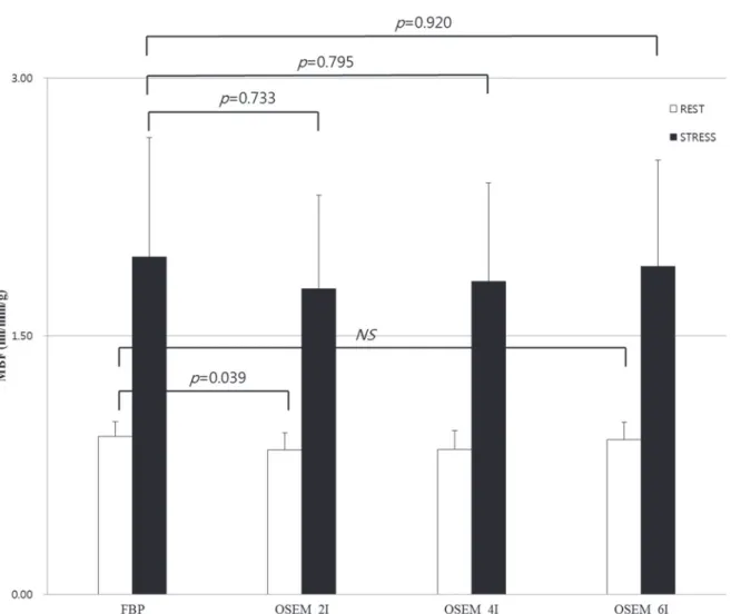
Figure5. MBFs, filtered by mm Gaussian filter, were compared in each reconstruction method. MBFs at stress were not significantly different; however, for OSEM_ ) at rest, MBF was significantly lower than that of FBP
visualization and measurement tasks are unclear. The selection of the number of iterations is a practical problem in every laboratory, using PET.
)ncreasing the number of iterations requires more time. For instance, eight OSEM iterations take times longer time than FBP. Moreover, the incremental change is insignificant beyond iterations. We compared the image quality in , , and OSEM iterations and , , and mm Gaussian filters, and found that any
reconstruction method with mm Gaussian
filter showed the best image quality Figure .
Chen et al compared rest/stress image
pairs, which were reconstructed by FBP and OSEM with subsets and , , and iterations . Regarding rest MBF, OSEM , OSEM , and OSEM iterations correlated well with FBP, but OSEM caused a significant underestimation of MBF. Considering stress MBF, both OSEM and OSEM correlated well with the standard FBP method. We tested , , and OSEM iterations
and compared them to FBP. We also found that
OSEM with mm Gaussian filter led to a
significant P= . underestimation of MBF.
OSEM and OSEM showed comparable data to that of FBP Figure . Therefore, we concluded that iterations are most appropriate for daily practice.
Chen et al also tested the size of Gaussian
filters in the measurement of MBF and CFR .
110 Asia Oceania J Nucl Med Biol. 2014; 2(2):104-110.
for the measurement of MBF and CFR. The increasing size of filters resulted in the gradual decrease of MBF, but no change in CFR.
Although N‐N( cardiac PET/CT imaging is
a robust tool to evaluate the pathophysiology of myocardial perfusion, preference of image characteristics and measurement of MBF are not
consistent in different laboratories.
Reconstruction algorithms do not result in consistent values in each vendor. Optimal protocol may vary from vendor to vendor and from laboratory to laboratory. Therefore, each laboratory should test each algorithm and select an optimal balance between image quality and accurate MBF measurement.
A limitation of this study was the small number of subjects, which did not cover a wide range of coronary artery diseases and healthy cohorts. The assessment of image quality could be affected by the narrow range of disease severity in this study group. (owever, we compared the results of FBP with various combinations of OSEM iterations and Gaussian filter size; therefore, the statistical power was significant with this number of comparisons.
)n conclusion, we recommend finding an optimal reconstruction algorithm in each laboratory in case of poor image quality. )n our setting, we found that four OSEM iterations with mm Gaussian filtering showed the best image quality without any change of quantitative
values of MBF in N‐N( myocardial perfusion
PET/CT imaging.
Conclusion
We analyzed images reconstructed by FBP and OSEM methods and filtered by Gaussian
filters in the N‐N( PET and obtained
images. The image quality of different images
was assessed by signal to noise ratio and visual analysis. We also compared the CFR of these images. Post‐reconstruction Gaussian filtering
with a filter size of mm significantly
enhanced the image quality of [ N]N( PET without changing the results of CFR calculation.
Acknowledgements
This study was supported by a grant
A from the Korea National Enterprise
for Clinical Trials.
References
. Sheep LA, Vardi Y. Maximum likelihood reconstruction for emission tomography. )EEE Trans Med )mag. ; : – .
. Dahlbom M, Eriksson L, Rosenqvist G, Bohm C. A Study of the Possibility of Using Multi‐Slice PET Systems for ‐D )maging. )EEE Trans Nuc Sci.
; : ‐ .
. (olte S, Schmidlin P, Linden A, Rosenqvist G, Eriksson L. )terative image reconstruction for positron emission tomography: a study of convergence and quantitation problems. )EEE Trans Nuc Sci. ; : ‐ .
. (udson (M, Larkin RS. Accelerated image reconstruction using ordered subsets of projection data. )EEE Trans Med )mag. ; :
– .
. Razifar P, Sandstrom M, Schnieder (, Langstrom B, Maripuu E, Bengtsson E, et al. Noise correlation in PET, CT, SPECT and PET/CT data evaluated using autocorrelation function: a phantom study on data, reconstructed suing FBP and OSEM. BMC Med )maging. ; : .
. Boellaard R, van Lingen A, Lammertsma AA. Experimental and clinical evaluation of iterative reconstruction OSEM in dynamic PET: quantitative characteristics and effects on kinetic modeling. J Nucl Med. ; : ‐ .
. Wang C, Snyder W, Bilbro G, Santago P. Performance evaluation of filtered backprojection reconstruction and iterative reconstruction methods for PET images. Comput Biol Med. ;
: ‐ .
. Khorsand A, Graf S, Pirich C, Muzi O, Kletter K, Dudczak R, et al. Assessment of myocardial perfusion by dynamic N‐ ammonia PET imaging: comparison of tracer kinetic models. J Nucl Cardiol. ; : – .
. Chen GP, Branch KR, Alessio AM, Pham P, Tabibiazar R, Kinahan P, et al. Effect of reconstruction algorithm on myocardial blood flow measurement with N‐ammonia PET. J Nucl Med. ; : ‐ .
. Krzywinski M, Sossi V, Ruth TJ. Comparision of FORE, OSEM and SAGE algorithms to DRP in D PET using phantom and human subject data. )EEE Trans Nuc Sci. ; : ‐ .
. Kitamura K, )ida (, Shidahara M, Miura S, Kanno ). Noise reduction in PET attenuation correction using non‐linear Gaussian filters. )EEE Trans Nuc Sci. ; : ‐ .