Full paper published online:May 31, 2007 ISSN 1678-9199.
CHARACTERIZATION OF AN ANTIBACTERIAL PEPTIDE FROM INDIAN COBRA (Naja naja) VENOM
SACHIDANANDA M. K. (1), MURARI S. K. (2), CHANNE GOWDA D. (3)
(1) Department of Chemistry, University of Mysore, Manasagangotri, Mysore, India; (2) Department of Biochemistry, University of Mysore, Manasagangotri, Mysore, India.
ABSTRACT: Due to the development of antibiotic resistance in microorganisms, antimicrobial peptides from natural sources have attracted attention in recent times. Several antimicrobial peptides have been isolated from a wide range of animal sources, particularly snake venoms. Naja naja venom showed antibacterial as well as direct and indirect hemolytic activities, and an antibacterial peptide was purified through gel permeation and ion exchange chromatography. Its molecular mass was 2491Da, which was determined using Matrix Assisted Laser Desorption/Ionization-Time-of-Flight (MALDI-TOF) mass spectrometry and the amino acids sequence of the N-terminus was DEQSTHGAYVWKL. The purified peptide showed potent antibacterial activity against Gram-negative and Gram-positive bacterial strains like
Escherichia coli, Pseudomonas aeruginosa and Vibrio cholerae, and Staphylococcus aureus, Enterococcus faecalis, Streptococcus pneumoniae, Streptococcus pyogenes, Bacillus subtilis, respectively. The most potent activity was towards Gram-negative bacteria. Activity was retained at concentrations as low as 100µg/ml. Minimum inhibitory concentrations (MIC; in μg) of Naja Antibacterial Peptide (NAP) and known antibiotics against Gram-positive and Gram-negative bacteria were determined using microdilution susceptibility test in sterile 96-well microdilution plates. However, the peptide did not show direct or indirect hemolytic activity.
KEY WORDS: Indian cobra, Naja naja, snake venom antibacterial peptide, hemolytic activity.
CONFLICTS OF INTEREST: There is no conflict.
FINANCIAL SOURCE: University grants commission, New Delhi.
CORRESPONDENCE TO:
INTRODUCTION
Antimicrobial Peptides (AMPs) are an extremely diverse group of small proteins that
have in common a native antimicrobial activity. The existence of AMPs has been
known for several decades, but only recently has their function been recognized as
essential to the animal immune response. They participate primarily in the innate
immune system and are used as a first line of immune defense by many organisms,
including plants, insects, bacteria and vertebrates (9). These molecules are peptides
with a high level of basic and hydrophobic amino acids. They present a broad
antimicrobial spectrum against bacteria, fungi or parasites,by acting through insertion
into the cell membrane or bind to receptors. Therefore, these molecules are
promising for the development of antibiotics especially for the treatment against
multiresistant microorganisms(7, 19). For this reason, significant commercial interest
and effort have been made to develop cationic peptides as potential antimicrobial
therapeutics. Recent studies have clarified that antimicrobial peptides are an
important component of the innate defense of all species (5).
More than 700 AMPs have already been identified in living species like bacteria,
fungi, amphibians, insects, reptiles and mammals (10). In the last years, several
AMPs have been found in different venoms from different animals and are
traditionally linked to defense mechanisms (6). Snake venoms are rich sources of
pharmacologically active polypeptides and proteins. Peptides from snake venoms are
of biological interest as a potential source of active compounds. These molecules
could act as (or be used as a prototype for)(i) therapeutic agents; (ii) research tools
for the diagnosis of several diseases; (iii) basic research about physiological and
pathologicalprocesses (22).
Snake venom has been established to show bactericidal activity (8) and the action of
its proteins on E. coli has been extensively studied (23). The action of snake venom AMPs on clinical bacterial strains has also been reported (24). Venoms from 30
different snake species were tested using disk diffusion test for antibacterial activity
(21). In the present study, the isolation and characterization of a peptide from N. naja
MATERIALS AND METHODS
Materials
Indian cobra (Naja naja) venom was purchased from Irulla Snake-Catchers Association, Chennai, Tamil Nadu, India. CM-Sephadex C-25, Sephadex G-25,
Sephadex G-75, and Bovine Serum Albumin (BSA) were purchased from Sigma
Chemical Company, St. Louis, MO, USA. The bacterial strains E. coli American Type Culture Collection (ATCC) 25922, E. coli ATCC 476, S. aureus National Cell Type Cell Culture (NCTCC) 6570, S. aureus NCTCC 6571, P. aeruginosa ATCC 26519, P. aeruginosa NCTCC 10662, V. cholerae Wild strain, E. faecalis MTCC 459, E. faecalis
MTCC 439, S. pneumoniae MTCC 497, S. pneumoniae MTCC 7978, S. pyogenes
NCTCC 7465, S. pyogenes NCTCC 7978, B. subtilis NCTCC 1040, and B. subtilis
NCTCC 8236 were purchased from American Type Cell Culture Institute, USA, and
Institute of Microbial Technology, Chandighar, India. Human blood samples were
collected from healthy volunteers from the Department of Biochemistry, University of
Mysore, Mysore, India. All other chemicals used were of analytical grade. All the
solvents were redistilled before use.
Protein Estimation
Protein concentration was determined according to the method of Lowery et al. (16) using BSA as standard.
Sephadex G-75 Column Chromatography
Lyophilized N. naja venom (300mg in 1ml) was dissolved in 10mM potassium phosphate buffer, pH 7.4, and centrifuged at 5000g for 5min. The supernatant was applied to a column (0.8cm X 120cm) of Sephadex G-75 equilibrated and eluted with
the same buffer at 20oC. The fractions from the column were eluted at a flow rate of
20ml/h and 2ml fractions were collected. Protein elution was monitored at 280nm
using a Shimadzu spectrophotometer (1601A). Alternate tubes were assayed for
antibacterial activity. Fractions presenting activity were individually pooled, desalted,
lyophilized and stored at -4oC.
CM-Sephadex C-25 Column Chromatography
Peak III (90mg in 3ml equilibrating buffer) from Sephadex G-75 column was loaded
potassium phosphate buffer, pH 7.4. The column was eluted by a stepwise gradient
of potassium phosphate buffer (10mM–150mM) and NaCl (0.01M–1M). Fractions
were eluted at 20oC at a flow rate of 25ml/h and 2.5ml fractions were collected.
Protein elution was monitored at 280nm using a Shimadzu spectrophotometer.
Fractions presenting antibacterial activity were pooled, desalted, lyophilized and
stored at -4oC.
Sephadex G-25 Column Chromatography
Peak IV (36mg in 1ml of equilibrating buffer) from CM-Sephadex C-25 column was
loaded onto a Sephadex G-25 column (0.75cm X 60cm) equilibrated with 10mM
potassium phosphate buffer, pH 7.4. The fractions from the column were eluted at a
flow rate of 20ml/h and 2ml fractions were collected. Protein elution was monitored at
280nm using a Shimadzu spectrophotometer (1601A). Alternate tubes were assayed
for antibacterial activity. Fractions presenting activity were individually pooled,
desalted, lyophilized and stored at -4oC.
High-Performance Liquid Chromatography
Purified NAP was subjected to Reverse Phase-High-Performance Liquid
Chromatography (RP-HPLC) on Vydac-C18 (5µm, 0.21cm X 25cm) column. The
column was first equilibrated with Solvent A (0.1% Trifluoroacetic Acid – TFA) until
the base line monitored at 220nm was stable. The peptide was then injected into the
column. Elution was carried out with a linear gradient (0%–100%) of Solvent B (70%
Acetonitrile in 0.1% TFA). Peptide elution was monitored at 220nm.
Mass Spectrometry
The molecular mass of NAP was determined using MALDI-TOF mass spectrometry
(Voyager Spec # 1 MC) in positive ionization mode. α-Cyano-4-hydroxycinnamic acid
was used as MALDI matrix.
N-Terminal Sequencing
The terminal sequencing of NAP was carried out in a fully automated Shimadzu
protein sequencer (PSQ-1) system that employs Edman’s degradation reaction for
Hemolytic Activity
Direct and indirect hemolytic activities were assayed as described by Boman and
Kalletta (4). The substrate for direct hemolytic activity was prepared by suspending
1ml of packed fresh human red blood cells (RBC) in 9ml of phosphate buffered saline
(PBS). For indirect hemolytic activity, 1ml of fresh hen’s egg yolk was included in the
above-mentioned suspension. One ml of the suspension was incubated with different
concentrations of NAP for 45min at 37°C. The reaction was stopped by adding 9ml of
ice-cold PBS. The suspensions were centrifuged at 2000g for 20min and the released hemoglobin was read at 530nm. The activity was expressed as percentage
(%) of hemolysis.
Evaluation of Antibacterial Activity
Antibacterial assay was described by Linzxing Zhong et al. (15). The microorganisms were grown in Muller-Hinton broth. After incubation for 16–18h at 37oC, the bacteria
were harvested by centrifugation (2000g for 10min), washed twice with 10mM sodium phosphate buffer, pH 6.0, and finally resuspended in 10ml buffer. Its density
was determined by measuring the absorbance at A600. The peptide MIC was
determined using a microdilution susceptibility test in sterile 96-well microdilution
plates. Microorganisms (1X10⁴ to 2X10⁴ CFU/50μl) were pipetted into the wells,
which consists of 20-300μg of peptide/ml. Assays for peptide were performed in
duplicate with each bacterium. After 24h incubation at the optimal growing
temperature, the optical density (OD) at 600nm was read on an absorption microtiter
plate reader (Biotek Instruments INC.). The percentage of inhibition was calculated
as {1-(a\b) X 100}, where a = OD 600nm of the bacteria with peptide, and b = OD
600nm of the control well containing only buffer, bacteria and media. The MIC
evaluation was defined as 100% inhibition. Control was run by replacing the peptide
solution with buffer solution. Respective antibiotics were used as standard drugs
replacing peptide solution.
Statistics
For all experiments, results were expressed as the mean ± SEM of at least 3
RESULTS
Purification of NAP
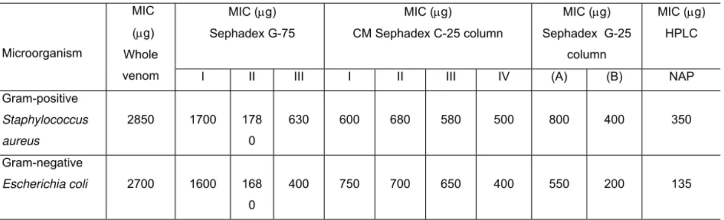
Naja naja venom (300mg) subjected to gel permeation chromatography on a Sephadex G-75 column resolved into three protein peaks. The antibacterial activity of
the whole venom and its fraction is shown in Table 1. When all the peaks were
screened for antibacterial activity, only peak III showed activity (Fig. 1). The peak III
from the Sephadex G-75 fraction was pooled, concentrated and desalted using
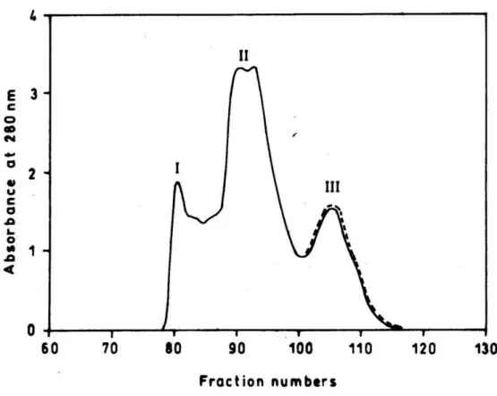
Sephadex G-10 column. The pooled peak III fraction was further resolved into four
peaks on CM-Sephadex C-25 column by applying NaCl gradient (Fig. 2). Only peak
IV exhibited potent antibacterial activity and contributed to 73.98% of the total activity
loaded, and 4.5% of the protein loaded on CM-Sephadex C-25 column was
recovered in this antibacterial fraction. The antibacterial activity of this fraction was
increased by 6-fold compared to the whole venom activity. Further, peak IV from the
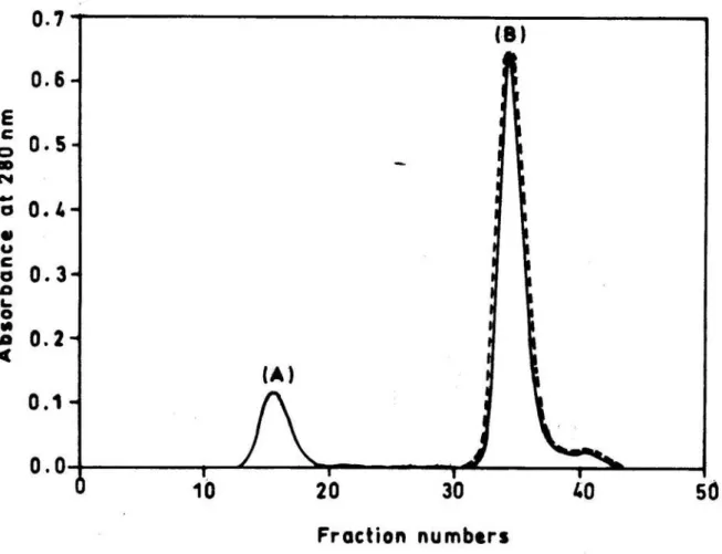
CM-Sephadex C-25 fraction was pooled, concentrated and loaded onto Sephadex
G-25 column. On fractionation, the peptide components resolved into two peaks, which
were designated as peak A and peak B (Fig. 3). Peak B showed significant
antibacterial activity and increased by 10-fold, compared to the whole venom activity.
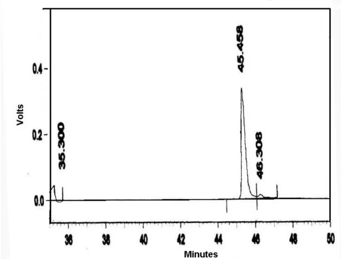
The homogeneity of the antibacterial peptide was examined by RP-HPLC using C18
column. The elution buffer contained 0.1% TFA and was eluted with acetonitrile
gradient. NAP eluted as single symmetrical sharp peak with a retention time of
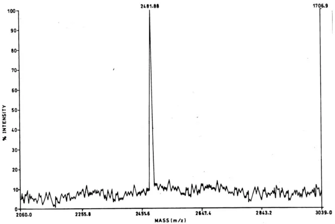
43.6min (Fig. 4). The molecular mass determined by MALDI-TOF mass spectrometry
was 2491Da (Fig. 5). The NAP N-terminal sequence analysis indicated the following
13 amino acids sequence: DEQSTHGAYVWKL. All these data clearly established
that the antibacterial peptide was purified to homogeneity.
Antibacterial Activity of NAP
The isolated peptide was tested against four species of Gram-positive and three
species of Gram-negative bacterial strains. NAP was also tested against a wide
variety of Gram-positive and Gram-negative bacteria collected from
immunosuppressed patients following disease or chemotherapy or from patients
suffering from other chronic diseases.
NAP exhibited antibacterial activity against a variety of bacterial clinical isolates; this
was evaluated by determining the MIC values. The difference in the activity of NAP
among Gram-negative bacteria in general, and not all species were susceptible
(Table 2). Staphylococcus aureus, Staphylococcus faecalies, Streptococcus pneumoniae, and Streptococcus pyogenes showed MIC values > 300μg/ml. On the other hand, E. coli, Pseudomonas aeruginosa, Vibrio cholerae, and Klebsilla pneumoniae were more susceptible to NAP. Hemolytic activity of whole venom exhibited direct and indirect lytic activity on human RBC causing 83% hemolysis.
Similarly, NAP was incubated with washed RBC at 37°C for 10min and increased in
dose-dependent manner. NAP did not show direct or indirect hemolytic activity even
at higher concentrations.
Table 1. Minimum inhibitory concentration (MIC) of peak fractions. MIC (μg)
Sephadex G-75
MIC (μg)
CM Sephadex C-25 column
MIC (μg)
Sephadex G-25 column MIC (μg) HPLC Microorganism MIC (μg) Whole
venom I II III I II III IV (A) (B) NAP
Gram-positive
Staphylococcus
aureus
2850 1700 178 0
630 600 680 580 500 800 400 350
Gram-negative
Escherichia coli 2700 1600 168 0
400 750 700 650 400 550 200 135
NAP: Naja antibacterial peptide
Table 2. Minimum inhibitory concentration (MIC) of Naja antibacterial peptide (NAP) and known antibiotics against Gram-positive and Gram-negative bacteria.
Microorganism Gram-negative bacteria
Strains Peptide MIC (μg)
Antibiotic MIC (μg)
ATCC 25922 130 80
Escherichia coli ATCC 476 120 Ciprofloxacin 100
ATCC 25619 100 80
Pseudomonas
aeruginosa NCTCC 10662 120 Gentamicin 80
Vibrio cholerae Wild strain 140 Tetracycline 50
Gram-positive bacteria
NCTCC 6570 >200 120
Staphylococcus
aureus NCTCC 6571 >250
Cloxacillin
150
MTCC 459 >220 80
Strptococcus faecalis MTCC 439 >250
Erythromycin
100
MTCC 497 >220 120
Streptococcus
pneumoniae MTCC 7978 >250
Ceftriaxone
120
NCTCC 7465 >300 100
Streptococcus
pyogenes NCTCC 7978 >280
Amoxicillin
120
NCTCC 1040 >200 60
Bacillus subtilis NCTCC 8236 >220
Penicillin G
80
ATCC: American Type Culture Collection NCTCC: National Cell Type Cell Culture
Figure 1. Sephadex G-75 column chromatography of N. naja venom.
Sephadex G-75 column (0.8cm X 120cm) was eluted with 10mM potassium
phosphate buffer, pH 7.4, at a flow rate of 20ml/h and 2ml fractions were collected.
The protein elution profile (─) was monitored at 280nm in a spectrophotometer.
Fractions showing antibacterial activity (dotted line) were pooled for further
Figure 2. CM-Sephadex C-25 column chromatography of peak III from G-75 column
chromatography.
The column (1.2cm X 40cm) was pre-equilibrated with 10mM potassium phosphate
buffer, pH 7.4, and eluted by a stepwise gradient of potassium phosphate buffer
(10mM–150mM) and NaCl (0.01M–1M) as indicated on the top of the Figure; 2.5ml
fractions were collected and the proteins elution profile (─) was monitored at 280nm in
a spectrophotometer. Fractions showing antibacterial activity (dotted line) were pooled
Figure 3. Sephadex G-25 column chromatography of peak IV from CM-Sephadex
C-25 column chromatography.
Sephadex G-25 column (0.75cm X 60cm) was eluted with 10mM potassium
phosphate buffer, pH 7.4, at a flow rate of 20ml/h and 2ml fractions were collected.
The protein elution profile (─) was monitored at 280nm in a spectrophotometer.
Figure 4. RP-HPLC elution profile of Naja antibacterial peptide (NAP).
NAP was run on a Vydac C18 RP-HPLC column. Solvent A was 0.1% Trifluoroacetic
acid (TFA) and Solvent B was 70% acetonitrile in 0.1% TFA. A gradient of 0%–100%
Solvent B was run from 0 to 60min, as indicated in the Figure. The elution profile
Figure 5. Matrix Assisted Laser Desorption/Ionization-Time of Flight (MALDI-TOF)
mass spectrum of Naja antibacterial peptide (NAP). MALDI-TOF mass spectrometry of NAP was carried out in positive ionization mode using α-cyano-4-hydroxycinnamic
acid as MALDI matrix.
DISCUSSION
Several AMPs have been found in different venoms from different animals and are
traditionally linked to defense mechanisms (6). Antimicrobial peptides have an ability
to kill or neutralize Gram-negative and Gram-positive bacteria, fungi (including
yeasts), parasites (including planaria and nematodes), cancer cells, and enveloped
viruses like HIV and herpes simplex virus (11). In this study, the low-molecular-weight
peptide from snake venom was referred as NAP.For the first time, purification and
N-terminal sequencing of a new potent antibacterial peptide from Naja naja snake venom was reported. The peptide was isolated from N. naja whole venom by subjecting it to gel permeation and ion exchange chromatography, which resulted in
10-fold purification. RP-HPLC, MALDI-TOF and N-terminal sequencing analysis
confirmed NAP homogeneity. Based on Basic Local Alignment Search Tool (BLAST)
of the primary N-terminal sequence of the antibacterial peptide, NAP is different from
It is generally accepted that different venoms have several thousands of proteins with
different properties. However, in recent years, more than 700 cationic peptides have
been isolated from mammals, amphibians, reptiles, arthropods, plants, bacteria and
viruses (3,7,19). Some of the first reports about antibacterial activity in snake venoms
were in 1948 and in 1968, involving Elapidae and Viperidae venoms (8, 25). Venoms
from snakes of the Viperidae family present antimicrobial activity against Sarcina
species, while in the Elapidae family, a lytic factor or cytotoxin composed of a basic
low-molecular-weight protein was found in Naja species, and Hemachatus haemachatus was shown to have antibacterial activity. They were able to disrupt S. aureus and E. coli phospholipid membranes, respectively (23, 25). In the present study, NAP displayed higher inhibitory activity against Gram-negative bacteria like E. coli, P. aeruginosa, V. cholerae, than against Gram-positive bacteria like S. aureus,
S. faecalis, S. pneumoniae, S. pyogenes and B. subtilis.
The peptide dissolves divalent cations that are essential for outer membrane and
consequently distorts the outer membrane bilayer (20). This allows access to the
cytoplasmic membrane where peptides channel formation has been proposed to
occur (13). It is an intermediate step in the uptake of peptides into the cytoplasm,
where it inhibits an essential function by binding to polyanionic DNA (18,26). It has
been argued that antimicrobial peptides provides organisms with molecules that are
rapidly synthesized because of small size, less costly to synthesize than antibodies
or specific phagocytic cells, and can be stored if necessary as processed
biologically-active components which are rapidly available for host defense (2). The ability of the
antibacterial peptide to lyse cells is the result of a complex interrelationship of factors
involving conformation, charge, hydrophobic and amphipathicity. The cationic
residues in an antimicrobial peptide are considered to be important in the initial
binding to the negatively charged phospholipids in the cell membranes of
microorganisms (27). It has been suggested that increasing the hydrophobic moment
of an antimicrobial peptide has a relatively modest effect on the ability to make the
microorganisms negatively-charged cell membrane permeable but not a marked
effect on the more zwitterionic phospholipid membrane of the erythrocyte (26). In
general, although it is accepted that a polypeptide chain of at least 20 amino acids is
necessary to span the membrane lipid bilayer to effect the formation of ion channels
(14), shorter cationic alpha-helical amphipathic peptides of 8–12 residues can also
In a conventional assay on human RBC, whole venom caused significant hemolysis,
but NAP did not cause a significant hemolysis. It was known that appearance of
numerous contiguous apolar residues in a helix is necessary for a significant
hemolysis to occur (17). Like other antimicrobial peptides, the polar residues in the
NAP might be well interspersed among the hydrophobic residues, interrupting the
contiguity of hydrophobicity, which gives the potential to form an amphipathic helix.
For this reason, NAP probably exhibits little hemolytic activity like many other
antimicrobial activities. The widespread use of antibiotics has caused numerous
antibiotic resistant strains to develop, resulting in the continuous need for new
antibiotics. Studies directed towards understanding the relationships between the
secondary structure and biological activities of these natural peptides have indicated
that amphipatic alpha-helical conformation plays an important role in their biological
activities (12). In conclusion, the present study on N. naja venom suggests the presence of a potent antibacterial peptide. Further studies on this peptide would be
interesting, the clinical isolates were investigated can cause infections at sites where
treatment with this type of peptide would probably help in the development as a
potential therapeutic agent applicable for clinical isolates.
ACKNOWLEDGMENTS
We acknowledge Department of Biochemistry and Biotechnology, University of
Mysore, Manasagangothri, Mysore, India, for providing instrumentation facility.
REFERENCES
1 AGAWA Y., LEE S., ONO S., AOYOGI H., OHNO M., TANNIGUCHI T., ANZAI K.,
KIRINO Y. Interaction with phospholipid bilayers, ion channel formation, and
antimicrobial activity of basic amphipathic alpha-helical model peptides of various
chain lengths. J. Biol. Chem., 1991, 266, 20218-22.
2 BOMAM HG. Antibacterial peptides: key components needed in immunity. Cell,
1991, 65, 205-7.
3 BOMAN HG. Antibacterial peptides: basic facts and emerging concepts. J. Int. Med., 2003, 254, 197-215.
4 BOMAN HG., KALLETTA U. Chromatography of rattlesnake venom: a separation
6 GALLO RL., MURKAMI M., OHTAJE T., ZAIOU M. Biology and clinical relevance
of naturally occurring antimicrobial peptides. J. Allergy Clin. Immunol., 2002,110, 823-31.
7 GANZ T. Defensins: antimicrobial peptides of innate immunity. Nat. Rev. Immunol., 2003, 3, 710-20.
8 GLASER HRS. Bactericidal activity of Crotalus venom in vitro. Copeia, 1948, 4, 245-7.
9 GOMES VM., CARVALHO AO., CUNHA M., KELLER MN., BLOCH JR C.,
DEOLINDO P., ALVES EW. Purification and characterization of a novel peptide with
antifungal activity from Bothrops jararaca venom. Toxicon, 2005, 45, 817-27.
10 HANCOCK RE., CHAPPLE D. Peptide antibiotics. Antimicrob. Agents
Chemother., 1999, 43, 1317-23.
11 HANCOCK RE., SCOOT MG. The role of antimicrobial peptides in animal
defences. Proc. Natl. Acad. Sci. USA, 2000, 97, 8856 -62.
12 KAISER ET., KEZDY FJ. Peptides with affinity for membranes. Ann. Rev. Biophys. Biophys. Chem., 1987, 16, 561-81.
13 LEAR JD., WASSERMAN ZR., GRADO WF. Synthetic amphiphilic peptide
models for protein ion channels. Science, 1988, 240, 1177-81.
14 LEHRER RI., GANZ T., SALTED ME. Defesins: endogenous antibiotic peptides of
animal cells. Cell, 1991, 64, 229-30.
15 LINZXIUG ZHONG., REBECCA PUTNAM J., CURTIS JOHNSON JR., GURAJ
RAO A. Design and synthesis of amphipathic antimicrobial peptides. Int. J. Pept. Protein Res., 1995, 45, 337-47.
16 LOWRY OH., ROSEBROUGH NJ., FARR AL., RANOALL RJ. Protein
measurement with the Folin-Phenol reagent. J. Biol. Chem., 1951, 193, 265-75.
17 NICOLAS P., MOR A. Peptides as weapons against microorganisms in the
chemical defense system of vertebrates. Ann. Rev. Microbiol., 1995, 49, 277-304. 18 PARK CB., KIM HS., KIM SC. Mechanism of action of the antimicrobial peptide
buforin II: buforin II kills microorganisms by penetrating the cell membrane and
inhibiting cellular functions. Biochem. Biophys. Res. Commun., 1998, 224, 253-7. 19 PEREZ-TRALLERO E., IGLESIAS L. Tetracyclines, sulfonamides and
20 PETERSON AA., FESIK SW., MCGROARTY EJ. Decreased binding of antibiotics
to lipopolysaccharides from polymyxin-resistant strains of Escherichia coli and
Salmonella typhimurium. Antimicrob. Agents Chemother., 1987, 31, 230-7.
21 STILES BG., SEXTON FW., WEINSTEIN SA. Antibacterial effects of different
snake venoms: purification and characterization of antibacterial proteins from
Pseudechis australis (Australian king brown or mulga snake) venom. Toxicon, 1991, 29, 1129-41.
22 STOCKER KF. Medical Use of Snake Venom Proteins. Boca Raton: CRC Press, 1990.
23 STOCKER JF., TRAYNOR JR. The action of various venoms on Escherichia coli.
J. Appl. Bacteriol., 1986, 61, 383-8.
24 TALAN DA., CITRON DM., OVERTURN GD., SINGER B., FROMAN P.,
GOLDSTEIN EJ. Antibacterial activity of crotalid venoms against oral snake flora and
other clinical bacteria. J. Infect. Dis., 1991, 164, 195-8.
25 WHITE J. Bites and stings from venomous animals: a global overview. Ther. Drug. Monit., 2000, 22, 65-8.
26 WIEPRECHT T., DATHER M., KRAUSE E., BEYERMANN M., MALOY WL.,
MACDONALD DL., BIENERT M. Modulation of membrane activity of amphipathic,
antibacterial peptides by slight modifications of the hydrophobic moment. FEBS Lett., 1997, 417, 135-40.
27 YEAMAN MR., YOUNT NY. Mechanisms of antimicrobial peptide action and