R E S E A R C H
Open Access
In vivo
evaluation of homeostatic effects of
Echis
carinatus
snake venom in Iran
Hossein Salmanizadeh
1, Mahdi Babaie
1and Hossein Zolfagharian
2*Abstract
Background:The venom of the family Viperidae, including the saw-scaled viper, is rich in serine proteinases and metalloproteinases, which affect the nervous system, complementary system, blood coagulation, platelet
aggregation and blood pressure. One of the most prominent effects of the snake venom ofEchis carinatus(Ec) is its coagulation activity, used for killing prey.
Materials and methods:Subfractions F1A and F1B were isolated from Ec crude venom by a combination of gel chromatography (Sephadex G-75) and ion exchange chromatography on a DEAE-Sepharose (DE-52). These subfractions were then intravenously (IV) injected into NIH male mice. Blood samples were taken before and after the administration of these subfractions. Times for prothrombin, partial thromboplastin and fibrinogen were recorded.
Results and conclusions:Comparison of the prothrombin time before and after F1A and F1B administrations showed that time for blood coagulation after injection is shorter than that of normal blood coagulation and also reduced coagulation time after Ec crude venom injection. This difference in coagulation time shows the intense coagulation activity of these subfractions that significantly increase the coagulation cascade rate and Causes to quick blood coagulation. The LD50 of the Ec crude venom was also determined to be 11.1μg/mouse. Different crude venom doses were prepared with physiological serum and injected into four mice. Comparison of the prothrombin times after injection of subfractions F1A and F1B showed that the rate of mouse blood coagulation increases considerably. Comparing the partial thromboplastin times after injecting these subfractions with this normal test time showed that the activity rate of intrinsic blood coagulation system rose sharply in mice. Finally, by comparing the fibrinogen time after subfraction injections and normal test time, we can infer intense activation of coagulation cascade and fibrin production.
Keywords:Snake venom, Procoagulant activity, Blood coagulation,Echis carinatus, Chromatography, LD50
Background
Snakebite affects around 2.5 million humans annually, accounting for more than 100,000 deaths. Coagulopathy is a significant cause of both morbidity and mortality in these patients, either directly, or indirectly. Clinical concerns in snakebite include types of coagulopathy (procoagulant, anticoagulant, fibrinogen clotting, fibrinolytic, platelet-active, thrombotic, hemorrhagic), diagnosis and treatment [1-3].
Where available, antivenom is the most effective treat-ment against snakebites, while standard treattreat-ments for
other forms of coagulopathy, such as factor replacement therapy and heparin, are either ineffective or dangerous, except in specific situations. Interference with aspects of the human hemostatic system is a common theme that encompasses all four families of venomous snakes includ-ingEchis carinatus(from Viperidae family) in Iran [4].
Among the many potential effects of envenoming by snakes in humans, only a few broad categories are of major clinical significance including paralysis and mild stroke; systemic myolysis; coagulopathy and hemorrhage; renal damage and failure; cardiotoxicity; and local tissue injury at the bite site [4,5].
Any single snake species may possess toxins that act in one or more of these categories, though rarely all six. In the past, it was wrongly assumed that a single
* Correspondence:[email protected]
2Department of Venomous Animals and Antivenom Production, Razi Vaccine
and Serum Research Institute, Karaj, Iran
Full list of author information is available at the end of the article
ophidian species would generally cause either local or systemic effects and that vipers caused local and/or hemorrhagic effects, while elapids caused purely sys-temic, non-hemorrhagic effects [6-9].
In this research, the effects of Echis carinatus crude venom and its fractions on mice were analyzed. Moreover, the results of coagulation tests on its venom were recorded.
Methods
Materials
Sephadex G-75, DEAE-Sepharose, was purchased from Pharmacia (Sweden). CaCl2, thromboplastin-D and APTT-XL kits were purchased from Fisher Diagnostics (Germany). The other reagents and chemicals of analyt-ical grade were purchased from Fluka and Merck.
Venom and animals
Sixty milligrams of Echis carinatus venom was obtained from the Venomous Animal Unit of Razi Vaccine and Serum Research Institute, Iran. Fifty-two NIH mice were supplied from the Laboratory Animal Breeding Unit of Razi Vaccine and Serum Research Institute, Iran. Mouse blood samples were centrifuged for ten minutes at 3,000 rpm. The plasma obtained was used for the pro-thrombin time (PT), partial thromboplastin time (PTT) and fibrinogen time (FT) tests.
Prothrombin time (PT) test
The thromboplastin-D vial was brought to the laboratory and equilibrated to room temperature. Two hundred micro-liters of the solution was poured into a hemolysis tube, and incubated for three minutes at 37°C. One hundred microli-ters of mouse plasma was poured into a hemolysis tube containing 200 μL of thromboplastin-D solution at the same moment that the chronometer was switched on. A glass pipe containing 200μL of thromboplastin-D solution and 100μL of the plasma was incubated for five minutes at 37°C. The process of plasma clotting was observed and the time recorded [10,11].
Partial thromboplastin time (APTT) test
The APTT-XL solution vial was equilibrated to room temperature in the laboratory. One hundred microliters of this solution was then poured into a hemolysis pipe; 100μL of mouse plasma was added to it and the mixture was incubated for three minutes at 37°C. Subsequently, 100μL of CaCl2was added and the chronometer was sim-ultaneously switched on. The preparation was shaken for 19 s inbain-marie(water at 37°C). The process of plasma clotting was observed and the time recorded [11].
Fibrinogen time (FT) test
Half an hour before conducting the test, the reagents were taken out of the refrigerator in order to equilibrate their temperature to room temperature. First step, dilu-tion: 0.1 mL of plasma was diluted with 0.9 mL of the test kit diluting buffer to achieve the plasma dilution 1:10. Incubation: 0.2 mL of the diluted plasma was poured into a hemolysis pipe for incubation for two minutes at 37°C. Clot formation: the thrombin containing reagent should have the lab temperature (25°C) through-out the test time. It should never be incubated at 37°C. Two minutes after incubation, 0.1 mL of the thrombin containing reagent was added to the diluted plasma and the chronometer simultaneously switched on. As soon as the first signs of clotting were observed, time was recorded and the fibrinogen level determined [12,13].
Measurement of the Ec crude venom coagulation activity For measuring the IranainEchis carinatus crude venom coagulation activity, 10 mg of the crude venom was ini-tially used to prepare different concentrations (1, 0.1, 0.01 mg/mL). These concentrations were them exposed in the PT test.
Isolation and purification of coagulation factors
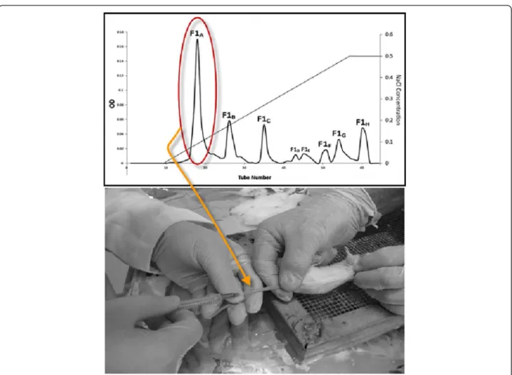
Isolation and purification of coagulation factors were performed using 50 mg of Ec crude venom using a com-bination of gel chromatography and ion exchange chro-matography. Ec crude venom was primarily isolated using gel chromatography (Sephadex G-75) column which initially gained equilibrium using 20 mM ammo-nium acetate buffer (pH 6.8). That is, the column input and output pH became the same. Fifty milligrams of Ec crude venom was dissolved in 4 mL of ammonium acet-ate buffer. The solution was then centrifuged for 15 min at 4°C at 14,000 rpm. The supernatant was isolated and gradually poured into the gel chromatography Sephadex G-75 (200 × 3 cm) column using a special syringe. The sample was then well absorbed by the column and was automatically eluted with ammonium acetate buffer using an automatic collector at the flow rate of 60 mL/h for 24 h. The absorption of the resulting solution was read using a spectrophotometer at 280 nm and relevant absorption curve was drawn in terms of the tube num-ber [10,14,15].
For taking the ammonium acetate buffer out of the solutions, each of the peaks was dialyzed for 24 h with distilled water. After dialysis, the fractions were concen-trated at 4°C with sucrose. The ion exchange chroma-tography (1.5 × 25 cm) column was equilibrated with Tris–HCl 0.05 mM buffer (pH 8.2), i.e., the input buffer
chromatography for further isolation and subfractionation. Initially, a certain amount of the chromatography first peak gradually entered the column (4°C) which was then eluted with Tris–HCl 0.05 mM buffer. Subsequently, the
column was eluted with Tris–HCl 0.05 mM buffer
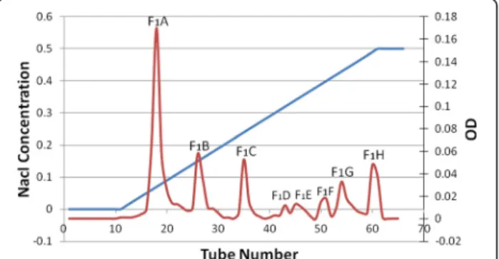
and gradient buffer; pH 8.2 (Nacl concentration 0.1 to 0.4 mM). The ion exchange chromatography out-put solution was collected by an automatic collector at a flow rate of 20 mL/h for 24 h. The absorption of collected tubes was read using a spectrophotom-eter at 280 nm and relevant optical absorption curve was drawn in terms of the tube number [10,14-16]. The subfractions were pooled and dialyzed like in gel chromatography.
Estimation of lethal dose venom (determination of LD50) This test was conducted according to the method by Meier and Theakston [17]. Different doses of crude venom were prepared in physiological serum and were each injected into four mice (2 mL/dose, 0.5 mL/mouse). The doses were chosen was so that no mouse would die at the lower dose, and all mice would die at the higher dose. Mouse mortality within 24 h was recorded and each sample LD50 was calculated. Upon recording of mortality, the Spearman-Karber statistical method was used for LD50 calculation [16,18,19].
Results
Echis carinatus crude venom decreases coagulation time of mouse plasma in relation to its normal levels. Thus, the venom shows coagulation properties. Based on the results of Table 1, it is clear that all Ec venom concentrations have coagulation properties. Therefore, as the venom concentration increases, its coagulation properties will also augment. The existence of coagula-tion factors in Ec venom was then established.
Ec crude venom Gel chromatography
By performing gel chromatography, five fractions were obtained according to Figure 1, respectively la-beled F1 to F5. As per the existing standards on gel chromatography in which protein molecules separate by size; larger molecules pass more freely, appearing
in the earlier fractions, F1 was considered the peak with the highest amount of protein. Regarding the gel chromatography isolation process based on molecular weight, peaks or fractions respectively containing less total protein will exit from the gel chromatography col-umn. Fractions F2to F5contain proteins with molecular weights lower than that of F1.
Study of the coagulation activity of the fractions from Gel chromatography
Regarding Table 2, by conducting the PT test on mouse plasma, it was shown that fraction F1 dimin-ished the coagulation time and that other fractions increased it.
Isolation of subfractions F1using Ion exchange chromatography
Among the fractions obtained from gel chromatog-raphy fraction F1 was selected for furhter isolation because of its lower coagulation time, and was taken to the DEAE-Sepharose ion exchange column. The eight fractions obtained were thus labeled F1A to F1H (Figure 2).
Study of the F1A and F1B subfractions coagulation activity The PT test was frequently conducted on human plasma using subfractions F1A and F1B. These results (p ≤ 0.05) showed a significantly more power-ful coagulation activity of these subfractions when compared with others. The mean PT obtained for subfractions F1A was 7 s and for subfractions F1B, 5 s. Compared with the normal time, this interval is lower, showing the intense coagulation properties of these subfractions. For more investigation into the coagulation activity, these subfractions were selected for injection into mice.
Injection of subfractions F1A and F1B
Subfractions F1A and F1B were intravenously (IV) injected into six NIH mice. Tables 3 and 4 show the results of the PT, PTT and FT tests before and after injection.
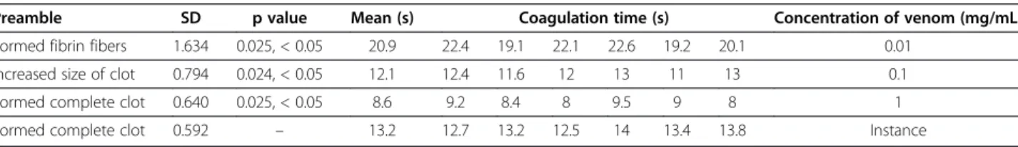
Table 1 Prothrombin time test for different concentrations of crude venom of Ec
Preamble SD p value Mean (s) Coagulation time (s) Concentration of venom (mg/mL)
Formed fibrin fibers 1.634 0.025, < 0.05 20.9 22.4 19.1 22.1 22.6 19.2 20.1 0.01
Increased size of clot 0.794 0.024, < 0.05 12.1 12.4 11.6 12 13 11 13 0.1
Formed complete clot 0.640 0.025, < 0.05 8.6 9.2 8.4 8 9.5 9 8 1
Estimation of LD50
The Spearman-Karber statistical method was used to calculate LD50as follows:
M¼X100d=nðΣr n=2Þ M¼Log LD50
X100¼ Log17:5 ¼ 1:24 n ¼ 4
t0:05 ¼ 2:20
For 4þ4þ 1 degrees of freedom
ð Þ
M ¼ 1:24 0:097=4 0ð þ1þ0þ0þ3þ2þ4–4=2Þ
M ¼ 1:24 0:194 M ¼ 1:046
LD50¼ Antilog 1:046 LD50¼ 11:1μg
½Antilog 1ð ;24þ0;and 194Þis notaproper number and will thus be rejected
Determination of the LD50range: Vm ¼0:0972=42 4
–1
ð Þ ð04þ13þ04þ0
4þ04þ31þ22þ40Þ
Vm ¼0:00196
Antilog 1:0462:20√0:00196
¼ Antilog 1ð :046þ0:097Þ
That is 8:89μgand 13:89μg
ð Þ
LD50¼ 11:1μg=mouse 8ð :89–13:89Þ
Discussion
It has been important for scientists to identify and study the compounds in snake venom. Nowadays, there are different manners to isolate and purify snake venom enzymes and proteins and study their effects. Chroma-tography is the most commonly used method for crude venom isolation.
Echis carinatuscrude venom fractions isolated by chro-matography showed that this method is useful for fraction separation. Viperidae venoms, including that of Ec, are rich in compounds that may be useful for medicine and pharmaceutics [20]. For measuring and confirming Ec crude venom coagulation activity, the PT test was con-ducted with different venom concentrations (Table 1). At lower concentrations, small clots are formed and coagula-tion time is longer, whereas at higher concentracoagula-tions, lar-ger clots are found and coagulation time is shorter [21].
the normal PT is equal to 13.2 s, the rate of the coagula-tion cascade activity will become 100%, with its inter-national normalized ratio (INR) equaling 1 (Table 1) [22].
For isolation, identification and investigation of the properties of Ec crude venom coagulation factors, a combination of gel chromatography and ion exchange chromatography was employed. Fifty milligrams of crude venom were subjected to gel chromatography (Sephadex G-75) and five fractions were obtained (F1 to F5). The isolation of subfractions was performed according to gel chromatography standards based on molecular weight. F1showed the highest level of proteins among the frac-tions. Therefore, the total protein level also decreased from peak 2 to peak 5 (Figure 1). After gel chromatog-raphy, the PT test was conducted to specify coagulation and anticoagulation properties of each fraction (Table 2). The total time of PT was obtained for fraction F1, with a mean of 17.08 s and its coagulation cascade activity was equal to 58.8% and INR to 1.5. Coagulation tests were performed with fraction F1and the coagulation cascade decreased, which could be due to venom toxic properties on the hemostatic system.
PT test showed that F1 was a coagulation fraction whereas other fractions were considered to be anticoa-gulation fractions. Then, fraction F1was subjected to ion exchange chromatography (Figure 2). F1 ion exchange chromatography led to the formation of eight subfrac-tions (F1A to F1H). The PT test was also conducted on mouse plasma using these subfractions. Regarding the PT test results, subfractions F1A (mean 6.8 s) and F1B (mean 3.8 s) were considered major coagulation frac-tions. Table 2 displays that the PT test using these
subfractions drastically increased the coagulation cas-cade activity level, extending it to over 100%. Thus, they were selected for injection into mice.
Another study, similar to ours, was conducted on snake venoms. Josephet al. [23] succeeded in purifying a prothrombin activator from Tropidechis carinatus venom using a combination of gel chromatography, ion exchange and HPLC methods. The purification phases were similar to our work.
A proteinase from Vipera lebetina snake venom, VLH2, has been similarly isolated using a combination of gel chromatography with Sephadex G-75 followed by ion exchange chromatography with Sepharose DEAE A-50 [24]. In another work, Agkistrodon acutus snake venom was exposed to ion exchange chromatography with Sepharose DEAE followed by gel chromatography on Sephacryl S-200 to isolate fractions with coagulation activities [25].
In our research, to further study in vivo the coagula-tion properties of these two subfraccoagula-tions, F1A and F1B were administered to male NIH mice. F1A was IV injected into six mice (concentration of 10 μg/mL), and F1B into other six animals. The mean PT before the F1A injection was 12.8 s which, according to Table 3, displays 100% activity of coagulation cascade and INR = 1. PTT and FT before F1A injection were also recorded. The mean PTT before administration was 31.7 s and the mean FT was 22.5 s.
About an hour after injection, blood samples were col-lected. The mean PT test after the F1A injection was 6.8 s, which enhanced the coagulation cascade more than 100% (Table 3). This rapid response of the coagula-tion cascade occurs in the animal body and generates clinical effects such as coagulopathy, which may provoke death. The mean PTT after injection was equal to 44.8 s. This value compared with the one before injection (31.7 s) is increased. With F1A, coagulation occurs with-out addition of the last test component, CaCl2(Table 3).
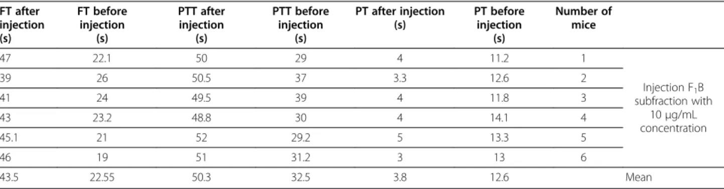
The mean FT before injection was 22.5 s and after in-jection was 43 s. This difference in coagulation time can be indirectly attributed to failure and dysfunction in blood coagulation factors by the presence of procoagula-tion compounds like prothrombin activators. Moreover, subfraction F1B was IV injected into six mice at a con-centration of 10 μg/mL (10 μg/mouse). Mouse plasma Figure 2Ion exchange chromatography of F1fraction.
Table 2 Prothrombin time test on mouse plasma by using fractions obtained from gel chromatography
Mean PT sample 6 (s) PT sample 5 (s) PT sample 4 (s) PT sample 3 (s) PT sample 2 (s) PT sample 1 (s) Fraction
17.08 16.7 17.3 16.8 17.4 16.5 17.8 F1
35.4 37.4 33.9 35 36.5 32 38 F2
– More than 5 min More than 5 min More than 5 min More than 5 min More than 5 min More than 5 min F3
– More than 5 min More than 5 min More than 5 min More than 5 min More than 5 min More than 5 min F4
PT, PTT and FT were determined before injection. The mean PT before injection was 12.6 s, and the coagula-tion cascade was 100% with INR = 1 (Table 4).
An hour after the injection of F1B, blood samples were collected. The mean PT after injection was 3.8 s, which shows an intense activity of the coagulation cascade (more than 100%), as seen in Table 4. The mean PTT before injection was 32.5 s and 42.9 s after injection, in-dicating an increase in PTT. After injection of the sub-fraction F1B, similarly with F1A, plasma coagulation occurred without addition of the last component, CaCl2 (Table 4). The mean FT before injection was 22.55 s and 43.5 s after injection.
This coagulation difference may be indirectly attributed to failure and dysfunction in blood coagulation factors by the presence of procoagulation compounds, such as pro-thrombin activators. Statistical results suggest that H0is rejected by both subfractions F1A and F1B and, hypothet-ically, H1is accepted by both. The p value will thus be sig-nificant, p≤0.05. In other words, according to H1, we will have Mμ1–Mμ2= 0. Other similar studies have also been conducted. For example, Gao et al.[26] fractionated the snake venom of Micropechis ikaheka into a few protein peaks with a combination of gel chromatography and ion exchange chromatography and tested relevant effects on blood coagulation. Their results corroborate ours concern-ing blood coagulation and anticoagulation factors [26].
Halys agkistrodon snake venom was analyzed by Ghorbanpuret al.[27] though gel chromatography. The crude venom was separated into five fractions (AH1 -AH5), all of which were exposed to the PT test in order to study the coagulation process. Then, fraction AH1 was positive in terms of coagulation. The PT assessment method showed that AH1had coagulation activities. Fur-ther purification steps were performed by ion exchange chromatography, generating five fractions (AH11-AH15), of which AH14showed coagulation properties [27].
In 2000 and 2003, Oyama and Takahashi [28,29] suc-ceeded in purifying a thrombin-like enzyme from Tri-meresurus eleganssnake venom in a three-phase method consisting of gel chromatography and two phases of ion exchange chromatography. This enzyme did not influ-ence human fibrinogen. However, it showed coagulation effects on rabbit fibrinogen [28,29].
In 2005, Howes et al.[30] isolated three metalloprotei-nases – EoVMP1, EoVMP2 and EoVMP3 – from the
venom ofEchis ocellatus. EoVMP2and EoVMP3provoked coagulation of human plasma and were considered pro-coagulation factors. They also led to disturbances in fibrin formation and caused systemic bleeding. All the three metalloproteinases were able to activate prothrombin and to convert it into different fractions [30].
Coagulation factors such as subfractions F1A and F1B–
which we successfully purified, determined their molecular
Table 4 Results of PT, PTT and FT tests before and after the injection of F1B subfraction FT after
injection (s)
FT before injection
(s)
PTT after injection
(s)
PTT before injection
(s)
PT after injection (s)
PT before injection
(s)
Number of mice
47 22.1 50 29 4 11.2 1
Injection F1B subfraction with
10μg/mL concentration
39 26 50.5 37 3.3 12.6 2
41 24 49.5 39 4 11.8 3
43 23.2 48.8 30 4 14.1 4
45.1 21 52 29.2 5 13.3 5
46 19 51 31.2 3 13 6
43.5 22.55 50.3 32.5 3.8 12.6 Mean
Table 3 Results of PT, PTT and FT tests before and after the injection of F1A subfraction FT after
injection (s)
FT before injection
(s)
PTT after injection
(s)
PTT before injection
(s)
PT after injection
(s)
PT before injection
(s)
Number of mice
47 25.2 48 30 6 11 1
Injection F1A subfraction with
10μg/mL concentration
33 22 51 32 5 13.6 2
50 19 52 37 7 12.2 3
42 22 49 33.4 8 14.1 4
49 23.1 50.5 34.2 9 13.9 5
37 24.2 53 33 6 12 6
weight and completely identified in our later studies –
may be used as important tools in laboratorial analysis, particularly related to liver diseases. Besides FXa, these enzymes act independently, eliminating the use of any cofactors, including FV, on carboxilated or nonocarboxi-lated prothrombin. Even in the presence of a FV disorder, the amount of prothrombin in patient blood may be mea-sured using these enzymes [26,29].
In the hemostatic system, precise control of blood co-agulation is mandatory for life.Echis carinatusvenom is a rich source of compounds that influence several pro-cesses that occur in this system of prey organism. Some of these molecules may bring benefits to human health. For example, cardiac arrest, arterial obstruction and other cardiovascular and cerebral diseases are important causes of mortality throughout the globe. Atheroscler-osis plays a major role in the pathophysiology of these diseases. Since blood clots consist of platelets and fibrin, treatment strategies have been developed based on co-agulation, fibrinolysis and platelet functions.
Ec venom compounds may be used as medicines to treat thromboembolic disorders. Prothrombin-like enzymes used in defibrillation are part of the thrombolytic treat-ment (clotting decomposition). These enzymes break down fibrinogen, yet they do not activate FXIII. Broken down fibrinogen peptides are somehow similar to fibrin without transverse links which are quickly removed from the blood circulation. Once fibrinogen is removed from the blood, its viscosity will decrease and the blood circula-tion will be optimized. Some of Ec venom compounds may also be purified and used as procoagulant medicines. That is, some Ec venom proteins act on blood coagulation and possess at the beginning a thrombin-like activity fol-lowed by a thromboplastin-like activity. The first enzyme provokes fibrinogen coagulation by breaking down fibri-nopeptide A, whereas the second activates FX [31].
These proteins accelerate platelet aggregation and, con-sequently, shorten coagulation time and reduce damage to blood, a property used to prevent and treat hemorrhage [2]. The lethal effects of snake venoms on different body systems have led to extensive studies on types of snake venom; differentiation and isolation of venoms into va-rious fractions and further investigation of their biochem-ical or pathophysiologbiochem-ical effects.
Enzymes obtained from snake venoms have been used to treat several diseases. Therefore, efforts to isolate frac-tions, purify different factors, analyze venom enzymes and toxicity properties have become more common. However, only a few actions have been taken so far in this regard in Iran. The Venomous Animal Unit of Razi Vaccine and Serum Research Institute is the main sup-plier of antivenom in the country. The scarce research has not only incited actions in this field, but also encour-aged efforts to isolate Echis carinatus crude venom into
different fractions and to study their enzymatic and toxic properties. This snake has been selected because of its massive presence in Iran and high incidence of snake-bites. This research was conducted in cooperation with the Venomous Animal Unit of Razi Vaccine and Serum Research Institute in the hope that other scientists may not ignore this valuable source of biological compounds and may conduct further studies on this issue.
Conclusions
The present study analyzed the venom ofEchis carinatus snake with regard to coagulation activities. Its coagulation proteins were isolated and evaluated using chromato-graphic methods. Ec, a native species in Iran, has been killed by people for a long time because of its bites. Now, it may be used as a rich source of proteins that may be employed in the pharmaceutical industry.
Ethics committee approval
The present study was approved by the Ethics Committee of Razi Vaccine and Serum Research Institute, Karaj, Iran.
Competing interests
The authors declare no conflicts of interests.
Authors’contributions
All authors read and approved the final manuscript.
Acknowledgements
We hereby express our thanks to the esteemed personnel of Razi Vaccine and Serum Research Institute, especially to the Serotherapy and Venomous Animals Unit. We also thank Mr. Dr. Jafari and Ms. Eng. Khamechian for their advice.
Financial source
The Razi Vaccine and Serum Research Institute, Karaj, Iran, provided the financial grants.
Author details 1
Young Researchers and Elites club, Science & Research Branch, Islamic Azad University, Tehran, Iran.2Department of Venomous Animals and Antivenom Production, Razi Vaccine and Serum Research Institute, Karaj, Iran.
Received: 19 June 2012 Accepted: 29 August 2012 Published: 27 February 2013
References
1. White J:Snake venoms and coagulopathy.Toxicon2005,45(8):951–967.
2. Sajevic T, Leonard A, Križaj I:Haemostatically active proteins in snake venoms.Toxicon2011,57(5):627–645.
3. Markland FS:Snake venoms and the hemostatic system.Toxicon1998, 36(12):1749–1800.
4. Bello CA, Hermogenes AL, Magalhães A, Veiga SS, Gremski LH, Richardson M, et al:Isolation and biochemical characterization of a fibrinolytic proteinase fromBothrops leucurus(white-tailed jararaca) snake venom.
Biochimie2006,88(2):189–200.
5. Gutiérrez JM, Rucavado A, Escalante T, Díaz C:Hemorrhage induced by snake venom metalloproteinases: biochemical and biophysical mechanisms involved in microvessel damage.Toxicon2005, 45(8):997–1011.
6. Guerranti R, Cortelazzo A, Hope-Onyekwere NS, Furlani E, Cerutti H, Puglia M, et al:In vitroeffects ofEchis carinatusvenom on the human plasma proteome.Proteomics2010,10(20):3712–3722.
metalloproteinase isolated fromBothrops jararacasnake venom.Toxicon 2008,51(4):488–501.
8. Petrovan RJ, Govers-Riemslag JW, Nowak G, Hemker HC, Rosing J, Tans G: Purification and characterization of multisquamase, the prothrombin activator present inEchis multisquamatusvenom.Thromb Res1997, 88(3):309–316.
9. Yamada D, Sekiya F, Morita T:Isolation and characterization of carinactivase, a novel prothrombin activator inEchis carinatusvenom with a unique catalytic mechanism.J Biol Chem1996,271(9):5200–5207.
10. Ghorbanpur M, Zare Mirakabadi A, Zokaee F, Zolfagharian H, Rabiei H: Purification and partial characterization of a coagulant serine protease from the venom of the Iranian snakeAgkistrodon halys.J Venom Anim Toxins incl Trop Dis2009,15(3):411–423.
11. Rizzo F, Papasouliotis K, Crawford E, Dodkin S, Cue S:Measurement of prothrombin time (PT) and activated partial thromboplastin time (APTT) on canine citrated plasma samples following different storage conditions.Res Vet Sci2008,85(1):166–170.
12. García-Manzano A, González-Llaven J, Lemini C, Rubio-Póo C: Standardization of rat blood clotting tests with reagents used for humans.Proc West Pharmacol Soc2001,44:153–155.
13. Assi AA, Nasser H:Anin vitroand in vivo study of some biological and biochemical effects ofSistrurus malarius barbourivenom.Toxicology1999, 137(2):81–94.
14. Masci PP, Whitaker AN, de Jersey J:Purification and characterization of a prothrombin activator from the venom of the Australian brown snake,
Pseudonaja textilis textilis.Biochem Int1988,17(5):825–835.
15. Serrano SM, Maroun RC:Snake venom serine proteinases: sequence homology vs. substrate specificity, a paradox to be solved.Toxicon2005, 45(8):1115–1132.
16. Kini RM:Serine proteases affecting blood coagulation and fibrinolysis from snake venoms.Pathophysiol Haemost Thromb2005,34(4–5):200–204.
17. Meier J, Theakston RD:Approximate LD50 determinations of snake venoms using eight to ten experimental animals.Toxicon1986, 24(4):395–401.
18. Rao VS, Joseph JS, Kini RM:Group D prothrombin activators from snake venom are structural homologues of mammalian blood coagulation factor Xa.Biochem2003,369:635–642.
19. Castro HC, Zingali RB, Albuquerque MG, Pujol-Luz M, Rodrigues CR:Snake venom thrombin-like enzymes: from Reptilase to now.Cell Mol Life Sci 2004,61(7–8):843–856.
20. Tans G, Govers-Riemslag JW, Van Rinj JL, Rosing J:Purification and properties of a prothrombin activator from the venom ofNotechis scutatus scutatus.J Biol Chem1985,260(16):9366–9372.
21. Nowak G:The ecarin clotting time, a universal method to quantify direct thrombin inhibitors.Pathophysiol Haemost Thromb2003,33(4):173–183.
22. Arif HK, Ayalew T, Rajiv KP:How to interpret and pursue an abnormal prothrombin time, activated partial thromboplastin time, and bleeding time in adults.Mayo Clin Proc2007,82(7):864–873.
23. Joseph JS, Chung MC, Jeyaseelan K, Kini RM:Amino acid sequence of trocarin, a prothrombin activator fromTropidechis carinatusvenom: its structural similarity to coagulation factor Xa.Blood1999,94(2):621–631.
24. Hamza L, Gargioli C, Castelli S, Rufini S, Laraba-Djebari F:Purification and characterization of a fibrinogenolytic and hemorrhagic
metalloproteinase isolated fromVipera lebetinavenom.Biochimie2010, 92(7):797–805.
25. Huang QQ, Teng MK, Niu LW:Purication and characterization of two fibrinogen-clotting enzymes from five-pace snake (Agkistrodon acutus) venom.Toxicon1999,37(7):999–1013.
26. Gao R, Kini RM, Gopalakrishnakone P:A novel prothrombin activator from the venom ofMicropechis ikaheka: isolation and characterization.Arch Biochem Biophys2002,408(1):87–92.
27. Ghorbanpur M, Zare Mirakabadi A, Zokaee F, Zolfagarrian H:Identification and partial purification of an anticoagulant factor from the venom of the Iranian snakeAgkistrodon halys.J Venom Anim Toxin incl Trop Dis2010, 16(1):96–106.
28. Oyama E, Takahashi H:Purification and characterization of a thrombin-like enzyme, elegaxobin, from the venom ofTrimeresurus elegans
(Sakishima-habo).Toxicon2000,38(8):1087–1100.
29. Oyama E, Takahashi H:Purification and characterization of a thrombin-like enzyme, elegaxobin II, with lys-bradykinin releasing activity from the
venom ofTrimeresurus elegans(Sakishima-habo).Toxicon2003, 41(5):559–568.
30. Howes JM, Kamiguti AS, Theakston RD, Wilkinson ML, Laing GD:Effects of three novel metalloproteinases from the venom of the West African saw-scaled viper,Echis ocellatuson blood coagulation and platelets.
Biochim Biophys Acta2005,1724(1–2):194–202.
31. Fox JW, Serrano SM:Approaching the golden age of natural product pharmaceuticals from venom libraries: an overview of toxin derivatives currently involved in therapeutic or diagnostic applications.Curr Pharm Des2007,13(28):2927–2934.
doi:10.1186/1678-9199-19-3
Cite this article as:Salmanizadehet al.:In vivoevaluation of homeostatic effects ofEchis carinatussnake venom in Iran.J Venom Anim Toxins incl Trop Dis.201319:3.
Submit your next manuscript to BioMed Central and take full advantage of:
• Convenient online submission
• Thorough peer review
• No space constraints or color figure charges
• Immediate publication on acceptance
• Inclusion in PubMed, CAS, Scopus and Google Scholar
• Research which is freely available for redistribution