INNOVATIVE DEVELOPMENTS IN DESIGN AND MANUFACTURING
Innovative Developments
in Design and Manufacturing
Advanced research in virtual
and rapid prototyping
Editors
Paulo Jorge da Silva Bártolo Ana Cristina Soares de Lemos António Mário Henriques Pereira Artur Jorge dos Santos Mateus Ausenda Luís Avelar Mendes Carla Sofia Monteiro de Moura Carlos Alexandre Bento Capela Catarina Sofia Gaspar da Silva Flávio André Carvalho Domingues
Helena Maria Coelho da Rocha Terreiro Galha Bártolo Henrique de Amorim Almeida
Irene Sofia Carvalho Ferreira João Manuel Matias
Nuno Manuel Fernandes Alves
Susana Cristina Serrano Fernandes Rodrigues
Centre for Rapid and Sustainable Product Development Polytechnic Institute of Leiria, Portugal
CRC Press/Balkema is an imprint of the Taylor & Francis Group, an informa business
© 2010 Taylor & Francis Group, London, UK
Typeset by Vikatan Publishing Solutions (P) Ltd., Chennai, India
Printed and bound in Great Britain by Antony Rowe (A CPI Group Company), Chippenham, Wiltshire
All rights reserved. No part of this publication or the information contained herein may be reproduced, stored in a retrieval system, or transmitted in any form or by any means, electronic, mechanical, by pho-tocopying, recording or otherwise, without written prior permission from the publisher.
Although all care is taken to ensure integrity and the quality of this publication and the information herein, no responsibility is assumed by the publishers nor the author for any damage to the property or persons as a result of operation or use of this publication and/or the information contained herein.
Published by: CRC Press/Balkema
P.O. Box 447, 2300 AK Leiden, The Netherlands e-mail: [email protected]
www.crcpress.com – www.taylorandfrancis.co.uk – www.balkema.nl
ISBN: 978-0-415-87307-9 (Hbk) ISBN: 978-0-203-85947-6 (Ebook)
V
Table of contents
Preface XIII
Sponsors XV
International scientific committee XVII
Invited lectures
New challenges for Reverse Engineering in facial treatments: How can the
new 3-D non invasive surface measurements support diagnosis and treatment? 3
L.M. Galantucci
Biomanufacturing
Scaffold micro-architecture optimization based on bio-mimetic principles 15
A.B. Bucklen, B.M. Wettergreen & C.M. Liebschner
A minibioreactor for developing “perfused” capillaries in cardiomyocyte aggregates 27
C.K. Chua, D. Liu, K.F. Leong, V. Mironov & V. Kasyanov
Spinning of biomaterial microfibers for tendon tissue engineering 31
C.K. Chua, J. An, K.F. Leong, C.M. Cheah & H. Chang
Stereolithographic rendering of low molecular weight polymer scaffolds
for bone tissue engineering 37
D. Dean, J. Wallace, K. Kim, A.G. Mikos & J.P. Fisher
Process flow for designing functionally graded tissue engineering scaffolds 45
C.K. Chua, N. Sudarmadji, K.F. Leong, S.M. Chou, S.C. Lim & W.M. Firdaus
Indirect fabrication of tissue engineering scaffolds using rapid
prototyping and a foaming process 51
J.Y. Tan, C.K. Chua & K.F. Leong
Fractal tool paths for layered manufacturing of scaffolds with matched bone properties 59
G.S. Kumar & P. Pandithevan
BioExtruder: Study of the influence of process parameters on PCL scaffolds properties 67
M. Domingos, F. Chiellini, A. Gloria, L. Ambrosio, P. Bartolo & E. Chiellini
The use of periodic minimal surfaces for scaffolds design 75
H.A. Almeida & P.J. Bártolo
Intelligent biopolymer selector system for medical applications 81
VI
CAD and 3D data acquisition technologies
Rapid Prototyping models of foetuses built from Ultrasound
3D and Magnetic Resonance files 89
J.R.L. Santos, J.R.L. Santos, R.C. Fontes, S. Campbell & H. Werner
A Computer Aided Design (CAD) support tool for parametric design
of products for Rapid Manufacture (RM) 95
P.C. Smith & A.E.W. Rennie
Comparison of CT and CBCT for fabrication of dentistry models
via rapid prototyping technology 101
H. Kheirollahi, F. Abesi & S. Rahmati
Global approach to design and manufacture Direct Parts 111
J. Kerninon, P. Mognol & J.Y. Hascoët
Novel methodology in design of custom-made hip prosthesis 117
F. Abbaszadeh, S. Rahmati, F. Farahmand & R. Fatollahzadeh
Use of BioCAD in the development of a growth compliant prosthetic
device for cranioplasty of growing patients 127
D.T. Kemmoku, P.Y. Noritomi, F.G. Roland & J.V.L. da Silva
Artificial teeth manufacturing: Inspection of mould and teeth
by contactless scanning systems 131
E. Atzeni, L. Iuliano, P. Minetola, A. Salmi & A. Gatto
Guided dental surgery based on integrating 3D image slicing and structured light scanning 137
S. Barone, A. Paoli & A.V. Razionale
A robotic system for 3D optical scanning of large surfaces 145
M. Maggini, S. Barone, A. Paoli & A.V. Razionale
A simple photogrammetric system for automatic capture and measurement
of facial soft tissues during movement 151
L.M. Galantucci, F. Lavecchia & G. Percoco
3D digitation of museum sculptures for model-making purposes: Difficulties
and possible solutions 157
G. Celani, L. Cancherini, A. Jardini, M. Oliveira, J.V.L. da Silva & V. Piccoli
The use of technologies as Rapid Prototyping and scanner inspection
in surgical planning to medical application 161
C.B.L. Ulbrich, H.A. Hermini & C.A.C. Zavaglia
Performance evaluation of non contact measuring systems considering bias 167
M. Cavallaro, G. Moroni & S. Petrò
A surgical training model manufacture using rapid prototyping technology 175
L. Queijo, J. Rocha, L. Barreira, T. Barbosa, P.M. Pereira & M.S. Juan
Image based modeling and morphological analysis of the human knee 181
G. Renner & L. Hajder
Design of customised bioceramic medical implants by layered manufacturing 187
S.F. Khan & K.W. Dalgarno
Reverse Innovative Design in Rapid Modelling and Reverse Engineering industrial applications 193
A.E. Sonn & D.M. Dimitrov
3D-Digitalization of ankle movement and 3D-CAD-method for patient
specific external ankle support development and Rapid Manufacturing 199
R. Björkstrand, J. Tuomi, M. Paloheimo, J. Lindahl & J. Salo
XIII
Preface
“Innovative developments in design and manufacturing”, contains papers presented at the 4th Interna-tional Conference on Advanced Research in Virtual and Physical Prototyping (VR@P 2009), held by the Centre for Rapid and Sustainable Product Development, Polytechnic Institute of Leiria, Portugal. The Centre for Rapid and Sustainable Product Developed is a centre of excellence hosted by the Polytechnic Institute of Leiria. Its mission is to contribute to the advancement of science and technology leading to more suitable, effective and efficient products, materials and processes, helping to generate added-value for Industry, and to promote the awareness of the role and importance of rapid and sustainable product development in society.
The International Conference on Advanced Research in Virtual and Physical Prototyping was designed to be a major forum for the scientific exchange of multi-disciplinary and inter-organisational aspects of virtual and rapid prototyping and related areas, making a significant contribution for further development of these fields. It joined participants from more than 20 countries. Such diversity was parallel to the vari-ous multi-disciplinary contributions to the conference, whose subjects enclose a wide range of topics like biomanufacturing, micromanufacturing, materials, advanced rapid prototyping technologies, rapid tool-ing and manufacturtool-ing, collaborative design and engineertool-ing, CAD and 3D data acquisition technologies, all of them making a significant contribution for future development. This research community has been strongly engaged in the development of innovative solutions to solve Industry’s problems, contributing to a more pleasant and healthy way of living. I hope that this Conference has been truly worthwhile and this book can represent a significant contribution to research in the field of virtual and physical prototyping.
I am deeply grateful to authors, participants, reviewers, the International Scientific Committee, Session chairs, student helpers and Administrative assistants, for contributing to the success of this conference. The conference was endorsed by:
− The Polytechnic Institute of Leiria (IPL)
− The Centre for Rapid and Sustainable Product Development (CDRsp) − Portuguese Foundation for Science and Technology
− The International Academy for Production Engineering (CIRP) − The Global Alliance of Rapid Prototyping Associations (GARPA) − The Rapid Manufacturing Platform
Paulo Jorge da Silva Bártolo
1 INTRODUCTION
In this study we will present Rapid Prototyping (RP) used as a tool to manufacture a biomedical solid model from a human spine with Lytic Spondylolis-thesis pathology for pre-surgical study procedures.
It is meant to do 3D reconstruction of a biomedi-cal model from a 2D image file obtained from Com-puterized Tomography (CT) scan. After this recon-struction, Rapid Prototyping technology – Three Dimensional Printing (3DP or TDP) is used to pro-duce the solid model.
1.1 Lytic Spondylolisthesis
The term spondylolisthesis refers to the slippage of a vertebra (and the spine above it) relative to the ver-tebra below. There are several aetiologies but the lytic or isthmic type is the most common. In a lytic spondylolisthesis there is a bilateral defect of the isthmus (pars interarticularis), which is the least re-sistant region of the posterior arch of the vertebra. The stress placed on this region by bipedal posture and loadings in extension may cause a fatigue (stress) fracture of the isthmus. With this fracture the vertebral body, pedicle and superior articular processes become separated from the inferior articu-lar processes and hence from the vertebra below. This condition creates the possibility of slippage be-tween the vertebrae.
The slippage between the vertebrae can cause the exiting nerve roots (the nerves exiting the spinal canal at this level, through the intervertebral forami-na) to be squeezed causing leg pain and difficulty walking. When this occurs, a surgical treatment may
be necessary to decompress the nerves and stabilize the spinal segment (to avoid further slippage) with or without reduction of the deformity.
1.2 CT images conversion to 3D models
In the conversion process of a computerized tomo-graphy in to a 3D model, it is needed a sequence of cross sections from the studied object. Using a 3D reconstruction software it is possible to transform these bi-dimensional images in a three-dimensional model that can be used to produce a solid model in rapid prototyping equipment (Foggiatto 2006).
Images obtained from computerized tomography obey to the international standards from DICOM (Digital Imaging and Communications in Medicine) pattern. Those are obtained from axial cuts of the study area and the equipment should be settled to the less possible thickness, as the lower this value is, the better will be the model quality (Foggiatto 2006).
1.3 Rapid prototyping and some medical applications
Rapid Prototyping is the automated manufacture of physical objects. It is an addictive-constructive process, layer by layer that allows complex form ob-jects direct production from three-dimensional data used to manufacture solid prototypes (Rocha & Alves 2000). The geometries needed can be obtained using some CAD software or obtained through the conversion of data proceeding from 3D Scanners, Computerized Tomography or Magnetic Resonance devices. The first techniques of Rapid Prototyping become available in the eighties and were used to
A surgical training model manufacture using rapid prototyping
technology
Luís Queijo, João Rocha, Luísa Barreira & Tiago Barbosa
Instituto Politécnico de Bragança, Bragança, Portugal
Paulo Miguel Pereira
Serviço de Neurocirurgia do Hospital de S. João, Porto, Portugal
Manuel San Juan
ESTII – Universidad de Valladolid; CIBER – Centro de Investigación Biomecánica y Ergonomía, Valladolid, Spain
produce models and prototype parts (Alves & Braga 2001).
One of the main applications of Rapid Prototyp-ing is the fast way that is allowed in verifyPrototyp-ing new concept projects in the earlier stages or even in ad-vanced phases of conception. In all Rapid Prototyp-ing processes, a 3D CAD model is used that is trans-lated into an STL (Stereolithography) format file, (Souza et al. 2003) where all the model surfaces are converted in a triangle mesh.
In Biomedical Engineering field, using Rapid Prototyping techniques it is possible to produce sev-eral types of anatomical models and implant replica with educational purposes or to better understand a specific patient pathology. The models, depending of available techniques, can be made of paper, wax, ceramic, plastic or metal (Antas & Lino 2008). These models can be produced without finishing or color or have these finishing operations done later to improve visualization. For educational purpose it is possible to manufacture implant replica with much lower cost than the implant value.
A great interest can be found in anatomical mod-els manufacture from patient tomographic images. These models allow students from biomedical field to have an easier view of a specific pathology and compare it with normal anatomical models. To bet-ter understand image techniques and anatomy, it is also possible to simultaneously compare the original image (TC or MRI) and 3D solid model.
Medical professionals cooperate with other field professionals to optimize pre-surgical pathology analysis, shorten surgical times, create personalized tools, facilitate the communication with patients and, simultaneously, to explore the capabilities this tech-nology offers in personalized prosthesis design (An-tas & Lino 2008).
Vertebral Spine replica are particularly useful to diagnose, plan and simulate surgical procedures as it also allows the patients to understand the nature of their pathologies as well the need for surgical proce-dures (Madrazo et al. 2008)
Several manufacturing processes are available to-day, as Fused Deposition Modeling (FDM), Stereoli-thography (SLA), Selective Laser Sintering (SLS), Tridimensional Printing (TDP or 3DP) and Lami-nated Object Manufacturing (LOM) among other specific processes.
A brief description of the most used Rapid Proto-typing processes is presented as follows:
• Fused Deposition Modeling (FDM): This prototyp-ing process build the prototypes by depositprototyp-ing an ex-truded thermoplastic material. The injection head draw transversal section perimeters and fills them building, this way, each layer. The most used ma-terial is ABS once it has good mechanical properties. More recently have been developed equipments that allow the used of materials such as polycarbonate
and polyphenilsulfone (PPSU) that have better me-chanical and thermic properties than ABS.
• Stereolitography (SLA): This system builds the prototype by polymerizing a photosensitive liquid resin by applying an ultraviolet light formed by a la-ser. The solidifying process is made layer by layer, allowing obtaining a good surface finished proto-type.
• Selective Laser Sintering (SLS): This process al-lows physical models building by using dust materi-als like ceramics or metal. These materimateri-als are proc-essed in an inert and thermally controlled environment inside a chamber. In here, the melting point (sintering) is achieved by action of a CO2
la-ser. After one layer being sinterized, another layer is deposited until the prototype is finished. This method demands a post-processing work to obtain a better surface.
• Three Dimensional Printing (TDP or 3DP): In this process, models are built from a dust material (which can be a blend using materials like compos-ite, cellulose among others) infiltrated with a liquid binder. This binder is applied through a printing head as used in traditional printing. The prototype is removed having the dust blended with the binder and needing operations of cleaning and medium consolidation.
• Laminated Object Manufacturing (LOM): In LOM, most of the times, the models are obtained by gluing successive layers of paper which are cut by a laser beam. All the paper not used in the model is cut in square or rectangle forms to make easier prototype remove. To ensure the needed rigidity a frame is also built. Model definition will result from paper thickness and quality. Sometimes, instead paper there can be, also, used glass fibres, ceramics or metal (Alves & Braga 2001).
2 METHODOLOGY
After patient’s authorization for TC images be used, these where transferred to the computer where would be done image processing and removed all personal information data.
The process to obtain the anatomic model is composed by the following steps:
• Pre-processing from bi-dimensional images and re-construction from the surface between the contours is done in image processing software ScanIP®. This step is done by using image processing operation such as threshold, floodfill and paint, which allow
through the color choice done by user allowing giv-ing the desired contrast degree to the model for an easier visualization as well to enlighten the desired elements.
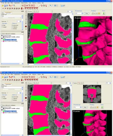
Figure 1. Image pre-processing done in ScanIP®.
The first step of conversion consisted of 3D re-presentation through the image processing applica-tion that allows closed volume visualizaapplica-tion, after a segmentation operation based in the signal intensity
–thresholding. This interactive application allow the
user to detect and select contours in the spondylolis-tehesis area by doing a redefinition of grey levels that led to a separation of the bone from soft tissues. This operation applies gray levels recognition algo-rithms allowing, this way, a bigger grade of automa-tization.
Figure 2. Images pre-processing done in ScanIP® software.
After obtaining the contours with the desired quality, those are enhanced in a manual way using
paint and floodfill operations. These operations
con-sist of adjusting the obtained contours to the shape of the elements intended to represent and model. This step is the most time consuming once the con-tours should be adjusted in more than one orienta-tion (with axis changes) and in a manual way in each image to be processed.
• Rendering and 3D visualization allows to follow the work development during the previous step, to detect and correct possible imperfections. 3D render-ing is done by the application of a consecutive pla-nar triangle mesh from the masks defined in pre-vious steps. Combining these two last phases it is possible to do an iterative process with the objective to present a model as close to the reality as possible.
By analyzing figures 1 and 3 it is possible to see the separation between masks corresponding to ver-tebrae bone tissue and intervertebral discs soft tis-sue. In figure 1 it is also possible to see model im-perfection in a phase previous to manual masks adjustment.
Figure 3. Rendering and 3D previewing in ScanIP® software.
• STL (Stereolitography) data generation allows combining all the active masks in a single file or the creation of several files with distinct masks.
In this kind of files data consist of the conversion and translation from both the 3D model mesh out-putted and the image processing software in to a printing format recognized by the rapid prototyping device. This format contains the model layer divi-sion allowing the layer by layer printing.
• Model manufacturing in Rapid Prototyping device Zprinter 310 from ZCorp.
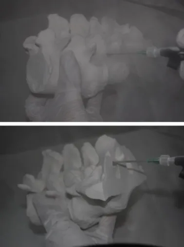
Figure 5. Complete model manufacturing.
In the images from figure 5 and 6 it is possible to visualize several phases of the manufacturing of a model, going from the layer impression until the cleaning of the residual dust.
• Finishing that includes removing and recycling of
excess material and model material consolidation.
Figure 6. Cleaning and recycling operations.
Cleaning operations consist in the global remove, through compressed air action, of the non used dust to obtain an irregular but non dusty surface. After that, model surface consolidation is done by apply-ing an epoxy resin or cyanoacrilate layer (Queijo & Rocha 2009)
3 CONCLUSIONS AND FURTHER WORK
3D replicas of vertebral spine sections are useful in diagnosing, planning and surgery simulation. The visualization and the possible manipulation, by pa-tients, from 3D replica allow them to understand their pathologies nature, surgical proceedings done by the surgeon as well to reduce anxiety facing sur-gery need.
With a multidisciplinary team cooperation it is possible to build, in a short period of time, vertebral spine section 3D models that fulfill all the require-ments.
symp-toms related to this pathology, like leg pain a walk-ing difficulty.
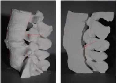
Figure 7. Problem areas highlighted in the printed models - vertebra misalignment and spinal canal estrangement.
By showing this model to the patient he can un-derstand the cause of his symptoms by comparing the two models shown above, and realize the need of actuate surgically to stabilize all that area, improving the well being of patients and avoiding further se-vere damages. On the other hand, the surgeon is able to start planning surgery by estimating the work to be done and by doing one first evaluation of the area he should intervene through this 3D replica model.
Figure 8. Estrangement in complete model and problem main area.
Further work will consist of 3D modeling of med-ical devices (screws, spacers and bars) and those will be inserted in the definitive positions assigned by medical staff allowing this way surgical planning.
Another model will be prototyped with vertebrae aligned due to medical devices application allowing the patient to get a better understanding of all surgi-cal procedures and to analyze which amount of
dis-placement that vertebrae’s new functional positions will lead.
REFERENCES
Alves, L. & Braga, F. 2001. Protoclick, prototipagem rápida. Porto. Protoclick, INEGI.
Alves L. & Rocha J. 2000. Desenvolvimento de moldações cerâmicas compósitas para o fabrico de ferramentas metáli-cas. O Molde.
Antas, A. F. et al. 2008. Utilização das Tecnologias de Prototi-pagem Rápida na Área Médica. 5º Congresso Luso-Moçambicano de Engenharia. Maputo. Moçambique. Arantes, J. A. A. 2006. Diretrizes do tratamento neurocirúrgico
das espondilolisteses degenerativas da Sociedade Brasileira de Neurocirurgia. (Projeto Diretrizes da Sociedade Brasilei-ra de Neurocirurgia)
Foggiatto, J. A. 2006. O Uso da Prototipagem Rápida na Área Médico-Odontológica. Tecnologia & Humanismo. v1, p. 60-68. Curitiba. Brasil.
Madrazo, I., et al. 2008. Stereolithography in spine pathology: a 2-case report. Surgical Neurology.
Netto, A. C. S. et al. 2003. Prototipagem rápida: uma ferramen-ta de projeto para a redução do tempo de desenvolvimento e melhoria de qualidade de produtos. IV Congresso. Brasilei-ro Gestão e Desenvolvimento de PBrasilei-rodutos. Gramado RS. Brasil.
Queijo, L. et al. 2008. A prototipagem rápida na modelação de patogenias. 3.º Congresso Nacional de Biomecânica. Bra-gança. Portugal.
Rocha, J. 2000. Moldações Cerâmicas Compósitas. FEUP. Por-to. Portugal
Rocha, J. & Alves, L. 2000. Utilização de moldações cerâmicas compósitas no fabrico de ferramentas metálicas. 2º Encon-tro nacional do colégio de engenharia mecânica da ordem dos engenheiros. Coimbra. Portugal