Braz. J. of Develop.,Curitiba, v. 6, n. 9, p. 65445-65458, sep. 2020. ISSN 2525-8761
Avaliação histomorfométrica do efeito de um biomodificador de dentina à base
de óleo de copaiba (Copaifera multijuga Hayne) na camada híbrida
Histomorphometric evaluation of the effect of a copaiba oil-based (Copaifera
multijuga Hayne) dentin biomodifier on the hybrid layer
DOI:10.34117/bjdv6n9-104
Recebimento dos originais: 08/08/2020 Aceitação para publicação: 04/09/2020
Joyce de Figueirado Meira
Mestr em Odontologia
Instituição: Faculdade de Odontologia da Universidade Federal do Amazonas - UFAM Endereço: Av. Ministro Waldemar Pedrosa, 1539- Praça 14 de Janeiro, Manaus- AM, Brasil
E-mail: joycefmeira@hotmail.com
Geisy Rebouças Lima
Doutoranda em Inovação Farmacêutica
Instituição: Faculdade de Ciências Farmacêuticas da Universidade Federal do Amazonas-UFAM Endereço: Av. General Rodrigo Octávio Jordão Ramos, 1200, Campus da UFAM, setor Sul -
prédio da Faculdade de Ciências Farmacêuticas - Coroado I, Manaus -AM, Brasil E-mail: geisylima@hotmail.com
Tatiana Nayara Libório-Kimura
Doutora em Patologia Bucal, Universidade de São Paulo - USP
Instituição: Departamento de Patologia e Medicina Legal, Faculdade de Medicina – UFAM Endereço: Rua Afonso Pena, 1053 - Praça 14 de Janeiro, Manaus- AM
E-mail: tatiana.liborio@gmail.com
Marne Carvalho de Vasconcellos
Doutora em Farmacologia pela Universidade Federal do Ceará
Instituição: Faculdade de Ciências Farmacêuticas da Universidade Federal do Amazonas-UFAM Endereço: Av. General Rodrigo Octávio Jordão Ramos, 1200, Campus da UFAM, setor Sul -
prédio da Faculdade de Ciências Farmacêuticas - Coroado I, Manaus -AM, Brasil E-mail: marne@ufam.edu.br
Fábio Correia Sampaio
Pós-Doutor em Odontologia pela Universidade de São Paulo
Instituição: Faculdade de Odontologia da Universidade Federal da Paraíba – UFPB Endereço: Campus I - Cidade Universitária - João Pessoa - PB - Brasil
E-mail: fcsampa@gmail.com
Carina Toda
Doutora em Reabilitação Oral pela Universidade Estadual Paulista Júlio de Mesquita Filho / Faculdade de Odontologia de Araraquara
Instituição: Faculdade de Odontologia da Universidade Federal do Amazonas- UFAM Endereço: Av. Ministro Waldemar Pedrosa, 1539 -Praça 14 de Janeiro, Manaus -AM, Brasil
Braz. J. of Develop.,Curitiba, v. 6, n. 9, p. 65445-65458, sep. 2020. ISSN 2525-8761
Nikeila Chacon de Oliveira Conde
Doutora em Estomatologia pela Universidade Federal da Paraíba
Instituição: Faculdade de Odontologia da Universidade Federal do Amazonas- UFAM Endereço: Av. Ministro Waldemar Pedrosa, 1539 -Praça 14 de Janeiro, Manaus -AM, Brasil
E-mail: nikeilaconde@gmail.com
Maria Fulgência Costa Lima Bandeira
Doutora em Dentística pela Universidade Estadual Paulista Júlio de Mesquita Filho/ Faculdade de Odontologia de Araraquara
Instituição: Faculdade de Odontologia da Universidade Federal do Amazonas-UFAM Endereço: Av. Ministro Waldemar Pedrosa, 1539 -Praça 14 de Janeiro, Manaus -AM, Brasil
E-mail: fulgencia@ufam.edu.br
RESUMO
Objetivo: Realizar avaliação histomorfométrica do efeito de um biomodificador de dentina (CM) à base de óleo de copaíba (Copaifera multijuga Hayne) na espessura do colágeno da matriz e na homogeneidade da camada híbrida.Materiais e métodos: Um total de 80 espécimes foram preparados a partir de trinta molares que foram divididos em cinco grupos: três deles com concentrações de conservante baixa (L), média (M) e alta (H) para o material testado (CM) e os outros dois com os grupos controle: negativo (água destilada - DW) e positivo (clorexidina 2% - CHX). Cada grupo foi subdividido de acordo com o substrato - dentina sadia (SD) ou dentina afetada por cárie (CAD). Após três meses de imersão nas soluções, a homogeneidade da camada híbrida foi analisada qualitativamente, enquanto a espessura dos casos com camada de colágeno exposta e hibridizada foi analisada quantitativamente por histomorfometria. Resultados: Em relação à avaliação histomorfométrica, dois CM testados (M e H) para SD apresentaram colágeno hibridizado mais espesso semelhante ao observado nos grupos controle positivo (SD e CAD), sem diferenças significativas entre eles (p> 0,05). A análise qualitativa mostrou que a melhor condição de hibridização, em termos de homogeneidade, foi identificada na concentração M da emulsão CM para SD. Conclusão: Nossos achados mostraram que a emulsão CM em concentração média apresentou a melhor homogeneidade para camada híbrida em SD entre todos os grupos.
Relevância clínica: Por se tratar de propriedades biológicas de uma planta medicinal da Amazônia, supõe-se que a emulsão CM provavelmente contribuiria para melhorar a interface dente / restauração.
Palavras-chave: dentina, fitoterapia, colágeno, adesivos. ABSTRACT
Objectives: To perform histomorphometric evaluation of the effect of a copaiba oil-based (Copaifera multijuga Hayne) dentin biomodifier (CM) on the matrix collagen thickness and the homogeneity of the hybrid layer. Materials and methods: A total of 80 specimens were prepared from thirty molars that were divided into five groups: three of them with a low (L), medium (M) and high (H) preservative concentrations for the tested material (CM) and the other two with the control groups: negative (distilled water - DW) and positive (2% chlorhexidine - CHX). Each group was subdivided, according to substrate – sound dentin (SD) or caries-affected dentin (CAD). After three months of immersion in the solutions, the homogeneity of the hybrid layer was qualitatively analysed, while the thickness of the cases with exposed and hybridised collagen layer was quantitatively analysed by histomorphometry. Results: Regarding the histomorphometric evaluation, two tested CM (M and H) for SD had thicker hybridised collagen similarly to that observed in the positive control groups (SD and CAD), with no significant differences between them (p>0.05). The qualitative analysis showed that the best hybridisation condition, in terms of
Braz. J. of Develop.,Curitiba, v. 6, n. 9, p. 65445-65458, sep. 2020. ISSN 2525-8761
homogeneity, was identified in the M concentration of CM emulsion for SD. Conclusion: Our findings showed that CM emulsion at medium concentration had the best homogeneity for hybrid layer in SD among all groups.
Clinical relevance: Due to it is biological properties of a medicinal plant from the Amazon, it is supposed that the CM emulsion would probably contribute to improving the tooth/restoration interface.
Keywords: dentin, phytotherapy, collagen, adhesives.
1 INTRODUCTION
The adhesion of polymeric materials to dentin is still considered a major challenge because dentin is a histologically complex substrate, predominantly tubular and intrinsically moist(Ricci et al., 2011).
Pathological processes such as sclerosis, caries and demineralisation of the dentin result in changes in the properties of the dentin substrate (Wang et al.,2007). These changes include activation of metalloproteinases (MMPs) in the dentin matrix after acid etching, and these enzymes can degrade the collagen present in the dentin, thus participating in the failure of adhesive restorations(Breschi et al., 2010).
The incomplete penetration and polymerisation of the resin inside the demineralised dentin allows nanometric channels to be established in the dentin/restorative material interface, a process that has been called nanoleakage. These nanoleakage pathways allow fluids to penetrate into the hybrid layer, which can lead to degradation of the interface, reducing the bond strength of restorative materials to the dentin substrate(Nakajima et al., 1995).
Studies have shown that when used after acid etching, chlorhexidine (CHX) inhibits the proteolytic activity of MMPs, slowing collagen degradation and improving the stability of the hybrid layer (De Campos et al., 2009; Cavalcanti et al., 2005; Kim et al.,2011; Pereira et al., 2006).
In addition to CHX, oxidising biomolecules are also being investigated, with possibly less deleterious effects to dental tissues. In vitro studies have shown the antibacterial activity, biological compatibility, activity on free radicals during the inflammatory and response acceptability in colour change tests of copaiba-based products in human teeth (Bandeira et al., 1999; Cavalcanti et al.,2005; Mendonça et al.,2009; de Araújo et al., 2020;Vasconcelos et al., 2008). These studies have shown that commonly used medicinal plants from the Amazon have the potential to be a source of natural products of industrial interest (Evangelista et al., 2013). In particular, essential oils stand out because they are biomolecules with various biological activities in cellular structures and have moderate or low toxicity(Bakkali et al.,2008).
Braz. J. of Develop.,Curitiba, v. 6, n. 9, p. 65445-65458, sep. 2020. ISSN 2525-8761
Thus, the present study aimed to evaluate morphometrically the in vitro effects of a 10%
copaiba oil-based (Copaifera multijuga Hayne) dentin biomodifier (CM) on the collagen of sound
dentin (SD) and caries-affected dentin (CAD) to identify the presence of exposed and hybridised collagen and the homogeneity of the hybrid layer.
2 MATERIAL AND METHODS
This research was approved by the Research Ethics Committee of the Federal University of Amazonas - UFAM under CAAE No. 13329213.9.0000.5020 and was conducted in the city of Manaus, state of Amazonas, at the Research Laboratory of the School of Dentistry, in the Department of Pathology and Legal Medicine - DPML of the School of Medicine and in the School of Pharmaceutical Sciences of UFAM.
The Copaifera multijuga Hayne oleoresin was collected and cataloged by National Institute of Amazonian Research (INPA) under number 270709. The copaiba oil emulsion of Copaifera
multijuga Hayne, formulated the following guidelines of Brazilian Pharmacopoeia (Brasil, 2010).
2.1 TEETH PREPARATION
Thirty sound third molars were randomly selected, provided by the Biobank of the School of Dentistry of UFAM. Teeth with cracks, fractures, restorations or hypoplastic areas were excluded from the study.
Using a Mecatome P100 precision cutting machine (PRESI, Grenoble - France) and a double-faced diamond disk (Extec Water Brade 4’x 0.12x ½, code 1010-584, Extec Corp., Enfield, CT, USA) under constant lubrication (300 rpm and 200 gf), the occlusal portions of the teeth were removed, and flat surfaces were created in the dentin.
The dentin surface was abraded in the occlusal surface using the AROTEC polishing machine (Aropol 2V, Arotec S.A. Indústria e Comércio, series 040865, Cotia, SP, Brazil) with silicon carbide abrasive of increasing granulations (180 - 240 – 320) under water cooling, aimed at producing a standardised smear.
2.2 INDUCTION OF ARTIFICIAL CARIES LESIONS
Fifteen teeth had their roots sealed with composite resin and were sealed with an epoxy adhesive layer (ARALDITE®, Ciba Especialidades Químicas Ltda, São Paulo, SP, Brazil) and an acid-resistant enamel layer (Colorama, CEIL Com. Exp. Ind. Ltda, São Paulo, Brazil), leaving only the dentin surface exposed.
Braz. J. of Develop.,Curitiba, v. 6, n. 9, p. 65445-65458, sep. 2020. ISSN 2525-8761
These teeth were suspended using an orthodontic wire in a beaker containing distilled water for sterilisation in an autoclave for 20 minutes at 121°C.
The cariogenic solution consisted of 3.7 g of BHI broth (Brain Heart Infusion, Becton Dickinson and Company, Sparks, MD, USA), 2 g of sucrose (Synth; LabSynth, São Paulo, SP, Brazil), 1 g of glucose (Synth; LabSynth, São Paulo, SP, Brazil) and 0.5 g of yeast extract (Becton Dickinson and Company, Sparks, MD, USA) for each 100 mL of distilled water. The solution was sterilised (20 minutes at 121°C), and subsequently, 2% volumes of Streptococcus mutans strain ATCC25175 were inoculated (inoculum at ≠ 0.5 overnight of the McFarland scale), provided by the Oswaldo Cruz Foundation - FIOCRUZ - RJ. The fifteen teeth were suspended in the cariogenic medium using a thread, and the set was incubated in a microaerophilic jar for 14 days.
During this period, the cariogenic solution was replaced every 48 hours, but no new microorganisms were inoculated. After the incubation period, the teeth were once again autoclaved. The biofilm was removed with gauze, and the isolating materials (epoxy adhesive and enamel) were removed manually with scalpel blades. The teeth were thoroughly washed with deionised water, showing a darkened dentin surface that was soft when touched with an exploratory probe (Sanabe et al., 2011).
2.3 SPECIMEN PREPARATION AND ADHESIVE PROCEDURES
The carious surface was removed using a spherical steel bur No. 4 (KG Sorensen, Barueri - SP, Brazil) at low speed, which was replaced by a new bur every four prepared teeth. Spherical burs were also used for 30 s on the sound dentin of the 15 remaining teeth so that the smear layer was created using the same type of cutting instrument. Subsequently, the roots of the 30 teeth were removed in the cutting machine. All procedures were performed by a single calibrated operator.
The preservative concentrations of the three tested copaiba oil-based (Copaifera multijuga
Hayne) dentin biomodifier (CM) emulsions prepared were L = low, M = medium and H = high. The
negative group was composed of distilled water (DW) and the positive group was of 2% chlorhexidine (CHX). The 30 teeth were divided into five groups (table 1), each one subdivided in two subgroups according to substrate – sound dentin (SD) or caries-affected dentin (CAD. Each subgroup of was composed of three teeth.
The dentin was conditioned for 15 s with 35% phosphoric acid (3M ESPE, St. Paul, MN, USA), washed for 15 s and dried with absorbent paper to obtain a moist surface. Subsequently, 20 µL of each solution was applied, according to the groups, for 60 s, and the dentin was dried again with absorbent paper.
Braz. J. of Develop.,Curitiba, v. 6, n. 9, p. 65445-65458, sep. 2020. ISSN 2525-8761
Two consecutive coats of the Single Bond® adhesive (3M ESPE, St. Paul, MN, USA) were applied, and each coat was individually subjected to mild air jets to remove the solvent. Finally, the coats were collectively cured with a light-curing unit (Dabi Atlante – DB686, Ribeirão Preto, SP, Brazil), whose irradiance was monitored with a radiometer (490 ± 10 mW/cm2). Subsequently, four additional adhesive coats were applied and light-cured at the end of the last coat to allow for specimen microtomy. The teeth were kept in an incubator with 100% relative humidity at 37°C for 24 hours(Sanabe et al., 2011).
The teeth were sectioned in the cutting machine to obtain specimens with 2 mm thickness, 2 mm height and 5 mm length for each tooth. Thus, each tooth provided three specimens, totalling 90 specimens. Ten were discarded, resulting in 80 specimens, which were immersed in specific test solutions for three months, maintaining a constant temperature of 37°C.
2.4 HISTOLOGICAL PROCESSING
After the storage period, the specimens were fixed in 10% formalin solution for 48 hours and demineralised in 10% Morse solution for two weeks without stirring. Subsequently, the specimens were washed, neutralised in 5% sodium sulphate solution for 24 hours and washed again in running water for 24 hours for subsequent dehydration in increasing alcohol solutions (70 to 100%), diaphanisation and vacuum inclusion in paraffin.
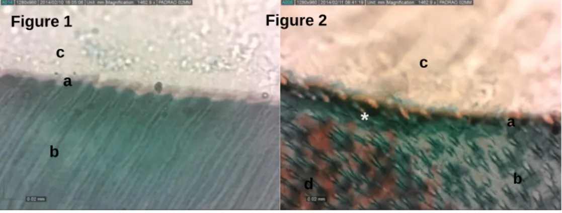
Cuts of approximately 4 μm thickness (820 Spencer Microtome - American Optical Corp, Buffalo, NY, USA) were stained by Goldner’s trichrome stain. In this staining, collagen not fully covered by the adhesive was available to react with the stain and was identified by the reddish colour. The demineralised dentin was stained in green, the adhesive layer in beige, and the adhesive-hybridised collagen in orange. One slide of each specimen was prepared, with 10 sections on each slide. Among these sections, one section of each slide was randomly selected and analysed under 400x magnification (Olympus BX51 – Olympus Corp, Tokyo, Japan).
2.5 QUALITATIVE AND QUANTITATIVE (HISTOMORPFOMETRIC) EVOLUATION The qualitative analysis was performed simultaneously by three experienced pathologists, who established a consensus regarding the disposition of the dentin tubules, hybridised collagen and exposed collagen layer, presence of adhesive and homogeneity of the hybrid layer. The pathologists also specified whether the layer was totally, partially or not hybridised (Erhardt et al., 2008).
Based on this analysis, three images from each section were collected (Dino-Eye Microscope Eye-Piece Camera – ANMO Electronics Corporation, Taiwan) for the morphometric evaluation of the thickness of the hybrid layer. Three measurements of the exposed and hybridised
Braz. J. of Develop.,Curitiba, v. 6, n. 9, p. 65445-65458, sep. 2020. ISSN 2525-8761
collagen zone (when present) were performed using the UTHSCSA Image Tool software (The University of Texas Health Science Center, Santo Antonio, TX, USA). The readings were performed twice by the same examiner, with a two-week interval between them.
2.6 STATISTICAL ANALYSIS
The mean of the two readings was computed for each specimen. For intraexaminer agreement, the weighted Kappa test (p<0.05) and Pearson’s correlation test (p<0.05) were used.
Data are presented using tables, and the medians and quartiles (Qi) of the quantitative data
were calculated. In the data analysis, the non-parametric Kruskal-Wallis test was applied because the normality hypothesis was rejected at the 5% significance level (p<0.05) by the Shapiro-Wilk test. The significance level was set at 5%.
3 RESULTS
The images obtained from the groups can be observed in Figures 1 to 10. Areas with exposed collagen were observed with greater frequency and intensity in the G5-B group, followed by groups G3-B and G3-A.
Figure 1 – Sound dentin specimen stained with Goldner’s trichrome. Negative control group: distilled water (Group 1-A); Figure 2 – Caries-affected dentin specimen stained with Goldner’s trichrome. Negative control group: distilled
water (Group 1-B); Figure 3 – Sound dentin specimen stained with Goldner’s trichrome. Positive control group: CHX (Group 2-A); Figure 4 – Caries-affected dentin specimen stained with Goldner’s trichrome. Positive control group: CHX (Group 2-B); Test CM solutions: Figure 5 – Sound dentin specimen stained with Goldner’s trichrome. Test solution with low concentration: CM-L (Group 3-A); Figure 6 – Caries-affected dentin specimen stained with Goldner’s trichrome. Test solution with low concentration: CM-L (Group 3-B); Figure 7 – Sound dentin specimen stained with Goldner’s trichrome. Test solution with medium concentration: CM-M (Group 4-A); Figure 8 – Caries-affected dentin specimen stained with Goldner’s trichrome. Test solution with medium concentration: CM-M (Group 4-B); Figure 9 – Sound dentin specimen stained with Goldner’s trichrome. Test solution with high concentration: CM-H (Group 5-A); Figure 10 – Caries-affected dentin specimen stained with Goldner’s trichrome. Test solution with high concentration: CM-H (Group 5-B). (*) hybridised collagen, (a) exposed collagen, (b) mineralised dentin, (c) adhesive, (d) demineralised dentin. Figure 1 a a Figure 2 c
*
b d b cBraz. J. of Develop.,Curitiba, v. 6, n. 9, p. 65445-65458, sep. 2020. ISSN 2525-8761
The dentinal tubules were arranged as follows: G1-A = 80% longitudinal and 20% cross-sectional, G1-B = 83.3% longitudinal and 16.7% cross-cross-sectional, G2-A = 100% longitudinal, G2-B = 87.5% longitudinal and 12.5% cross-sectional, G3-A = 100% longitudinal, G3-B = 25% longitudinal and 75% cross-sectional, G4-A = 60% longitudinal and 40% cross-sectional, G4-B =
Figure 3
*
* Figure 4 a b b d c c Figure 5 Figure 6 a a c b c b * b*
Figure 8 b Figure 7 c c Figure 9 Figure 10 c a c b b*
aBraz. J. of Develop.,Curitiba, v. 6, n. 9, p. 65445-65458, sep. 2020. ISSN 2525-8761
71.4% longitudinal and 28.6% cross-sectional, G5-A = 100% longitudinal and G5-B = 100% longitudinal.
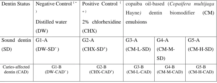
Table 1 – Group and subgroup distribution according to negative and positive controls and the three tested materials in
different emulsions concentrations. DW-SD¯: negative control in sound dentin; DW-CAD¯: negative control in caries-affected dentin; CHX-SD+: positive control in sound dentin; CHX-CAD+: positive control in caries-affected dentin; CM-L-SD: copaiba based dentin biomodifier with concentration low in sound dentin; CM-L-CAD: copaiba oil-based dentin biomodifier with concentration low in caries-affected dentin; CM-M-SD: copaiba oil-based dentin biomodifier with concentration medium in sound dentin; CM-M-CAD: copaiba oil-based dentin biomodifier with concentration medium in caries-affected dentin;CM-H-SD: copaiba oil-based dentin biomodifier with concentration high in sound dentin; CM-H-CAD: copaiba oil-based dentin biomodifier with concentration high in caries-affected.
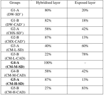
The percentage of hybridised and exposed collagen in the groups is show in table 2.
No significant differences were observed between G3-A, G3-B, G4-A and G5-A for the thickness of exposed collagen. There were also no significant differences when comparing G4-A, G4-B, G5-A and G5-B with G2-A and G2-B. Groups G1-A and G1-B exhibited exposed collagen with lower thickness (Table 2).
Dentin Status Negative Control ( ¯ ) Distilled water (DW) Positive Control ( + ) 2% chlorhexidine (CHX)
copaiba oil-based (Copaifera multijuga
Hayne) dentin biomodifier (CM)
emulsions Sound dentin (SD) G1-A (DW-SD¯) G2-A (CHX-SD+) G3-A (CM-L-SD) G4-A (CM-M-SD) G5-A (CM-H-SD) Caries-affected dentin (CAD) G1-B (DW-CAD¯) G2-B (CHX-CAD+) G3-B (CM-L-CAD) G4-B (CM-M-CAD) G5-B (CM-H-CAD)
Braz. J. of Develop.,Curitiba, v. 6, n. 9, p. 65445-65458, sep. 2020. ISSN 2525-8761
Table 2 – Results of the percentage of hybridized and exposed collagen for each group.
Groups G4-A and G5-A exhibited hybridised collagen with greater thickness; however, there were no significant differences between them. Groups G1-A and G1-B exhibited hybridised collagen with lower thickness. Groups G2-A, G2-B and G4-B did not show significant differences between them or between the other groups mentioned (Table 3). The total number of specimens with hybridised collagen fibres was too low to allow statistical analyses in the following groups: G3-A, G3-B and G5-B.
Table 3 - Distribution according to the median of the measures, in µm, of the exposed collagen in relation to the different
groups. p<0.001 (Kruskal-Wallis non-parametric test); Qi = Quartile.
Groups Hybridised layer Exposed layer G1-A (DW-SD¯) 80% 20% G1-B (DW-CAD¯) 82% 18% G2-A (CHX-SD+) 58% 42% G2-B (CHX-CAD+) 87% 13% G3-A (CM-L-SD) 40% 60% G3-B (CM-L-CAD) 22% 78% G4-A (CM-M-SD) 100% - G4-B (CM-M-CAD) 58% 42% G5-A (CM-H-SD) 87% 13% G5-B (CM-H-CAD) 27% 83% Groups n Q1 Median Q3
G3-A – Sound Dentin + CM-L 3 26.65 32.32a 42.38 G3-B - Carious Dentin + CM-L 5 24.18 28.54a 37.16 G4-A - Sound Dentin + CM-M 4 20.25 26.64ab 30.67 G5-A - Sound Dentin + CM-H 6 12.17 21.49ab 26.15 G4-B - Carious Dentin + CM-M 7 17.19 21.47bc 22.95 G2-A - Sound Dentin + CHX 6 15.15 16.29bc 19.22 G5-B - Carious Dentin + CM-H 7 12.72 15.48c 17.31 G2-B - Carious Dentin + CHX 8 12.01 13.36c 16.12 G1-A - Sound Dentin + Water 4 6.52 7.12d 9.90 G1-B - Carious Dentin + Water 6 6.37 7.02d 9.71
Braz. J. of Develop.,Curitiba, v. 6, n. 9, p. 65445-65458, sep. 2020. ISSN 2525-8761
4 DISCUSSION
The Amazon region has the world’s largest biome, with economic potential for the development of new herbal medicines, especially in dentistry. Thus, studies related to the processing of raw materials from medicinal plants into products to be used in dentistry are extremely relevant and timely for scientific and technological development, especially those studies aimed at improving the stability and homogeneity of the hybrid layer, favouring the adhesion quality of restorative materials to the dental structure.
In the present study, when analysing the arrangement of dentinal tubules in relation to favouring collagen hybridisation, there was no relationship between the two variables, given that, despite the similarities in the results of groups G1-A (DW-SD¯), G1-B (DW-CAD¯), G2-B (CHX-CAD+) and G3-B (CM-L-CAD), in which the higher percentage of longitudinal tubules was directly proportional to the percentage of hybridised collagen, the remaining groups exhibited opposite proportions when compared with each other, demonstrating the possible clinical use of CM in occlusal and proximal cavities.
The best hybridisation condition for both substrates was observed in specimens stored in G4-A (CM-M-SD) (Figure 7). This result emphasises that CM is oily, with similar results reported by Sanabe et al (2011), who showed that the hybridisation zone stained in orange was better viewed in the control group (stored in mineral oil). In regard to copaiba oil, there is still no evidence in the literature that supports these findings. However, according to Sanabe et al(2011), this storage medium may prevent enzymatic and hydrolytic degradation mechanisms from manifesting on the adhesive interface, which was confirmed when these results were compared to those of water-storage groups. Due to the hydrophobic characteristic of CM, the water present in the adhesive union is gradually removed, preventing the hydrolytic degradation of the hybrid layer.
Storage in DW has often been used as a negative control in methods that investigate the longevity of the adhesive interface because it is responsible for the hydrolytic degradation in the adhesion (Hebling et al., 2005; Nakajima et al., 1995; Pereira et al., 2006; Sanabe et al.,2011; Sano et al., 1995; Tjaderhane et al., 1998). However, in the present study, it was observed that even after three months of immersion in DW, 80% and 82% of specimens in groups A (DW-SD¯) and G1-B (DW-CAD¯) had hybridised collagen, respectively.
The activation of metalloproteinases (MMPs) depends on a decrease in pH to values below 5.5, which causes mineral loss and conversion of latent MMPs into the active form. Once activated, these MMPs would have to be deactivated through the use of solutions to allow the collagen to be impregnated by the adhesive system(Pashley et al.,2004). Among the solutions tested, only the emulsions of groups G3-A (CM-L-SD) and G3-B (CM-L-CAD) had pH values of 5. This factor
Braz. J. of Develop.,Curitiba, v. 6, n. 9, p. 65445-65458, sep. 2020. ISSN 2525-8761
would explain why the number of specimens with hybridised collagen was not significant for these groups. The remaining solutions had the following pH values: DW and CHX - pH 7, CM-M and CM-H - pH 6.
Several studies have reported that the application of CHX in dental cavities improves the stability of the hybrid layer. We believe that this improvement in the durability of the resin/dentin union occurs because CHX inhibits the activity of MMPs, which are responsible for collagen autodegradation (De Campos et al.,2009; Cavalcanti et al., 2005; Franco et al., 2007; Kim et al.,2011; Maveric et al.,2017).The exact mechanism of action of this cationic agent is not yet fully understood; however, it has been suggested that due to its positive charge, CHX molecules compete with MMP sites that should be activated by metallic ions such as calcium and zinc, thus inactivating their proteolytic action(Sanabe et al., 2011).
Considering the thickness of the exposed collagen, the present study results differ from those presented by Haj-Ali et al (2006) and Sanabe et al (2011) Using the same histological staining, these authors observed significantly thinner exposed collagen zones in both caries-affected dentin and sound dentin. Haj-Ali et al (2006) showed that the mean thickness of the exposed collagen zone observed for the caries-affected dentin was 8.6 µm, whereas for sound dentin, the mean thickness was 6.01 µm (p=0.042).
Sanabe et al(2011) obtained lower mean thicknesses of exposed collagen, 2.99 µm and 2.30 µm for caries-affected and sound dentin, respectively. The different values between the studies can be attributed to the fact that Haj-Ali et al(2006) used a chemical storage method for specimens in buffered acid solution (pH 4.5) for dentin demineralisation, in addition to 38% phosphoric acid. However, Sanabe et al(2011) used a microbiological method for the production of artificial caries lesions and 35% phosphoric acid as the dentin demineralisation agent.
In the present study, during the histological processing, the specimens were immersed in Morse solution (decalcifying solution) for approximately 10 days, until reaching an adequate hardness. In this regard, the method of Sanabe et al (2011) recommended the immersion of specimens in Morse solution for 48 hours. It is believed that the longer immersion time in the aforementioned solution provided greater exposure of collagen fibrils.
Considering the thickness of the exposed and hybridised collagen and the quality of the hybrid layer, it can be suggested that the homogeneity of the hybrid layer is more important than its thickness and that it may provide greater adhesive resistance in restorations. This layer homogeneity was clearly observed histologically in G4-A (CM-M-SD).
Braz. J. of Develop.,Curitiba, v. 6, n. 9, p. 65445-65458, sep. 2020. ISSN 2525-8761
5 CONCLUSION
Our findings showed that of copaiba oil-based (Copaifera multijuga Hayne) dentin
biomodifier (CM) emulsion at medium concentration had the best homogeneity for hybrid layer in
SD among all groups. Additionally, it had a similar thickness measurement compared to the standard CHX. Taking these results together, we suggest that CM should be a promising and alternative option as dentin adhesive material use in dentistry; however, further studies are required to elucidate its clinical application in regard to the interference in the properties of restorative materials.
REFERENCES
BAKKALI, F.; AVERBECK, S.; AVERBECK, D.; IDAOMAR, M. Biological effects of essential oils - A review. Food and Chemical Toxicology. Vol. 46; 446–75,2008.
BANDEIRA, M. F. C. L.; OLIVEIRA, M. R. B.; BENATTI NETO, C.; LIA, R. C. C. Comparative study of biocompatibility of essential oil and resin of Copaifera multijuga (copaiba oil) associated with calcium hydroxide in rat molars. J Bras Clín Estét Odontol. 3(16):42-49, 1999.
BRASIL. Farmacopeia Brasileira, volume 2. Agência Nacional de Vigilância Sanitária. Brasília: Anvisa, 2: 546p, 2010.
BRESCHI, L.; MAZZONI, A.; NATO, F.; CARRILHO, M.; VISINTINI, E.; TJÄDERHANE, L.; RUGGERI JR, A.; TAY, F. R.; DORIGO, E. D.; PASHLEY, D. H. Chlorhexidine stabilizes the adhesive interface: A 2-year in vitro study. Dent Mater. Apr;26(4):320–5,2010.
CAVALCANTI NETO, A. T.; ARRUDA, T. E. P.; ARRUDA, T. T. P.; PEREIRA, S. L. S.; TURATTI, E. Comparative analysis of copaíba oil-resin and chlorhexidine gluconate in tissue healing process. Histological study in rats. Rev Odontol UNESP. 34(2):107-112,2005.
DE ARAÚJO, L. C. R.; LINS, M. A.; LIMA, G. R.; MORESCHI, A. R. C.; LIMA, E. S.; HANNAN, S. A.; TODA, C.; BANDEIRA, M. F. C. L. Atividade do óleo de copaíba sobre radicais livres formados durante a resposta inflamatória. Braz. J. of Devolop. Jul. 6(7): 53538-53553, 2020.
DE CAMPOS, E. A.; CORRER, G. M.; LEONARDI, D. P.; PIZZATTO, E.; MARAIS, E. C. Influence of chlorhexidine concentration on microtensile bond strength of contemporary adhesive systems. Braz Oral Res. 23(3):340–5,2009.
ERHARDT, M.C.G.; OSORIO, R.; TOLEDANO, M. Dentin treatment with MMPs inhibitors does not alter bond strengths to caries-affected dentin. J Dent. Dec;36(12):1068–73,2008.
EVANGELISTA, S. S.; SAMPAIO, F. C.; PARENTER, R. C.; BANDEIRA, M. F. C. L. Phytotherapics in odontology: ethnobotanical study in Manaus. Rev Bras Pl Med. 15:513-519, 2013.
FRANCO, A. P. G. O.; DOS SANTOS, F. A.; MARTINS, G. C.; PILATTI, G. L.; GOMES, O. M. M.; GOMES, J. C. Desinfecção de cavidades com clorexidina cavity desinfection using chlorexidine. Biol. Saúde, Ponta Grossa. Vol.13(1/2):53-58, 2007.
Braz. J. of Develop.,Curitiba, v. 6, n. 9, p. 65445-65458, sep. 2020. ISSN 2525-8761
HAJ-ALI, R.; WALKER, M.; WILLIAMS, K.; WANG, Y.; SPENCER, P. Histomorphologic characterization of noncarious and caries-affected dentin/adhesive interfaces. J Prosthodont. Mar;15(2):82–8, 2006.
HEBLING, J.; PASHLEY, D. H.; TJÄDERHANE, L.; TAY, F. R. Chlorhexidine arrests subclinical degradation of dentin hybrid layers in vivo. J Dent Res. 84(8):741-746,2005.
KIM, D. S.; KIM, J.; CHOI, K. K.; KIM, S. Y. The influence of chlorhexidine on the remineralization of demineralized dentine. J Dent. Dec;39(12):855–62,2011.
MAVERIC, T.; COMBA, A.; MAZZONI, A.; CADENARO, M.; SCOTTI, N.; CHECCHI, V.; BRESCHI, L. Effect of a chlorhexidine-based adhesive on dentin hybrid stability. Dental
Materials.33: e51, 2017.
MENDONÇA, D. E.; ONOFRE, S. B. Atividade antimicrobiana do óleo-resina produzido pela copaiba - Copaifera multijuga Hayne (Leguminosae). Brazilian J Pharmacogn. Apr;19(2 B):577– 81, 2009.
NAKAJIMA, M.; SANO, H.; BURROW, M. F.; TAGAMI, J.; YOSHIYAMA, M.; EBISU, S.; CIUCCHI, B.; RUSSELL, C. M.; PASHLEY, M. F. Tensile Bond Strength and SEM Evaluation of Caries-affected Dentin Using Dentin Adhesives. J Dent Res.74(10):1679–88,1995.
PASHLEY, D. H.; TAY, F. R.; YIU, C.; HASHIMOTO, M.; BRESCHI, L.; CARVALHO, R.M.; ITO, S. Collagen degradation by host-derived enzymes during aging. J Dent Res. 83(3):216 – 221,2004.
PEREIRA, P. N. R.; NUNES, M. F.; MIGUEZ, P. A.; SWIFT JR, E. J. Bond strengths of a 1-step self-etching system to caries-affected and normal dentin. Oper Dent. Nov;31(6):677–81,2006. RICCI, H. A.; SCHEFFEL, D. L. S.; BALDAN, L. G.; SANTOS, F. J.; JAFELICCI JR, M.; HEBLING, J. Influence of chlorhexidine on the wettability of sound and caries-affected dentin by an adhesive system. Rev Odontol Bras Central. 20(53):119-124,2011.
SANABE, M. E.; COSTA, C. A.; HEBLING, J. Exposed collagen in aged resin-dentin bonds produced on sound and caries-affected dentin in the presence of chlorhexidine. J Adhes
Dent.13(2):117-124,2011.
SANO, H.; YOSHIYAMA, M.; EBISU, S.; BURROW, M. F.; TAKATSU, T.; CIUCCHI, B.; CARVALHO, R.; PASHLEY, D. H. Comparative SEM and TEM observations of nanoleakage within the hybrid layer. Oper Dent. 20(4):160-167,1995.
TJADERHANE, L.; LARJAVAL, H.; SORSA, T.; UITTO, V.; LANNAS, M.; SALOL, T. The Activation and Function of Host Matrix Metalloproteinases in Dentin Matrix Breakdown in Caries Lesions. J Dent Res. Vol.77(8):1622-1629, 1998.
VASCONCELOS, K.R.F.; VEIGA JUNIOR, V.F.; ROCHA, W.C.; BANDEIRA, M.F.C.L. In vitro assessment of antibacterial activity of a dental cement constituted of a Copaifera multijuga Hayne oil-resin. Rev bras de Farmacogn.18:733-738,2008.
WANG, Y.; SPENCER, P.; WLAKER, M. P. Chemical profile of adhesive/caries-affected dentin interfaces using Raman microspectroscopy. J Biomed Mater Res- Part A. 81(2): 279-86,2007.