CASO CLÍNICO
Revista Científica da Ordem dos Médicos www.actamedicaportuguesa.com 779 Donmez E, et al. Incidental asymptomatic accessory mitral valve, Acta Med Port 2014 Nov-Dec;27(6):779-782
Tabela 2 - Exemplos de relatos clínicos recentes reportando os dois métodos tradicionais e um método alternativo para o fabrico e colo-cação de próteses de dedos
Autor Relato clínico
Doppen et al11 Reporta o resultado de três doentes com amputações ao nível da falange média ou distal num total de 4 dedos
que foram tratados com implantações osteointegráveis para colocação de próteses de dedos em silicone. É reportada uma adaptação às técnicas tradicionais de osteointegração com o objectivo de reduzir o risco de infeccão baseada nos principios de rotação regular do metal e limpeza com um agente de secagem após colocação de brincos ou outros pierciengs corporais. Todos os doentes reportam satisfação cosmética e funcional, inclusivé osteopercepção. Não são indicados detalhes de fabrico da prótese.
Kamble et al12 Reporta a produção manual de próteses de dedos em dois casos clínicos de doentes com amputação de
múltiplos dedos. É detalhado o possesso de produção impressões em gesso odontológico, moldes em cera, próteses finais em silicone e unhas em resina acrílica colorida. Para atenuar a linha de separação das próteses aos dedos foram colocados anéis largos e/ou adesivos de cianoacrilato (super cola). As próteses foram colocadas por sucção. Não ouve avaliação funcional ou de retenção da prótese.
Leow et al13 Avalia um método alternativo de retenção de próteses baseado na colocação de um tubo de Micropore™-PVC
para a extensão do comprimento de segmentos residuais de dedos amputados menores do que os 15 mm necessários para retenção de próteses de silicone. O estudo compara directamente este novo método com o método de colocação de prótese por sucção com e sem adesivo cutâneo num total de 10 doentes. O nível de retenção das próteses foi quantificado de acordo com a avaliação dos doentes. Não são indicados detalhes de fabrico da prótese.
REFERÊNCIAS
1. Goldner RD. Replantation. In: Wolfe SW, Pederson WC, Kozin SH, editors. Green’s Operative Hand Surgery. 6th ed. Philadelphia: Elsevier
Churchill Livingstone; 2011. p. 1585-601.
2. Aydin C, Karakoca S, Yilmaz H. Implant-retained digital prostheses with custom-designed attachments: a clinical report. J Prosthet Dent. 2007;97:191-5.
3. Tanner P, Leachman S, Boucher K, Ozcelik TB. Depigmented skin and phantom color measurements for realistic prostheses. Skin Res Tech-nol. 2014;20:37-42.
4. Colombo G, Facoetti G, Rizzi C. A digital patient for computer-aided prosthesis design. Interface Focus. 2013;3:20120082.
5. Marques M, Correia-Sa I, Festas MJ, Silva S, Silva AI, Silva A, et al. Six years of follow-up after bilateral hand replantation. Chir Main. 2013;32:226-34.
6. Wang P, Hu J, Ma PX. The engineering of patient-specific, anatomically shaped, digits. Biomaterials. 2009;30:2735-40.
7. Verdonck HW, Poukens J, Overveld HV, Riediger D. Computer-assisted maxillofacial prosthodontics: a new treatment protocol. Int J Prostho-dont. 2003;16:326-8.
8. Eggbeer D, Bibb R, Evans P. Toward identifying specification require-ments for digital bone-anchored prosthesis design incorporating
sub-structure fabrication: a pilot study. Int J Prosthodont. 2006;19:258-63. 9. Wu G, Bi Y, Zhou B, Zemnick C, Han Y, Kong L, et al. Computer-aided
design and rapid manufacture of an orbital prosthesis. Int J Prosthodont. 2009;22:293-5.
10. Eggbeer D, Evans P. Computer-aided methods in bespoke breast pros-thesis design and fabrication. Proc Inst Mech Eng H. 2011;225:94-9. 11. Doppen P, Solomons M, Kritzinger S. Osseointegrated finger
prosthe-ses. J Hand Surg Eur Vol. 2009;34:29-34.
12. Kamble V, Desai R, Arabbi K, Mahajan k, Patil S. Fingers prosthesis for multiple finger amputations: two case reports. National Journal of Medi-cal and Dental Research. 2013;1:38-42.
13. Leow M, Chong A, Peng Y, Pho R. Fitting very short finger stumps with silicone prosthesis: a nonsurgical method. Prosthet Orthot Int. 2013;37:415-20.
14. Arazpour M, Mardani MA, Ahmadi Bani M, Zarezadeh F, Hutchins SW. Design and fabrication of a finger prosthesis based on a new method of suspension. Prosthet Orthot Int. 2013;37:332-5.
15. Leow ME, Kour AK, Ng WK, Pho RW. Creating a model for fabricating a partial hand glove prosthesis using the realigned casts of the contralat-eral digits. Prosthet Orthot Int. 1999;23:72-4.
Incidental Asymptomatic Accessory Mitral
Valve: a Differential Diagnosis for Intracardiac
Mass
Achado Acidental de Válvula Mitral Acessória: um Diagnóstico Diferencial de
Massa Intracardíaca
Esra DONMEZ1, Taner SEKER1, Yahya Kemal ICEN1, Zafer ELBASAN1, Mustafa GUR1, Murat CAYLI1
Acta Med Port 2014 Nov-Dec;27(6):779-782
ABSTRACT
Accessory mitral valve is a rare entity which is commonly detected in children. It might present as a cause for left ventricular outflow obstruction, aortic regurgitation or a source for thromboembolic event. Diagnosis is based on echocardiographic findings, transesophageally when possible. Usually treatment is not required however treatment decisions are based on associated abnormalities
1. Department of Cardiology. Adana Numune Research and Training Hospital. İstanbul. Türkiye.
Recebido: 20 de Novembro de 2013 - Aceite: 19 de Setembro de 2014 | Copyright © Ordem dos Médicos 2014
anos
35
35 ano s a p rom ov er as ciê ncia s bio méd ica s A CT A MÉ DI CA P ORT UGUE SA 1979 - 2014CASO CLÍNICO
780
Revista Científica da Ordem dos Médicos www.actamedicaportuguesa.com Donmez E, et al. Incidental asymptomatic accessory mitral valve, Acta Med Port 2014 Nov-Dec;27(6):779-782
RESUMO
A válvula mitral acessória constitui uma entidade rara, sendo habitualmente detectada em crianças. Pode surgir associada a um quadro de obstrução do tracto de saída do ventrículo esquerdo, de regurgitação aórtica ou como ponto de origem de um evento tromboembólico. O diagnóstico baseia-se nos achados do ecocardiograma, sempre que possível por via trans-esofágica. Não requer habitualmente tratamento; a decisão terapêutica deve contudo basear-se nas alterações e sintomas que lhe estão associados. É descrito um caso clínico que nos foi referenciado no contexto de uma massa intracardíaca suspeita.
Palavras-chave: Deficiências Cardíacas Congénitas; Válvula Mitral/anómalias congénitas.
INTRODUCTION
The accessory mitral valve (AMV) is a rare congenital cardiac anomaly and its incidence is one per 26000 echo-cardiograms.1 It was first described in 1842 by Cheevers
while the first surgical intervention is made in 1963 by McLean.2 The etiology of the disease is not completely
understood but it is thought to arise from an endocardial cushion defect. Occasionally AMV is accompanied by con-genital abnormalities of the heart (ventricular septal defect, patent ductus arteriosus, Ebstein anomaly) and transposi-tion of great vessels. The degree of left ventricular outflow obstruction is associated with the symptoms such as chest pain, exercise intolerance or syncope. When symptomatic, it is generally diagnosed in childhood. We are presenting an adult case who presented with systolic murmur and a suspected intracardiac mass on transthoracic echocardiog-raphy.
CASE REPORT
A 55 year-old male without any history of cardiac diseases was assessed by an anesthesiologist prior to a pilonidal sinus operation. Upon his physical examination systolic murmur was detected. Afterwards he was
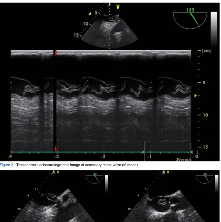
counselled to the cardiologist in the same state hospital. A 2/6 systolic murmur was confirmed while a suspected intracardiac mass related with the aortic valve was detected on transthoracic echocardiography (Fig.s 1, 2). He was referred to our clinic for further investigation.
On physical examination 2/6 mid-systolic murmur on left sternal border was detected. Electrocardiographic findings and laboratory tests were within normal range. On transthoracic echocardiography (TTE) ejection fraction was 0.60 while the cardiac chambers were of normal size. No left ventricular hypertrophy was observed. A mass like structure which was attached to the ventricular part of the anterior mitral laeflet was discovered. No aortic regurgitation and left ventricular outflow obstruction (LVOT) obstruction were detected (resting and exercise peak pressure gradient across LVOT was 8 mmHg). During transeosaphageal echocardiography a mobile, echogenic, chorda tendinea like structure which was attached to the ventricular side of the anterior mitral valve was observed (Fig. 3). It was thought to be an AMV. No other congenital heart disease and aortic regurgitation was found. Annual echocardiographic follow-up was planned since the patient was symptom free.
Figure 1 - Transthoracic echocardiographic image of accessory mitral valve
and symptoms. Our aim is to report a case which was referred to our clinic as a suspected intracardiac mass.
CASO CLÍNICO
Revista Científica da Ordem dos Médicos www.actamedicaportuguesa.com 781 Donmez E, et al. Incidental asymptomatic accessory mitral valve, Acta Med Port 2014 Nov-Dec;27(6):779-782
DISCUSSION
Accessory mitral valve tissue is a rare congenital cardiac malformation.3 The widespread use of transthoracic
echocardiography in the last decades brought up easier diagnosis upon first description by Cheevers in 1842. The patients are usually asymptomatic however they are referred to echocardiography because of systolic murmur. The mechanisms of LVOT obstruction are thought to be due to mass effect of the accessory valve tissue or fibrous tissue deposition resulting a turbulent flow. Symptomatic cases are
usually diagnosed in the early childhood. Symptoms include exercise intolerance, chest pain on exertion, syncope or transient ischemic attack.1,4,5 Obstruction and other
co-existing cardiac anomalies are observed in patients with symptoms regardless from age.3,6,7
Transthoracic echocardiography is the key step in diagnosis. The mass like structure is usually observed in the subaortic area. The Doppler echocardiography should be used to demonstrate the gradient between the left ventricule and the aorta.1 Other LVOT masses like myxoma,
Figure 2 - Transthoracic echocardiographic image of accessory mitral valve (M mode)
CASO CLÍNICO
782
Revista Científica da Ordem dos Médicos www.actamedicaportuguesa.com Dores LA, et al. Parésia do nervo motor ocular comum em contexto de sinusite esfenoidal, Acta Med Port 2014 Nov-Dec;27(6):782-786 papillary fibroleastoma, thrombus or vegetations should be
differentiated from AMV by TTE and transeaosophageal echocardiography.8
Embryologically, AMV is the result of incomplete seperation of the mitral valve tissue from the endocardial cushion.1,9,10 Two types of accessory valve tissue were
defined by Faggian et al in 1983.7 The mobile type is a
parachute-like leaflet floating in the ventricular outflow tract. LVOT obstruction may be caused by mobile type. The fixed type is attached to the interventriular septum firmly and this type may sometimes decrease the size of interventricular defect.
Surgical resection of the AMV is needed when it is symptomatic and related to LVOT obstruction. Surgery usually resolves the symptoms. There is insufficient data regarding the follow-up of asymptomatic cases. The surgical approach for obstructed cases is not well established in the literature.
With regard to this case, AMV should be kept in mind in
the differential diagnosis for intracardiac masses. Careful transechocardiography is needed in order to detect such entity. Further imaging modalities are seldomly needed. No intervention or medication is necessary if the patient is symptom free. Although some authors recommend antibiotic prophylaxis in AMV, it is not indicated in European Society of Cardiology (ESC) Valvular Heart Disease Guidelines 2012. Current ESC guidelines do not recommend prophylaxis for thromboembolic events in AMV specifically. However there are case reports against this recommendation. Authors suggest individual decision for antiaggregant/antiplatellet prophylaxis.
CONFLICTS OF INTEREST
The authors declared that they have no conflicts of interest.
FUNDING SOURCES
None stated.
REFERENCES
1. Rovner A, Thanigaraj S, Perez JE. Accessory mitral valve in an adult population: the role of echocardiography in diagnosis and management. J Am Soc Echocardiogra. 2005;18:494-8.
2. McLean LD, Kane DJ. Subaortic stenosis due to accessory mitral valve. J Thorac Cardiovasc Surg. 1963;45:383-8.
3. Yasui H, Kado H, Tokunaga S, Kanegae Y, Fukae K, Masuda M, et al. Trans-ventricular septal defect approach for resection of accessory mitral valve tissue. Ann Thorac Surg. 1993;55:950-3.
4. Prifti E, Frati G, Bonacchi M, Vanini V, Chauvaud S. Accessory mitral valve tissue causing left ventricular outflow tract obstruction: case reports and literature review. J Heart Valve Dis. 2001;10:774-8. 5. Yetkin E, Turhan H, Atak R, Senen K, Cehreli S. Accessory mitral valve
tissue manifesting cerebrovascular thromboembolic event in a 34-year-old woman. Int J Cardiol. 2003;89:309-11.
6. Gomes AS, Nath PH, Singh A, Lucas RV Jr, Amplatz K, Nicoloff DM, et
al. Accessory flaplike tissue causing ventricular outflow obstruction. J Thorac Cardiovasc Surg. 1980;80:211-6.
7. Faggian G, Frescura C, Thiene G, Bortolotti U, Mazzucco A, Anderson RH. Accessory tricuspid valve tissue causing obstruction of the ventricular septal defect in tetralogy of Fallot. Brit Heart J. 1983;49:324-7.
8. Panduranga P, Al-Mukhaini M. Isolated non-obstructive accessory mitral valve tissue in an adult mimicking ruptured chordae. Indian Heart J. 2013;65:334-6.
9. Rozo JC, Medina D, Guerrero C, Calderon AM, Mesa A. Accessory mitral valve without left ventricular outflow tract obstruction in an adult. Texas Heart Institute journal / from the Texas Heart Institute of St Luke’s Episcopal Hospital, Texas Children’s Hospital. 2008;35:324-6. 10. Uslu N, Gorgulu S, Yildirim A, Eren M. Accessory mitral valve tissue:
report of two asymptomatic cases. Cardiology. 2006;105:155-7.
Parésia do Nervo Motor Ocular Comum em
Contexto de Sinusite Esfenoidal
Third Cranial Nerve Palsy in Sphenoid Sinusitis
1. Departamento de Otorrinolaringologia, Voz e Perturbações da Comunicação. Hospital de Santa Maria. Faculdade de Medicina de Lisboa. Lisboa. Portugal. Recebido: 01 de Fevereiro de 2014 - Aceite: 09 de Abril de 2014 | Copyright © Ordem dos Médicos 2014
Luís Almeida DORES1, Marco Alveirinho SIMÃO1, Marta Canas MARQUES1, Óscar DIAS1
Acta Med Port 2014 Nov-Dec;27(6):782-786
RESUMO
A patologia do seio esfenoidal é particular não só na sua apresentação clínica, mas também pelas suas complicações. Apesar de raras estas podem apresentar-se como défices dos pares cranianos, sendo importante ter um elevado índice de suspeição e estar familiarizado com o seu diagnóstico e abordagem terapêutica. Os sintomas são por vezes muito inespecíficos, contudo os mais fre-quentes são as cefaleias, alterações da acuidade visual e diplopia no contexto de disfunção de um ou mais nervos oculares motores. Reporta-se o caso de um indivíduo do sexo masculino, 59 anos de idade, que foi referenciado ao Serviço de Urgência Otorrinolarin-gologia por quadro de cefaleias frontotemporais de carácter progressivo com uma semana de evolução e agravamento clínico nas últimas 12 horas associando-se diplopia. O exame neurológico sumário não releva outras alterações para além da parésia do nervo motor ocular comum esquerdo. Na rinoscopia anterior/posterior não se verificava presença de massas ou rinorreia mucopurulenta. A Tomografia Computadorizada revelava preenchimento por componente tecidual de partes moles dos seios esfenoidais e erosão do
anos
35
35 anos a p rom ov er as ciê ncia s bio méd ica s A CT A MÉ DI CA P ORT UGUE SA 1979 - 2014Esra DONMEZ, Taner SEKER, Yahya Kemal ICEN, Zafer ELBASAN, Mustafa GUR, Murat CAYLI