421
Pakistan Veterinary Journal
ISSN: 0253-8318 (PRINT), 2074-7764 (ONLINE)
Accessible at: www.pvj.com.pk
Quantification of Mitral Regurgitation in Anatolian Shepherd Dogs with Asymptomatic
Degenerative Mitral Valve Disease
Kursad Turgut1*, Yilmaz Koc2, Hasan Guzelbektes1,3, Amir Naseri1, Mehmet Ege Ince1 and Ismail Sen1
1Selcuk University, Faculty of Veterinary Medicine, Department of Internal Medicine; 2Department of Surgery, Konya/Turkey; 3Kyrgyz Turk Manas University, Faculty of Veterinary Medicine, Department of Internal Medicine, Bishkek/Kyrgyzstan
*Corresponding author: [email protected]
ARTICLE HISTORY (16-069) A B S T R A C T
Received: Revised: Accepted: Published online:
March 20, 2016 May 19, 2016 June 19, 2016 August 23, 2016
Degenerative mitral valvular disease (DMVD) is the most frequent cardiac disease, causing mitral regurgitation (MR) in dogs. The purpose of this study was to compare the ratio of the regurgitant jet area (RJA) to the left atrial area (LAA) (RJA/LAA) with subtracting method to quantify regurgitant volume (RegV) and regurgitant fraction (RF) in asymptomatic Anatolian Shepherd Dogs (ASHs) with DMVD. Thirty-eight ASHs with DMVD were used as experimental group. The control group consisted of 35 healthy ASHs. In 38 ASHs with DMVD (20 B1 dogs and 18 B2 dogs), the severity of MR was assessed by RJA/LAA and subtraction method. No differences were noted between the assays measuring the severity of
MR by χ2 analysis. The observed agreement between the assays was 81% for
RJA/LAA vs RegV and was 73% for RJA/LAA vs RF, and the kappa statistic values for RJA/LAA vs RegV and for RJA/LAA vs RF were 0.63 (substantial agreement) and 0.50 (moderate agreement), respectively. Our results indicate that each quantification method was valuable to estimate the acuteness of the disease in ASHs with MR and all were in good accordance with the echocardiographic heart size and N-terminal Pro-Brain Natriuretic Peptide (NT-proBNP) measurements. Therefore, the each of these non-invasive methods may be functional to serially estimate the acuteness of MR in DMVD in order to monitor the progression of disease. Future studies have to evaluate, if these will be useful to anticipate the risk or time of decompensation in asymptomatic dogs.
©2016 PVJ. All rights reserved Key words:
Anatolian shepherd dog Canine
Cardiac
Degenerative mitral valve disease
Quantification of mitral regurgitation
To Cite This Article: Turgut K, Koc Y, Guzelbektes H, Naseri A, Ince ME and Sen I, 2016. Quantification of mitral regurgitation in Anatolian Shepherd dogs with asymptomatic degenerative mitral valve disease. Pak Vet J, 36(4): 421-424.
INTRODUCTION
Degenerative mitral valvular disease (DMVD) is the most frequent cardiac disease, causing mitral regurgitation (MR) in dogs (Terzo et al., 2009; Borgarelli and Buchanan, 2012; Nakamura et al., 2014). Myocardial
function, mostly normal or is mildly compromised because of increased preload and decreased afterload in dogs with MR secondary to DMVD, even in the presence of cardiac failure (Haggstroöm et al., 2009). Therefore, the assessment of the regurgitated blood volume into the left atrium (LA) in systole is the major determinant of the degree of left heart volume overload (Kittleson and William, 2003).
Non-invasive estimation of the acuteness of MR is valuable for therapeutic decisions and monitoring of progression. One method used to assess the
semi-quantification MR severity consists of calculating the ratio of the regurgitant jet area (RJA) to the LA area (LAA) (RJA/LAA) using color-flow Doppler mode. The source and course of the jets are also evaluated by this method (Marcello et al., 2014; Sargent et al., 2015a). The subtraction method, where the regurgitant volume (RegV) is determined by subtracting the effective systemic stroke volume (SV) from the total left ventricular SV is the other method to determine regurgitant volume (RegV) quantitatively (Paiva et al.,
2012).
There is a debate about the value of each of these quantification methods and only a few studies have reported the comparative value. The purpose of this study was to compare these methods to quantitate RegV and regurgitant fraction (RF) in asymptomatic Anatolian Shepherd Dogs (ASHs) with DMVD.
Pak Vet J, 2016, 36(4): 421-424. 422
MATERIALS AND METHODS
This study was accepted by ethics committee of the Faculty of Veterinary Medicine (permit number: 2012/053). Adult ASHs dogs (n=38) with DMVD and 35 adult healthy ASHs dogs were used as the materials. All the ASHs dogs were referred to the Small Animal Hospital both for a cardiology consultation and for a regular check-up.
The severity of cardiac disease was assigned as specified by the American College of Veterinary Internal Medicine consensus statement (Matos and Glaus, 2010). The vertebral heart scale (VHS) and echocardiographic left atrial/aortic root (LA/Ao) ratio were used for class B animals to differentiate between class B1 and B2. Dogs (n=18) from the experimental group had both VHS scores >10.5 and LA/Ao ratios >1.7 (stage B2, enlarged atrium
and ventricle), whereas 20 had both VHS scores ≤10.5 and LA/Ao ratios ≤1.7 (stage B1, normal cardiac size).
Inclusion criteria in the experimental group were the recognition of mitral valve leaflet thickening and/or mitral valve prolapse (MVP) and the determination of MR. The control dogs were regarded as normal according to the normal findings of cardiologic examinations. Dogs were excluded if they had concomitant non-cardiac diseases or cardiac diseases other than DMVD.
Clinical and cardiologic examination: The systolic and diastolic blood pressures of each dog were determined with an oscillometric technique (Ware, 2010). The VHS of each dog was measured, and the cardiac enlargement was graded according to the VHS method (Buchanan and Bucheler, 1995).
Echocardiography: A complete transthoracic echocardio-graphic examination, including 2-D, M-mode and Doppler was performed in all the dogs in accordance with techniques (Boon, 2011). The presence mitral valve lesions and MVP, and LA/Ao, end-diastolic volume index (EDV-I), mitral valve early wave velocities (E) and peak early
diastolic velocity in the left ventricle mitral annulus (E’) were determined. The transmitral E/E’ ratio as a filling
pressure index was calculated (Höllmer et al., 2016). The mitral valve insufficiency jets and its direction (central, eccentric) were assessed using color-flow Doppler study. The severity of mitral valve insufficiency jets (%) was subjectively assessed. The largest regurgitant jet found in any plane was recorded as RJA/LAA. MR was assigned as mild, moderate or severe if the RJA/LAA ratio was <20%, 20-40% and >50%, respectively (Boon, 2011).
The quantitation of RegV and RF were assessed by subtracting method where the RegV is determined by subtracting the effective systemic SV from the total left ventricular SV described by Boon (2011). Double measurements were performed to avoid mismeasurements. The plasma N-terminal Pro-Brain Natriuretic Peptide (NT-proBNP) concentrations were measured by enzyme immunoassay.
Statistical analysis: All data are reported as the mean±SE. The values for NTpro-BNP, EDV-I and E/E’ were evaluated with ANOVA and Tukey’s test. Independent
sample t-test was performed to compare the RJA/LAA, RV and RF in the B1 and B2 dogs. To examine univariate associations among RegV, RF, NTpro-BNP and LA/Ao, Pearson correlation test was used. To examine univariate associations between ARJ/LAA and RegV, along with, RF, NTpro-BNP and LA/Ao, the Spearman rank correlation test was used. The agreement among the methods of assays was
assessed by McNemar’s chi-square analysis and kappa calculation (SPSS 19.0). Significance level was set P<0.05.
RESULTS
The cardiologic examination findings including the regurgitant jet properties, ARJ/LAA, RegV and RF in the control and the experimental dogs (the B1 dogs, the B2 dogs) are summarized in Table 1 and 2.
The increase in EDV-I was different (P<0.05) in the B2 dogs when compared with the B1 dogs and the control dogs. There was no difference between the B1 dogs and the control dogs (Table 1). The mean plasma NT- proBNP concentration was more elevated (P<0.05) in the B2 dogs (2985±218pmol/L) when compared with the B1 dogs (1588±212pmol/L) and the control dogs (759±47pmol/L). The differences between the B2 dogs and the B1 dogs were
also significant (P<0.05) (Table 1). The E/E’ showed no
difference among the groups (Table 1).
Table 1: Cardiologic examination parameters in the control and
experimental (B1, B2) groups of dogs
Parameters Control group (n= 35)
Experimental group B1 (n=20) B2 (n=18) EDV-I (ml/m2)
(mean and range)
55.7±2.4
(29.9-78.1)b 52.2±3.6
(31.4-73.7)b 75.7±2.2
(54.2-85.2)a
NT-proBNP (pmol/L) (mean and range)
759±47 (250-2050)c
1588±212 (900-4650)b
2985±218 (1250-4300)a E/E’ (septal)
(mean and range)
4.84±0.27 (2.50-8.00)
5.28±0.49 (3.20-11.76)
4.90±0.40 (3.00-7.40) EDV-I=end-diastolic volume index; NT-proBNP=N-terminal Pro-brain Natriuretic Peptide; E/E’=Mitral valve E wave velocity/peak early
diastolic velocity in the left ventricle mitral annulus ratio; abc
= values in each row marked with different character are statistically different with P<0.05.
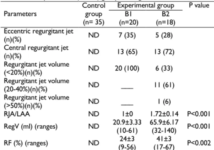
The 26 dogs (68%) in the experimental group with mitral valve leaflet thickening presented with central regurgitation jet, whereas the 12 dogs (32%) in the experimental group with MVP presented with eccentric regurgitation jet (Table 2). On the color-flow Doppler examination, all the B1 dogs consisting of 20 dogs (100%) had mild mitral regurgitation (<20%), whereas in the B2 dogs consisting of 18 dogs; 6 of which (33%) had mild (<20%), 11 of which (61%) had moderate (20-40%) and 1 of which (6%) had severe (>50%) MR. The severity of mitral regurgitation was more elevated (P<0.05) in the B2 dogs than the B1 dogs (Table 2). According to the results of subtracting method, the RegV and RF values were significantly (P<0.05) higher in the B2 dogs than the B1 dogs (Table 2).
Pak Vet J, 2016, 36(4): 421-424. 423
Table 2: The regurgitant jet properties, RJA/LAA, RegV and RF in the
control and experimental (B1, B2) groups of dogs.
Parameters
Control group (n= 35)
Experimental group P value B1
(n=20)
B2 (n=18) Eccentric regurgitant jet
(n)(%) ND 7 (35) 5 (28) Central regurgitant jet
(n)(%) ND 13 (65) 13 (72) Regurgitant jet volume
(<20%)(n)(%) ND 20 (100) 6 (33) Regurgitant jet volume
(20-40%)(n)(%) ND ___ 11 (61) Regurgitant jet volume
(>50%)(n)(%) ND ___ 1 (6)
RJA/LAA ND 1±0 1.72±0.14 P<0.001 RegV (ml) (ranges) ND 20.9±3.33
(10-61)
65.9±6.17
(32-140) P<0.001
RF (%) (ranges) ND 24±3 (9-56)
41±3
(17-67) P<0.002 RJA/LAA=the ratio of the regurgitant jet area (RJA) to the left atrial area (LAA); RegV=regurgitant volume; RF=regurgitant fraction; ND=not determined.
Table 3: Spearman rank correlations test with RJA/LAA in the B2 and B1
dogs
Variable t P value RegV 5.155 0.000***
RF 2.794 0.008**
LA/Ao 3.624 0.000*** NT-proBNP 3.199 0.017* RegV=regurgitant volume; RF=regurgitant fraction; LA/Ao=left atrial to aortic root ratio; NT-proBNP=N-terminal Pro-brain Natriuretic Peptide; *=P<0.05; **=P<0.01; ***=P<0.001
Table 4: Pearson correlations test with RF in the B2 and B1 dogs
Variable Pearson test P value RegV 0.735 0.00** LA/Ao 0.388 0.019* NT-proBNP 0.708 0.000*** RegV=regurgitant volume; LA/Ao=left atrial to aortic root ratio; NT-proBNP=N-terminal Pro-brain Natriuretic Peptide; *=P<0.05; **=P<0.01; ***=P<0.001
Table 5: Pearson correlations test with RegV in the B2 and B1 dogs
Variable Pearson test P value
RF 0.735 0.000***
LA/Ao 0.577 0.000*** NT-proBNP 0.643 0.000*** RF=regurgitant fraction; LA/Ao=left atrial to aortic root ratio; NT-proBNP=N-terminal Pro-brain Natriuretic Peptide; ***=P<0.001
Table 6: Comparison of kappa statistics and observed agreement for
RJA/LAA vs RegV and for RJA/LAA vs RF for determining MR severity Test
compared
No. of samples
Observed agreement (%) χ
2 valuea
Kappa statistic ARJ/LAA vs RegV 38 81 1.29* 0.630
ARJ/LAA vs RF 38 73 3.60* 0.500 a
=McNemars χ2value for correlated proportions. *
=insignificant difference (P>0.05). RJA/LAA=the ratio of the regurgitant jet area (RJA) to the left atrial area (LAA); RegV=regurgitant volume; RF=regurgitant fraction.
No differences were noted between the assays measuring the severity of MR by chi-square analysis. The observed agreement between the assays was 81% for RJA/LAA vs RegV and was 73% for RJA/LAA vs RF, and kappa statistic values for RJA/LAA vs RegV and for RJA/LAA vs RF were 0.63 (substantial agreement) and 0.50 (moderate agreement), respectively (Table 6).
DISCUSSION
The RJA/LAA has both advantage and disadvantage (Marcello et al., 2014; Sargent et al., 2015b). The major advantage of the RJA/LAA is the rapidity, repeatability and
reproducibility. It has also been suggested that the determined RJA/LAA corresponded very well to the different degrees of MR, the severity of the clinical symptoms and the other quantitative values (Mele et al.,
1995; Lopez-Alvarez et al., 2014). However, RJA/LAA is a semi-quantitative method to detect the regurgitant volume which limits its application (Marcello et al., 2014). In addition to this, the factors such as systemic blood pressure, LA pressure, pulse repetition frequency, angle between jet and ultrasound beam, gain settings and spatial orientation of the jet may effect RJA/LAA evaluations (Muzzi et al.,
2003). All the ASHs dogs in this study had no more than 18 mmHg systolic blood pressure, and the difference for septal
E/E’ among the groups was not significant (Table 1). The
26 dogs (68%) of the experimental group had central regurgitation jet, whereas the remaining 12 dogs (32%) with MVP had eccentric regurgitation jet (Table 2). We agreed that ARJ/LAA method was rapid and reproducible. The observed ARJ/LAAs were mostly comparable with the stage of heart failure. Because, all the B1 dogs had mild mitral regurgitation (<20%) and most of the B2 dogs had moderate mitral regurgitations (20-40%). So, the severity of mitral regurgitation was higher (P<0.05) in the B2 dogs than the B1 dogs (Table 2). The observed ARJ/LAAs were also comparable with LA/Ao and NT-proBNP (Table 3). In this study, more severe mitral regurgitation found in the B2 dogs might be the result of the left atrial and ventricular dilatation, because of high LA/Ao, VHS, NT-proBNP and EDV-I (Table 1).
The other methods to determine RegV and RF quantitatively are a) Proximal Isovelocity Surface Area method, and b) subtraction method (Chetboul and Tissier, 2012; Paiva et al., 2012; Sargent et al., 2015a). Significant correlation has been obtained between the two methods (r2=0.94) in the determination of RegV (Doiguchi and Takahashi, 2000). RJA/LAA is a semi-quantitative method. However, the quantification of MR severity by calculation of the RegV, and assessment of the RF is the main advantage of subtracting method (Gouni et al., 2007). Calculation of the cross-sectional area of the aorta by measuring diameter is the main disadvantage of the subtracting method. Any error in measurement is compounded in the calculation because of squaring the radius (Kittleson et al., 2003). For this reason, measurements in subtracting method were double checked to avoid mismeasurements, in this study.
Mitral insufficiency is usually graded as moderate and severe, according to RF values higher than 30-50% and 75%, respectively (Chetboul et al., 2009). The results of this study showed that RF for the B1 dogs was 24±3% and for the B2 dogs was 41±3%. The difference for RF between the B1 dogs and the B2 dogs was significant (P<0.002) (Table 2). So, in another word, we may inform that the B1 dogs had mild mitral insufficiency, whereas the B2 dogs had moderate mitral insufficiency.
Pak Vet J, 2016, 36(4): 421-424. 424
study showed that asymptomatic ASHs with left heart enlargement (the B2 dogs) or without left heart enlargement (the B1 dogs) might have mild to moderate MR, indicating that the assessment of MR severity could be relevant at this stage of the disease. Therefore, the RegV and RF values may give meaningful results to make decision in the severity of MR in ASHs dogs even at B stage of the disease.
Correlation of the RJA/LAA in mitral regurgitation with both RegV and RF is controversial. Chetboul and Tissier (2012) suggested that the RJA/LAA method has been found to be significantly correlated to RF. The RJA/LAA method might therefore be used for both to assess MR severity and for follow-up the DMVD progression. It may also compensate some limitations of quantitative methods. Muzzi et al. (2003) also informed that the RJA/LAA method in MR correlated with both RegV and RF, determined by subtracting method (r=0.81). On the other hand, the studies performed by Grossmann et al. (1995) in humans were contrary to this. They have found poor correlation coefficients due to erratum in the Doppler method. It might contribute to the differences between the RJA/LAA and RegV and RF by Doppler method. Kittleson and Brown (2003) informed that this was also true in dogs. Only a portion of the LA can be filled with regurgitant jet in a dog with severe regurgitation and, conversely, complete of the LA can be filled with regurgitant jet in a dog with mild regurgitation. The size of the color-flow Doppler jet not only reflects the blood flow ejected into the LA but also the blood around it. Consequently, the RJA/LAA method correlates poorly with RegV. However, it was determined that RJA/LAA was positively correlated to RegV and RF in this study (Table
3). We also found no differences between the assays by χ2 analysis in this study. The observed agreement between the assays was 81% for RJA/LAA vs RegV and 73% for RJA/LAA vs RF, and kappa statistic values for RJA/LAA vs RegV and for RJA/LAA vs RF were 0.63 (substantial agreement) and 0.50 (moderate agreement), respectively (Table 6).
Conclusions: The RJA/LAA method was easy to obtain as opposed to subtraction method. Our results indicate that the quantification method was valuable to estimate the acuteness of the disease in ASHs dogs with MR and all were in good accordance with echocardiographic heart size and NT-proBNP levels. Therefore, the each of these non-invasive methods may be useful to serially assess the severity of MR in DMVD in order to monitor the progression of the disease. Future studies are proposed to valuate, if these will be useful to predict the risk or time of decompensation in asymptomatic dogs.
Acknowledgements: This study was supported by both The Scientific and Technical Research Council of Turkey (TUBITAK) and Selcuk University Scientific Research Project Council (SUBAP). We are thankful to Dr. Şeref Inal for his excellent assistance to statistical analyses.
Author’s contribution: KT performed echocardiography,
designed and supervised the study, approved the final version of the manuscript. IS, AN and MEI performed echocardiography and drafted parts of the manuscript. YK
and HG performed statistical analysis of the data, created the figures and tables, drafted parts of the manuscript. All authors approved the manuscript.
REFERENCES
Boon JA, 2011. Veterinary Echocardiography, 2th Ed, Blackwell Publishing, Ames, Iowa 50014-8300, USA, pp: 264-308.
Borgarelli M and Buchanan JW, 2012. Historical review, epidemiology and natural history of degenerative mitral valve disease. J Vet Cardiol, 14: 93-101.
Buchanan J and Bucheler J, 1995. Vertebral scale system to measure canine heart size in radiographs. J Am Vet Med Assoc, 206: 194-199. Chetboul V, Serres F, Tissier R, Lefebvre HP, Carlos SC, et al., 2009.
Association of plasma N-terminal pro-B-type natriuretic peptide concentration with mitral regurgitation severity and outcome in dogs with asymptomatic degenerative mitral valve disease. J Vet Intern Med, 23: 984-994.
Chetboul V and Tissier R, 2012. Echocardiographic assessment of canine degenerative mitral valve disease. J Vet Cardiol, 14: 127-148. Doiguchi O and Takahashi T, 2000. Examination of quantitative analysis and
measurement of the regurgitation rate in mitral valve regurgitation by
the ‘‘proximal isovelocity surface area’’ method. J Vet Med Sci, 62:
109-112.
Gouni V, Serres F, Pouchelon JL, Tissier R, Lefebvre HP, et al., 2007. Quantification of mitral valve regurgitation in dogs with degenerative mitral valve disease by use of the proximal isovelocity surface area method. J Am Vet Med Assoc, 231: 399-406.
Grossmann G, Giesler M and Schmidt A, 1995. Assessment of severity of mitral insufficiency. Value of various color Doppler echocardiographic methods. Z Kardiol, 84: 190–197.
Haggstroöm J, Hoöglund K, Borgarelli M, Haggstrom J and Hoglund K, 2009. An update on treatment and prognostic indicators in canine myxomatous mitral valve disease. J Small Anim Pract, 50: 25-33. Höllmer M, Willesen JL, Tolver A and Koch J, 2016. Comparison of four
echocardiographic methods to determine left atrial size in dogs. J Vet Cardiol, 18: 137-145.
Kittleson MD and William A, 2003. Regurgitant fraction measured by using the proximal isovelocity surface area method in dogs with chronic myxomatous mitral valve disease. J Vet Intern Med, 17: 84-88. Lopez-Alvarez J, Boswood A, Moonarmart W, Hezzell MJ, Lotter N, et al.,
2014. Longitudinal electrocardiographic evaluation of dogs with degenerative mitral valve disease. J Vet Intern Med, 28: 393–400. Marcello MDi, Terzo E, Locatelli C, Palermo V, Sala E, et al., 2014.
Assessment of mitral regurgitation severity by Doppler color flow mapping of the vena contracta in dogs. J Vet Intern Med, 28: 1206-1213. Matos JM and Glaus TM, 2010. Medical treatment of canine heart failure.
European J Comp Anim Pract, 20: 171-176.
Mele D, Vandervoort P and Palácios I, 1995. Proximal jet size by Doppler color flow mapping predicts severity of mitral regurgitation – clinical studies. Circulation, 91: 746-754.
Muzzi RA, de Araujo RB, Muzzi LA, Pena JL and Silva EF, 2003. Regurgitant jet area by Doppler color flow mapping: quantitative assessment of mitral regurgitation severity in dogs. J Vet Cardiol, 5: 33-38. Nakamura K, Osuga T, Morishita K, Suzuki S, Morita T, et al., 2014.
Prognostic value of left atrial function in dogs with chronic mitral valvular heart disease. J Vet Intern Med, 28: 1746-1752.
Paiva RM, Garcia-Guasch Lain, Manubens J and Montoya-Alonso JA 2012. Proximal isovelocity surface area variability during systole in dogs with mitral valve prolapse. J Vet Intern Med, 29: 1280-1289. Sargent J, Connolly DJ, Watts V, Mõtsküla P, Volk HA, et al., 2015a.
Assessment of mitral regurgitation in dogs: comparison of results of echocardiography with magnetic resonance imaging. J Small Anim Pract, 56: 641–650.
Sargent J, Muzzi R, Mukherjee R, Somarathne S, Schranz K, et al., 2015b. Echocardiographic predictors of survival in dogs with myxomatous mitral valve disease. J Vet Cardiol, 17: 1-12.
Serres F, Pouchelon JL, Poujol L, Lefebvre HP, Trumel C, et al., 2009. Plasma N-terminal pro-B-type natriuretic peptide concentration helps to predict survival in dogs with symptomatic degenerative mitral valve disease regardless of and in combination with the initial clinical status at admission. J Vet Cardiol, 11: 103-121.
Terzo E, Di Marcello M, McAllister H, Glazier B, Lo Coco D, et al., 2009. Echocardiographic assessment of 537 dogs with mitral valve prolapse and leaflet involvement. Vet Radiol Ultrasound, 50: 416-422. Ware VA, 2010. Cardiyovascular disease in small animal medicine. Manson