2011/2012
Lídia Sousa
Catecholaminergic Polymorphic
Ventricular Tachycardia
Mestrado Integrado em Medicina
Área: Cardiologia
Trabalho efectuado sob a Orientação de: Doutor Manuel Joaquim Lopes Vaz da silva
Lídia Sousa
Catecholaminergic Polymorphic
Ventricular Tachycardia
MESTRADO INTEGRADO EM MEDICINA
Ano letivo: 2011/2012
Nome do Estudante: Lídia Sousa
Orientador : Doutor Manuel Joaquim Lopes Vaz da Silva Área do Projeto: Cardiologia
Título do Projeto: Catecholaminergic Polymorphic Ventricular Tachycardia Resumo:
A Taquicardia Ventricular Polimórfica Catecolaminérgica é uma doença hereditária arritmogénica rara, mas altamente letal quando não tratada. Constitui um problema de saúde importante, pois pode causar morte súbita em indivíduos jovens e aparentemente saudáveis. Caracteristicamente é induzida pelo exercício físico ou stress emocional e manifesta-se geralmente como uma síncope acompanhada de taquicardia ventricular, polimórfica, bidireccional.
Nas últimas décadas tem havido uma crescente consciencialização sobre distintas canalopatias hereditárias, que são potencialmente letais, o que levou a um desabrochar da investigação neste campo. O objetivo principal desta revisão é resumir o conhecimento atual e destacar as novas descobertas sobre as características clínicas, o diagnóstico, as bases genéticas e fisiopatológicas, a terapêutica e o prognóstico da Taquicardia Ventricular Polimórfica Catecolaminérgica.
Catecholaminergic Polymorphic Ventricular Tachycardia
Sousa, Lídia
Aluna da Faculdade de Medicina da Universidade do Porto
Contact: [email protected]
Faculdade de Medicina da Universidade do Porto
Index
List of Abbreviations ... 2 List of Figures ... 3 List of Tables ... 3 Abstract ... 4 Keywords... 4 Resumo ... 5 Palavras-chave ... 5 Introduction ... 6Material and Methods ... 7
Clinical Manifestations ... 8
Diagnosis and ECG Pattern ... 9
Pathophysiology of Arrhythmias ... 11
Genetic Basis ... 11
RyR2 Mutations ... 11
CASQ2 Mutations ... 13
Ionic and Cellular Mechanisms ... 15
RyR2 Mutations and CPVT ... 17
CASQ2 Mutations and CPVT... 18
Effects of ß -Adrenergic Stimulation ... 19
Current and Future Therapies for CPVT ... 20
Acute Treatment... 20
Chronic Treatment ... 21
ß- Blocker Therapy... 21
Other Options for Chronic Treatment ... 21
Future Developments ... 24 Genetic Testing ... 26 Conclusions ... 27 Acknowledgements ... 29 Conflicts of Interest... 29 References ... 30
2
List of Abbreviations
ACC-American College of Cardiology AHA-American Heart Association CAMKII- Calmodulin Kinase II CASQ2- Calsequestrin 2
CPVT- Catecholaminergic Polymorphic Ventricular Tachycardia DADs- Delayed Afterdepolarizations
ECG- Electrocardiogram
ESC-European Society of Cardiology HRS-Heart Rhythm Society
ICD- Implantable Cardioverter Defibrillators LSD- Left Sympathetic Denervation
LTCC-L-Type Calcium Channels PKA- Phospho- Kinase A
RyR2- Ryanodine Receptor 2 SCD- Sudden Cardiac Death SR- Sarcoplasmatic Reticulum VF- Ventricular Fibrillation VT- Ventricular Tachycardia
3
List of Figures
Figure 1- Typical ECG of a patient with CPVT.
Figure 2- Transmenbranar structure of the RyR2 channel, with localization of a new mutation. Figure 3- CASQ2 gene sequentiation.
Figure 4- Excitation-contraction coupling mechanism.
Figure 5- Schematic sequence: Cytoplasmatic calcium overload- Delayed Afterdepolarizations- Polymorphic Ventricular Tachycardia.
List of Tables
Table 1- RyR2 mutations.4
Abstract
Catecholaminergic Polymorphic Ventricular Tachycardia (CPVT) is a rare, although highly lethal, inherited arrhythmogenic disease. It constitutes an important health burden as may cause sudden cardiac death in young otherwise healthy subjects. Characteristically it manifests as exercise or emotion-induced syncopal events and a distinctive pattern of reproducible, stress- related, bi-directional ventricular tachycardia.
Clinical evaluation by exercise stress testing and holter monitoring can facilitate early identification, even though molecular genetic screening is critical to confirm uncertain diagnosis of CPVT.
An autossomal-dominant form (related to mutations in the gene that codifies the Ryanodine Receptor 2) and an autossomal- recessive form (related to mutations on the cardiac isoform of calsequestrin 2) have been well described, and a mutation on either of these genes is found in 50 to 70% of patients with this syndrome.
The basic arrhythmogenic mechanism of CPVT is thought to be deregulated leak of calcium from the sarcoplasmatic reticulum causing intracytoplasmatic overload and thus potentiating delayed afterdepolarizations and triggered activity.
It is generally accepted that ß-blocker agents are the cornerstone of chronic CPVT therapy, nonetheless, many patients still remain symptomatic; recurrence and mortality remain high and many patients require an implantable cardioverter defibrillator. A great number of innovative therapeutic options have been described and seem promising but more investigation is still warranted.
In the past decade there has been an increasing awareness of distinct, potentially lethal, heritable channelopathic syndromes, which led to a boom of investigation in this field. The main objective of this review is to summarize the current knowledge and highlight the new discoveries on the clinical characteristics, diagnostic, genetics, pathophysiologic mechanisms, therapeutic and prognostic features of CPVT.
Keywords
Catecholaminergic Polymorphic Ventricular Tachycardia; Delayed afterdepolarizations; Lethal arrhythmias; RyR2 Mutation; CASQ2 Mutation; Treatment
5
Resumo
A Taquicardia Ventricular Polimórfica Catecolaminérgica (TVPC) é uma doença hereditária arritmogénica rara, mas altamente letal quando não tratada. Constitui um problema de saúde importante, pois pode causar morte súbita em indivíduos jovens e aparentemente saudáveis. Caracteristicamente é induzida pelo exercício físico ou stress emocional e manifesta-se geralmente como uma síncope acompanhada de taquicardia ventricular, polimórfica, bidireccional.
A avaliação clínica através de um teste de stress ou monitorização com Holter pode facilitar a identificação precoce, ainda que uma investigação genética molecular seja fundamental para confirmar diagnósticos incertos.
Estão descritas duas variantes desta doença, uma forma autossómica dominante que se deve a mutações no Receptor Rianodínico Tipo 2, e uma forma autossómica recessiva associada a mutações na isoforma cardíaca da proteína Calsequestrina 2. Mutações em qualquer um destes genes são encontradas em 50-70% dos pacientes com esta síndrome.
O mecanismo arritmogênico básico consiste na desregularão da libertação de cálcio pelo reticulo sarcoplasmático, causando uma sobrecarga intracelular deste ião e potenciando pós-despolarizações tardias e actividade deflagrada.
É geralmente aceite que os ß-bloqueadores são a pedra angular da terapêutica crónica da TVPC, no entanto, muitos pacientes ainda permanecem sintomáticos; a recorrência e mortalidade permanecem elevadas e em muitos casos é necessário um cardioversor desfibrillador implantável. Têm vindo a ser descritas inúmeras terapêuticas inovadoras, que parecem promissoras, mas ainda é necessária muita investigação nesta área.
Nas últimas décadas tem havido uma crescente consciencialização sobre distintas canalopatias hereditárias, que são potencialmente letais, o que levou a um desabrochar da investigação neste campo. O objetivo principal desta revisão é resumir o conhecimento atual e destacar as novas descobertas sobre as características clínicas, o diagnóstico, as bases genéticas e fisiopatológicas, a terapêutica e o prognóstico da TVPC.
Palavras-chave
Taquicardia Ventricular Polimórfica Catecolaminérgica; Pós-despolarização tardia; Arritmias Letais; Mutação RyR2; Mutação CASQ2; Tratamento
6
Introduction
A sudden death is always dramatic, especially when it concerns young apparently healthy people. Inherited isolated arrhythmogenic disorders, the so called channelopathies, are believed to cause about a third of unexplained deaths in children and adolescents, thus traducing a major health problem. Among these disorders, Catecholaminergic Polymorphic Ventricular Tachycardia (CPVT), a recently discovered and less known condition, is thought to be responsible for approximately 14% of sudden, unexpected death1, 2.
CPVT is a highly lethal form of inherited arrhythmogenic disease, characterized by adrenergically mediated polymorphic ventricular tachycardia3. Despite being rare, with an estimated prevalence at 1/10.000, the mortality rate of this syndrome reaches up to 50% by the age of 30 in untreated individuals, thus making early diagnosis crucial4.
Clinical evaluation by exercise stress testing and holter monitoring can facilitate early identification, even though molecular genetic screening is critical to confirm uncertain diagnosis of CPVT.4
Signs and symptoms usually begin in childhood, and the clinical presentation encompasses exercise or emotion- induced syncopal events, seizures or sudden cardiac death (SCD), that can be the first manifestation of the disease, together with a distinctive pattern of reproducible, stress related, bidirectional ventricular tachycardia (VT) in the absence of both structural heart disease and prolonged QT interval3, 5, 6
In 1961 Horan and Venables described the case of a 6 years old Maltese girl, in whom emotional upset and effort precipitated unusual rhythms of chaotic heart action , bi-directional tachycardia and ventricular fibrillation (VF), in the absence of any cardiovascular abnormality7.
Only 14 years after there was a similar report, when Reid et al. reported a case of bidirectional ventricular tachycardia precipitated by effort and emotional stress in a child with no evidence of any structural abnormality of the heart8.
The first comprehensive description of CPVT was provided in 1995 by Leenhardt et al., who described a 7 years follow up of 21 children suffering from stress or emotion induced syncope, with no evidence of structural heart disease and normal QT interval. It was the first time the term CPVT was used, but no genetic analysis was reported, despite a family history of syncope or sudden death in seven patients suggesting a genetic origin for this syndrome 5.
In 1999, Swan et al. described an arrhythmic disorder mapped to the chromosome 1q42-q43 causing an exercise induced malignant polymorphic ventricular tachycardia in
7 structurally normal hearts; however this study failed to find any mutations or polymorphisms, but pointed Ryanodine Receptor-2 (RyR2) as a candidate target for gene mutation9. Two years latter Priori et al. demonstrated that RyR2 gene mutations are in fact responsible for an autossomal dominant form of CPVT10 (CPVT1). In the same year, Lahat et al. showed that a mutation in the gene for calsequestrin 2 (CASQ2) was the cause of a rare autosomal recessive form of CPVT11 (CPVT2).
More recent studies showed that in approximately 50 to 70% of patients with CPVT a mutation in RyR2 or CASQ2 was identified3, 12. Mutations in these two genes destabilize the RyR2 Ca2+ release channel complex in sarcoplasmatic reticulum (SR) and result in spontaneous Ca2+ release, leading to intracellular accumulation of this ion which potentiates delayed afterdepolarizations (DADs) and triggered activity3, 5, 13-15.
Traditionally, adrenergic ß-blockers and implantable cardioverter defibrillator (ICD) have been used to treat patients with CPVT, either symptomatic or asymptomatic and can substantially decrease the arrhythmia burden and mortality despite being less than ideal treatments16. Other drugs (i.e. verapamil, flecainide, propofenone, dantrolene), and other therapeutic approaches (i.e. left sympathetic denervation and pulmonary vein isolation) have been recently proposed and seem promising17, 18.
During the past decade there has been an increasing awareness of distinct, potentially lethal heritable channelopathic syndromes as they are related to SCD in infants and children. The aim of this review is to highlight and summarize the current knowledge on the clinical characteristics, diagnostic, genetics, pathophysiologic mechanisms, therapeutic and prognostic features of CPVT, a primary electrical heart disease characterized by severe arrhythmias in young patients with apparently normal hearts3.
Material and Methods
This monograph was based on a literature search using the databases: PubMed, Scopus and ISI Web of Science. The former was based on research of MeSH terms
Catecholaminergic Polymorphic Ventricular Tachycardia, to search for articles without any
restriction by type of article, author or publication year. The present research was finished on 14th October 2011.
8
Clinical Manifestations
The first clinical manifestation of CPVT is usually a syncope or cardiac arrest triggered by emotional or physical stress or while swimming. Other manifestations include palpitations, dizziness and convulsions3, 5. In more than a third of cases syncope is associated with seizure, thus explaining delays in diagnosis as often the initial treatment is an anti- epileptic drug. Increasing evidence reinforces that SCD can be the first manifestation of this disease6.
In the first population of CPVT patients described in 1995 by Leenhardt et al. the mean age at first event was 7.8 (±4) years ,which is remarkably similar to the results of a recent study from Priori et al. that reported a mean age of presentation of 8 (±2) years3, 5. The pioneer study of Leenhardt et al. also showed a clear relationship between the age of first syncope and the severity of the disease, concluding that the earlier the first syncope occurred the worse was the prognosis. Nonetheless SCD before the age of 10 was extremely rare , fact that can be explained by the rarity of true VF in small size hearts5.
Patients often have an unremarkable resting electrocardiogram (ECG), without QT prolongation, atrioventricular conduction defects or Brugada-like ST/T- segment pattern; cardiac imaging examinations are also normal3, 5. Therefore, as syncopal events occur in otherwise healthy children with a normal cardiac examination, the diagnosis is frequently missed unless an exercise stress test or holter is performed to document the arrhythmia. During this long delay, between the first symptom and the diagnosis, the patients are obviously under a high risk of sudden death6.
According to Nian et al., the correct diagnosis is established after an average delay of 2 years from the first syncope, and frequently the cardiac events are considered vasovagal events or syncopal events caused by neurologic factors. This group estimated (based on a population of 60 probands, which is so far the largest population of CPVT reported in the world) that 23% of the probands came to the clinical observation for stress induced cardiac arrest, 72% for documentation of polymorphic or bi-directional VT, and 5 % for stress induced syncope. The study performed by this group also demonstrated that 62% of all sudden cardiac death occurred as the first manifestation of CPVT19.
Among patients with mutations in the RyR2 gene the clinical presentation is even more varied and, importantly, 20% show no ventricular arrhythmia during stress test and are completely asymptomatic. In this healthy carrier group there is a predominance of females (80%) thus pointing male gender as a risk factor for cardiac events3.
9
Diagnosis and ECG Pattern
Collecting a careful clinical history is always essential in the approach to any patient. In the particular setting of an arrhythmogenic disorder resembling CPVT it is crucial to establish the relationship between syncope and physical activity or stress; a detailed family history, including questions about any SCD in the family and unexplained syncope, is also of major interest, as approximately 30% of CPVT patients have a positive family history of stress – related syncope, seizures or SCD 3, 5, 20.
As already stated, it is usual that CPVT patients have normal physical examination, normal resting ECG, normal structure in the echocardiography and normal electrophysiological studies, thus making the diagnosis easily overlooked3, 5. Nonetheless, some recent investigations have shown that patients with CPVT may have sinus bradycardia and prominent U-waves in resting ECG, but there is no general agreement on this issue21, 22.
The most reliable way to diagnose CPVT is a standardized exercise stress test 21, 23-25 that induces ventricular arrhythmias when the sinus rate exceeds an individual threshold, usually at rates of 110-130 bpm 23, 25. It is advisable to perform the exercise stress test with a defibrillator readily available in case a VT degenerates into VF 26 (Figure 1).
The arrhythmias become more complex when the sinus rate further increases and consist of scattered premature ventricular complexes, couplets, bigeminy and bidirectional or polymorphic VT , often appearing in this order5, 9.
Bidirectional VT is characterized by the R-wave axis rotation of 180 ° from stroke to stroke3 and is also a typical finding in patients with digitals intoxication and heart failure, both states with increased intracellular Ca2+ concentration19, 27.
Intravenous infusion of adrenaline can also induce progressive ventricular arrhythmias and is mainly used to diagnose CPVT in patients with contraindications for exercise3, 5, 21, 23,
25
.
The arrhythmias can also be detected during Holter monitoring, especially in those where the arrhythmias are mainly triggered by emotional stress 26.
For the CPVT diagnosis it is necessary to exclude structural heart disease and electrolyte abnormalities as well as the use of drugs known to trigger arrhythmias 20.
Important differential diagnosis to be considered are vasovagal or neurocardiogenic syncope, epilepsy/seizures, hypertrophic cardiomyopathy, arrhythmogenic right ventricular cardiomyopathy, long QT syndrome, Andersen-Tawil syndrome and Brugada syndrome. Resting ECG, echocardiography, tilt testing, electroencephalogram and possibly
10 electrophysiological studies with flecainide/ajmaline can be useful for differentiation of these aetiologies 2.
Diagnosis of CPVT can be verified by means of genetic engineering, by identifying precise mutations that have been associated with this syndrome. Post-mortem analysis is relevant in SCD in children and young adults, when no cause of death is found at the autopsy. This may be important to get an explanation of the death and to discover other family members who may be at risk 20.
Figure 1- Holter electrocardiogram recorded during an exercise-related syncopal episode in a young girl with CPVT showing a bidirectional VT degenerating into self-limiting VF. (From Richter et al.28, with permisson.)
11
Pathophysiology of Arrhythmias
Genetic Basis
Since the initial descriptions of CPVT, a familial transmission was reported in about 30% of cases of this disease, fact that conducted to exploration of its genetic basis 19.
Genetic investigations have identified two variants of CPVT: an autosomal dominant form (CPVT1), associated with mutations in the gene encoding the RyR2 and a much less common recessive form (CPVT2), associated with homozygous mutations in the gene encoding the cardiac isoform of CASQ2, this latter being a simple explanation for sporadic CPVT cases without familial involvement 29.
However, in about 30 to 50 % of the patients, it was not identified any disease-causing mutation on either RyR2 or CASQ2 genes, therefore making it likely that other CPVT related genes exist despite not yet discovered3.
Importantly, in 2002, Priori et al. demonstrated that the natural history of the disease does not significantly differ between affected individuals with or without RyR2 mutations. However, 90% of patients with nongenotyped CPVT were females and became symptomatic later in life, i.e.20 ±12 years vs. 8 ± 2 years in patients with RyR2- associated CPVT3.
Recently, cases of mosaicism in CPVT were described30, 31 and this can lead to tragic consequences, since silent mutation carriers are at risk for arrhythmic events12. The manifestations and severity of symptoms of the disease in a carrier of somatic and germline mosaicism depend on several aspects, such as the degree of mosaicism and the gene involved
32
.
Roux-Buisson et al. reinforced the fact that the possibility of germline mosaicism in asymptomatic parents justifies systematic genetic screening of sibling of a sporadic proband, even if the standard methods failed to clearly detect the mutation in the parents31.
Additionally, germline mosaicism also carries a risk of recurrence that despite thought to be <1% should not be underestimated31
RyR2 Mutations
In approximately 50% 3, 27 to 70%33 of the patients with CPVT, mutations on RyR2 gene can be identified, and they have a mean penetrance of 80%33. RyR2 is a tetrameric
12 intracellular Ca2+ release channel located in the SR and required for cardiac excitation-contraction coupling.
The identified mutations are mostly single-base-pair substitutions leading to the replacement of highly conserved amino acids 34, and appear to cluster in three hot spot regions of the gene : N-terminus (77-466), the central domain (2246-2534) and the C- terminal pore region (3778-4959) 15.
In 1999, Swan et al. demonstrated for the first time, the linkage of CPVT to chromosome 1q42-q439.Two years later, RyR2 mutations were identified in four families with the typical pattern of CPVT and history of sudden cardiac death, thus confirming RyR2 as a causative gene for autossomal dominant CPVT form10.
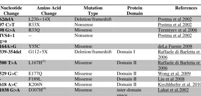
This data has been shortly after confirmed by many other investigators, and a huge number of mutations on the RyR2 gene were identified since then. Those mutations are summarized on Table 1 (see appendix).
Nowadays, the knowledge about RyR2 mutation is growing really fast and this could be illustrated by the fact that just in the first 6 months of the present year of 2011, three new RyR2 mutations were described.
Lobo et al. described a very severe form of CPVT caused not by a simple amino acid substitution, but, by the entire deletion of the exon 3 of RyR2 gene that encodes secondary structure elements that are crucial for folding of the N-terminal domain35.
Kazemian et al. revealed a novel missense mutation in exon 90 of the ryanodine receptor gene resulting in substitution of arginine for serine at residue 4153 (S4153R)36 (Figure 2).
Meli et al. reported a novel CPVT-linked RyR2 mutation that results in the substitution of cysteine for glycine at position 230, RyR2-G230C, located in the “hot-spot” N-terminal domain of cardiac RyR237.
13
Figure 2- Schematic representation of the cardiac RyR2 channel topology. Mutations cluster in RyR2 and are distributed in 3 “hot-spot” regions, called domains I (N-terminal), II (central), and III (channel region). It is also identified the location of the novel S4153R substitution missense mutation. (Adapted from Kazemian et al. 36, with permission.)
CASQ2 Mutations
Identification of CASQ2 mutations is a much more rare event and occurs in only about 2% 3, 27 to 7%33 of the patients with CPVT.
In 2001, Lahat et al. published the sequentiation of the CASQ2 gene (Figure 3) and described a missense mutation, in a highly conserved region of CASQ2, associated with an autossomal recessive form of CPVT, thus confirming CASQ2 (the major Ca2+ reservoir within the SR of cardiac myocytes and a part of RyR2 complex) as a causative gene of CPVT11. It was classically accepted that CASQ2 mutations were responsible for a recessive form of CPVT and thus only homozygous carriers would manifest pathologic clinical phenotype, while heterozygous carriers would be silent3. However, in 2006, Di Barletta et al.
14 reported the first CPVT patient carrying two distinct CASQ2 mutations, demonstrating that compound heterozygous mutations also lead to CPVT phenotype38.
Until now, a limited number of CASQ2-genotyped patients have yet been reported, therefore a significantly smaller number of CASQ2 mutations where identified, when comparing to RyR2 mutations. The former are summarized on Table 2 (see appendix).
Despite CASQ2 related CPVT still awaits more clinical and genetic data, the research in this area is flourishing, as revealed in a study published in 2011 by Roux-Buisson et al., that reported the first splicing abnormalities in CASQ2 caused by intron mutation (c.939+5G>c) or synonymous change (c.381C>T, p.Gly127Gly). This investigation also reported two single nucleotide deletions (c.546delT; c.03delA) and three single nucleotide substitution (c.320-24>g; c.7.37+1g>A; c.101+1G>A), thus contributing significantly for a better understanding of CASQ2 related CPVT genetic mechanisms 39.
Figure 3
a- Physical map of the CPVT locus. Landmark genes are shown as red boxes with their HUGO nomenclature
symbols. Microsatellite markers are indicated by arrows pointing to their exact position on the genomic bar map. b- Expanded view of a 75-kb segment, showing the genomic organization of the CASQ2 interval. The interval is
covered by two genomic clones that have 5-kb overlap. AL449264 contains the first four exons, and AL450389 contains the other seven exons, depicted by blue boxes. The lengths of the exons are shown below the boxes, and those of the introns are shown above the line. The translation-initiation site and the stop codon are indicated. (From Lahat H et al.11, with permission).
15
Ionic and Cellular Mechanisms
The plateau phase of ventricular action potential is the result of balanced Ca2+ influx trough the L-type Ca2+ channels (LTCC) and K+ efflux (critically depending on the delayed rectifier K+ channels: IKr (hERG) and IKs (KCNQ1/KCNE1)40, 41.
Following an action potential, a small influx of Ca2+ through the L-type voltage dependent channels, rises intracellular Ca2+ concentration, thus activating the RyR2 Ca2+ release channel located in the SR, a mechanism known as Ca2+ induced Ca2+ release 40. Calcium ions will then bind to the troponin complex and activate the contractile apparatus 40,
41
.
Relaxation of the cardiac muscle will follow Ca2+ removal from the cytosol, in the rabbit, cat, dog, guinea pig, and humans most of the Ca2+ (70%) is recycled into the SR via SR Ca2+ -ATPase (SERCA), while 28% is extruded from the cell through Na+ / Ca2+ exchanger (NCX), about 1% is removed by the sarcolemmal Ca2+-ATPase, and an additional 1% is transported into the mitochondria 42, 43. Reduction of cytosolic calcium [Ca2+] to the basal level allows Ca2+ dissociation from the myofilaments and mechanical relaxation.
The ability to store a large amount of Ca2+ into the SR is due to the activity of CASQ2, a selective Ca2+ buffering protein, located in specialized areas of the SR, where it forms junctions with the sarcolema of the myocytes 43, 44.
Further, CASQ2 forms a quaternary complex with the RyR2 channel and with the junctional SR membrane proteins Triadin-1 and Junctin, thus being also an important regulator of RyR2 channel function apart of its role for global Ca2+ binding and buffering 45.
It is easy to understand that, playing a central role in the contraction/relaxation cycle of the heart, a tight regulated release of Ca2+ from the SR is essential to maintain normal cardiac activity 40, 42, 44 (Figure 4).
Deregulation of function of either RyR2 channel or CASQ2 will, as one can expect, destabilize the mechanism of Ca2+ intracellular currents from the SR resulting in spontaneous excessive release of this ion 43.
When cardiac cells are overloaded with Ca2+, Ca2+ sparks (the ascent and descent in [Ca 2+] during a cardiac cycle is called Ca2+transient; an elementary unit of Ca2+release from the SR, generated by Ca2+
influx from a single LTCC and adjacent RyR2, is regarded as calcium spark) lead to propagating Ca2+ waves that potentiate DADs, which in turn can lead to triggered activity and bidirectional/polymorphic VT, mimicking the toxic effect of digitals 13, 43 (Figure 5).
16 Delayed afterdepolarizations are enhanced by adrenergic stimulation, which can explain why CPVT manifest during stress 10, 43.
Figure 4- Protein complexes, cardiomyocyte architecture, and intracellular organelles involved in excitation– contraction coupling. (From Watanabe et al. 43 with permission.)
17
a- Intracellular calcium overload caused by RyR2 and CASQ2 mutations and potentiated by B-adrenergic stimulation. (Adapted from Farwell et al.46, with permission.)
b- Delayed afterdepolarizations caused by intracellular calcium overload.
c- Polymorphic Ventricular Tachycardia related to delayed afterdepolarizations as showed in b.
RyR2 Mutations and CPVT
There are various proposed mechanisms underlying leaky RyR2 channel as a result of RyR2 mutations.
Open probability of RyR2 channel is increased by Ca2+ concentration in the cytosol, and spontaneous Ca2+ release by RyR2 channel occurs when Ca2+ concentration reaches a certain level (the Ca2+ release threshold) 47.
Meli et al. recently showed that some RyR2 mutations (including RyR2-S2246L, RyR2-R2474s, RyR2-R4497c, RyR2-P2328S, RyR2-Q42o1R and RyR2-V4653F), linked to
18 CPVT, shift the sensitivity for cytosolic Ca2+ dependent activation of the channel, allowing the channel to be opened even at low diastolic levels of cytosolic Ca2+, thereby causing a diastolic SR Ca2+ leak. Importantly, this alteration in sensitivity was only observed after PKA (phosphokinase A) phosphorylation, condition that mimics the effect of exercise or stress on the channel activity37. More recently, it was proved that caffeine also has the ability to lower the threshold at which luminal Ca2+can activate spontaneous opening of RyR2 channel 48.
Normally, when the cytosolic Ca2+ level rises too high, there is a Ca2+ dependent inhibition of RyR2 channels, stopping the leak of Ca2+ from the SR. Thomas et al. reported that RyR2-L433P and RyR2-N23861 mutant channels exhibit impaired sensitivity to Ca2+ dependent channel inhibition49.
Investigations using the CPVT mutant model RyR2-R2474S suggests that CPVT linked mutations induce defective interdomain conformational changes that destabilize the closed state of RyR2 channel and enhance the probability of opening50.
The importance of SR Ca2+ load in the genesis of Ca2+ waves and arrhythmias is also supported by the investigations of Sedej et al. With the same mice model (RyR2-R4496) they demonstrated that, using ouabain (an inhibitor of Na+/K+-ATPase) to produce Na+ dependent rise of SR Ca2+ load (Na+ - Ca2+ exchange), it is possible to reproduce the Ca2+ waves leading to arrhythmias as those seen in CPVT patients 51.
CASQ2 Mutations and CPVT
In what concerns to Calsequestrin 2 mutations, the best described mechanisms leading to CPVT is the reduction or complete loss of activity of this selective Ca2+ buffering protein 43. Several studies using CASQ2 null mice have shown that a modest reduction of calsequestrin protein is sufficient to increase arrhythmia susceptibility even in the absence of any change in other SR proteins41, 43.
Mutations in CASQ2 gene can also impair interaction of Calsequestrin with RyR2 protein complex, despite a normal amount of this protein, thus impairing the regulation of Ca2+ release from the SR. A good example is the point mutation D307H that leads to a profoundly altered conformation of calsequestrin that no longer responds normally to Ca2+ and fails to bind to Triadin and Junctin 52.
A pioneer work from Eckey et al., showed that CASQ2 is also a functional modulator of the hERG K+ repolarizing channels and that some CASQ2 mutants (including R33Q and
19 F189L) show changes in modulations of these channels leading to abnormalities in the plateau phase of the cardiac action potential41.
Finally, the most recent study about the way CASQ2 mutations lead to CPVT suggests that some mutations modify calsequestrin behavior, including folding (L167H mutation), aggregation/ polymerization (R33Q mutation) and selectivity towards Ca2+ (D307H and P308L mutations44.
Effects of ß -Adrenergic Stimulation
Sympathetic stimulation of ß-adrenergic receptors leads to activation of adenylate cyclase through a Gs protein, ultimately inducing cyclic AMP synthesis. The rise in cyclic AMP activates PKA to phosphorylate its target proteins, mediating the well known inotropic and lusitropic sympathetic effects. Phosphorylation of LTCC increases Ica currents and Phospholamban phosphorylation increases Ca2+ uptake into the SR, enhancing relaxation and increasing [Ca2+] SR 27.
RyR2 phosphorylation leads to its dissociation of Calstabin 2 (also called FKBP12.6, a channel stabilizing protein that binds to RyR2 and stabilizes it in a closed state), thus facilitating Ca2+ release from the SR 27. Phosphorylation of Calstabin 2 by PKA further reduces its ligation to RyR2, resulting in increased Ca2+ leak 43, 53.
One of the most recent publications about the mechanisms leading to CPVT aimed to further clarify why arrhythmias only develop during ß-adrenergic stimulation. Mice carrying a human CPVT mutation (RyR2-R4496) were stimulated with isoprotenerol (ß1 and ß2 agonist) and it was verified that the Ca2+ load of SR and the diastolic Ca2+ waves significantly rose, which typically reproduced the CPVT pathophysiology. Those data allowed several authors to postulate that ß-adrenergic stimulation, besides lowering the threshold for RyR2 channel opening, as already stated, also contributes to the arrhythmias by raising the SR Ca2+ load54.
Cumulatively these mechanisms result in increased RyR2 open probability and Ca2+ leak, thereby explaining increased predisposition to Ca2+ induced arrhythmia during sympathetic stimulation.
20
Current and Future Therapies for CPVT
Since the first descriptions of the disease it has been recognized that the first step in the approach to a patient with CPVT should be: careful information about the importance of the therapeutic compliance; advising against participation in competitive sports and clarifying the contraindication to use sympathomimetic agents5, 16.
Until now there is a lack of information to identify patients with such a low risk of arrhythmic events that would make treatment unnecessary. Therefore, all phenotypically and/ or genotypically diagnosed patients are considered at risk and should receive appropriate therapy16.
CPVT therapy is challenging because it interferes with excitation-contraction coupling and must therefore be compatible with adequate contractility and physiological electrical activity.
Acute Treatment
In the acute management of a sustained VT or VF, in the setting of CPVT, the most critical aspect is to recognize that it concerns in fact a CPVT patient and thus standard adrenaline infusion, for resuscitation, should be avoided 16.
Intravenous ß-blocker therapy is considered as Class I recommendation and should be the first choice for acute management. General anesthesia can be used as last resort when there is no response to ß-blockers agents 4.
Despite the general previous statements, acute treatment of polymorphic VT as provided in the ACC/AHA/ESC guidelines4 could be summarized as follows: 1) if hemodynamic compromise is present, immediate direct current cardioversion should be performed; 2) for recurrent polymorphic VT, intravenous beta blockers should be administered, especially if ischemia is suspected or cannot be excluded; 3) for recurrent polymorphic VT in the absence of congenital or acquired QT interval prolongation, intravenous amiodarone should be administered.
Adenosine triphosphate and verapamil can be considered as future options for the management of these acute situations, since they were effective terminating adrenaline-induced VT during a recent CPVT electrophysiological study 55.
21
Chronic Treatment
ß- Blocker Therapy
The relationship between VT and adrenergic activation, as well as the efficacy of ß-blocker therapy were recognized since the pioneer clinical descriptions of CPVT 5, 7, 8.
In the first comprehensive CPVT series published by Leenhardt et al. in 1995 it became apparent that ß-blockers agents were the most effective drug therapy reducing ventricular arrhythmias during exercise testing or Holter monitoring, as well as preventing arrhythmic events. Particularly, the efficacy of nadolol was documented in all the patients, both clinically and on ECG studies 5.
In subsequently published series the results have been variable, with the range of arrhythmic events on ß-blocker therapy (acebutulol, atenolol, bisoprolol, metoprolol, nadolol, or propanolol) ranging from 0 to 55% in 10 studies conducted until now (including between 14 and 101 young patients, with a follow up duration between 1.8 to 7.9 years). It was also reported a proportion of nearly fatal and fatal arrhythmic events ranging from 0 to 40% and 2 to 20% respectively 3, 5, 9, 12, 21, 23, 24, 56-58 .
Despite these different results it is generally accepted that ß-blocker agents are the cornerstone of chronic CPVT therapy4. However, inadequate response or an “escape” phenomenon, is not uncommon; recurrence and mortality remain high and many patients require an ICD27 fact that conducted to the exploration of other management options.
Other Options for Chronic Treatment
Implantable cardioverter-defibrillators
Class I recommendation was given for implantation of an ICD in addition to ß-blocker therapy in CPVT patients who were survivors of aborted cardiac arrest4. Patients with CPVT who develop sustained VT or syncope (for which causes other than VT are excluded) while taking a beta blocker are likely to be at higher risk of future SCD. Electrophysiological testing is generally not helpful in patients with CPVT since the arrhythmia is usually not inducible with programmed ventricular stimulation. In such patients, ICD placement is a reasonable approach (class II recommendation according the 2006 ACC/AHA/ESC Guidelines for
22 Management of patients with Ventricular arrhythmias and the Prevention of Sudden Cardiac Death4. In 2008 ACC/AHA/HRS Guidelines For Device-Based Therapy of Cardiac Rhythm Abnormalities, reinforced those indications59.
Nonetheless, ICDs have been implanted in a more liberal way before and after these guidelines were published. This might be a dangerous practice, because ICDs can have a harmful effect in CPVT patients. There were reported cases showing that appropriate and inappropriate ICD shocks can trigger catecholamine release, subsequently resulting in multiple shocks, arrhythmic storm, and death. Thus, the implantation of an ICD does have a potential proarrithmic effect60-62 .
More recently , the Hearth Rhytm UK Statement on Clinical Indications for ICD in adult Patients with Familial Sudden Cardiac Death concurred with the previous recommendations, while adding left sympathetic denervation (LSD) as a therapeutic consideration before ICD implantation63.
Verapamil
The rationale for treating CPVT patients with a L-type Ca2+ channel blocker is easy to accept, because, as it was already stated, Ca2+ influx through the L-type Ca2+ channels, triggers Ca2+-dependent Ca2+ release thought defective or inappropriately regulated RyR2 channel, leading to the ionic and electrical abnormalities that characterize CPVT 38.
Three recent studies 64-66 have evaluated and proved a beneficial effect of verapamil in treating CPVT selected patients, but just in combination with ß-blocker therapy, thus the true role of verapamil has not yet been well assessed. However the fact that in none of these three studies was reported a negative ionotropic effect of verapamil is a very positive indication.
Flecainide
Recently, it was discovered that the antiarrhytmic agent and Na+ channel blocker flecainide directly blocks RyR2 channels, prevents RyR2-mediated premature Ca2+ release, and suppresses triggered beats in myocytes isolated from mouse hearts lacking calsequestrin, an animal model of CPVT67. This effect is not mediated by Na+ channel block, the conventional mode of action thought to underlie flecainide activity, but rather can be attributed to open state block of RyR2 channels ( that is, flecainide directly targets the molecular defect responsible for the arrhythmogenic Ca2+ waves that trigger CPVT in vivo )68.
23 Additionally, it was also reported that flecainide completely prevented CPVT arrhythmic events in two human subjects who remained highly symptomatic on conventional drug therapy, indicating that it might be promising in the management of CPVT 67.
The efficacy of flecainide was also retrospectively evaluated in a relatively large multicenter study including 33 CPVT patients, refractory to ß-blocker therapy, alone or combined with Ca2+ channel blockers. The results were encouraging, as in 76% of the patients flecainide (the median daily dose in responders was 150 mg - range 100 to 300 mg; follow-up 12-40 months) suppressed exercise induced ventricular arrhythmias (partially or completely), and importantly, proarrhytmia as a result of flecainide was not observed 17.
Currently is ongoing a clinical trial comparing ß-blocker therapy alone and with flecainide, to test the effect of flecainide prospectively [more information can be accessed at (http:77clinicaltrials.gov:NTC01117454)].
Left sympathetic denervation
LSD (also known as left stellate cardiac ganglionectomy) consists in a transection of the left sympathetic chain at the levels of T1 to T5. Moss et al. described it for the first time in 1971 for the management of Long QT syndrome, and since than it has been used, with good results, in patients with this syndrome that are refractory to other therapies, fact that encouraged its use in other arrhythmogenic diseases69.
In CPVT the sympathetic nervous system plays a crucial role in the genesis of life-threatening arrhythmias. Thus, it was predictable that LSD would increase ventricular refractoriness threshold 70.
Since LSD was first applied to CPVT patients refractory to ß-blocker therapy and receiving recurrent ICD discharges, two case series 71, 72 and some isolated cases 73, 74 were reported, using video-assisted thoracoscopic LSD, with excellent results.
Unlike ß-blockers, LSD does not impair the physiological increase in ventricular function with exercise, neither decreases the resting ventricular function, because the contralateral sympathic chain is intact70.
The drawbacks of LSD include rare potential complications such as transient or persistent Horner´s syndrome and pneumothorax, and the fact that it is not a universally available procedure , requiring an expert surgeon and delicate instrumentation75.
24 Recently, it has been postulated that LSD should be performed simultaneously with ICD implantation, as it does not increase the length of hospital stay or procedure related mortality, and can prevent the potential proarrhytmogenic effect of ICD shocks 18.
Future Developments
Apart from the previously described options for chronic management, that are already being introduced into clinical practice, there are also some other treatment modalities that have more recently being reported, with good results, in some selected cases of CPVT, and therefore may soon become available in a larger scale.
Propafenone
Propafenone, a class Ic antiarrhytmic agent that also contains ß-receptor blocking properties was clinically proved to be effective in suppressing appropriate ICD shocks and exercise induced CPVT in a Turkish patient, who remained severely symptomatic after maximal drug therapy and LSD, and thus may be an ideal drug in CPVT management. Propafenone was attempted because flecainide is not available in Turkey, and subsequent in vitro and in vivo studies showed that propafenone has similar RyR2 blocking properties as flecainide76.
Dantrolene
Dantrolene proved recently to prevent exercise and adrenaline induced ventricular arrhythmias in a CPVT mouse model77. This action is thought to be due to ligation and stabilization of the mutated RyR2 78 and restoration of the affinity of calmodulin to bind RyR279. Dantrolene also improved substantially the contractile function in pacing induced failing hearts, fact that is quite advantageous for its clinical use78.
Calmodulin kinase II inhibition
The Calmodulin kinase II (CAMKII) inhibitor KN93 was proven to completely prevent catecholaminergic-induced sustained VT in RyR2 mutant mice. Mechanistically CAMKII inhibition acts on several elements of the excitation-contraction coupling cascade, including attenuation of SR Ca2+ leak and blunting catecholaminergic-mediated SERCA activation, and thus may represent a novel therapeutic target for patients with CPVT 80.
25 Magnesium supplementation
The nonspecific RyR2 blocking effect of Mg2+ was rather remarkable in CASQ2 mutant mice and this electrolyte has a proven added value in numerous types of ventricular arrhythmia. Katz et al.27 suggest that preserving normal levels of Mg2+ and supplementations to increase intracellular Mg2+ should be recommended to CPVT patients unless otherwise contraindicated.
Selective serotonin reuptake inhibitors
Chen et al. described recently the case of a patient, in whom, the selective serotonin reuptake inhibitor paroxetine, combined with ß-blocker therapy, prevented ICD shocks during two years, while the patient had received two shocks in the previous 6 months, when treated with ß-blocker agents alone. However the exact mechanism underlying this action remains unclear81.
Pulmonary vein isolation
Pulmonary vein isolation was performed in a patient with CPVT that received inappropriate ICD shocks due to atrial fibrillation. This procedure did not just reduce this supraventricular arrhythmia, but also, decreased the number of VTs and premature ventricular complexes on Holter registration82.
Pulmonary vein isolation may have decreased the sympathetic enervation, and thus, may be an additional treatment option in patients resembling the case described.
26
Genetic Testing
Genetic testing is of major interest since CPVT is one of the rare cases when lifestyle modification and even drug therapy appear to be indicated even in the absence of clinical phenotype, based only on genetic diagnosis83.
RyR2 is the prevailing candidate gene, and thus, in a cost-contained environment, should be the first gene to be screened, unless disease transmission is incompatible with autosomal dominant inheritance. Screening of CASQ2 gene is warranted after excluding RyR2 mutations, in particular, when CPVT is familial and the history is suggestive, or possibly compatible with recessive inheritance (i.e., consanguinous, similar ethnic background, etc). Because RyR2 is a huge gene, a cost effective strategy was developed restricting the screening to 20% of gene length but covering the regions encompassing 95% of the mutations described until now 84.
If the genetic study is negative for both RyR2 or CASQ2 gene mutations, screening for Long QT genes, starting with KCNJ2, may be considered, particularly in females with bidirectional/polymorphic VT85.
27
Conclusions
CPVT is a rare, although highly lethal, inherited arrhythmogenic disease. Affecting predominantly young otherwise healthy subjects it represents an important health burden.
Patients experience catecholamine induced ventricular arrhythmias with modest exercise or emotional stress, fact that creates a significant limitation to their normal lives.
SCD can be the first presentation and thus early diagnosis and institution of therapy can drastically change the prognosis and be lifesaving.
Just in the last 12 months 58 articles were published, presenting novel information on this disease, thus, it seemed important to summarize the current knowledge and highlight the new discoveries on the clinical characteristics, diagnostic, genetics, pathophysiologic mechanisms, therapeutic and prognostic features of CPVT, constituting this, the main objective of this review.
Since the first case reports of this disease, the knowledge about clinical characteristics and diagnosis has not improved a lot, as proved by the fact that a definitive CPVT diagnosis is nowadays still made with an average delay of about two years.
In what concerns to understanding the pathophysiology of the arrhythmias, numerous explanations have been proposed, but, since there is often more than one possible interpretation of experimental findings, several controversies have evolved and there is still no consensus in this field. There is although general agreement that the base of the electrical abnormalities is intracellular Ca2+ overload, which potentiates DADs and triggered activity.
The field with more information being published is the genetic basis of CPVT. An increasing number of CASQ2 and mainly RyR2 gene mutations have been identified. To date, there are more than 100 known mutations in RyR2 causing CPVT1 and 12 CASQ2 mutations reported to cause CPVT2. This represents a major advance since, identifying a disease causing mutation, not only confirms and refines the clinical diagnosis, facilitating the most appropriate behavioral counseling and therapy, but also, creates the possibility of genotyping and risk-stratifying family members of the proband. Ilustrating this, the recent description of mosaicism in CPVT justifies genetic screening of sibling of a sporadic proband even when standard methods fail to detect mutations in the parents.
In what concerns to the management of CPVT, the recent advances have also been impressive. ß-Blocker agents are still considered the cornerstone of therapy, and ICD implantation is recommended for survivors of cardiac arrest but a great number of new agents and management procedures have been reported with good results in selected patients.
28 The generation of animal models for arrhythmogenic channelopathies is well underway, and analyses of these models will continue to provide insights into molecular processes leading to various diseases. Ultimately, mouse model lines will offer the potential for testing therapeutic approaches that might help combat these disorders.
Summing up, CPVT is a recently discovered and not well known disorder; a lot of work still remains to be done in order to a better understanding of the ionic and cellular arrhythmogenic mechanisms and the genetic subtract of the disease, providing clues for an early diagnosis as well as new possible therapeutic targets. In what concerns to the latter, specifically tailored therapy for a given molecular defect is still a long way off, but seems a promising possibility in the treatment of CPVT.
29
Acknowledgements
I would like to thank my tutor, Dr. Manuel Vaz Silva, for the unconditional support; to all the staff of the library at Faculty of Medicine of Oporto University for helping me with the bibliographic research, specially to Dr. Filipa Torres; to all my friends who read and criticised my monograph and finally to all the authors that gently gave me the permission to use their images to illustrate my work. To all this people, my best accomplishments.
Conflicts of Interest
There are no conflicts of interest to declare.30
References
1. Morentin B, Suarez-Mier MP, Aguilera B. Sudden unexplained death among persons 1-35 years old. Forensic Sci Int. 2003;135(3):213-7.
2. Tester DJ, Ackerman MJ. Cardiomyopathic and channelopathic causes of sudden unexplained death in infants and children. Annu Rev Med. 2009;60:69-84.
3. Priori SG, Napolitano C, Memmi M, Colombi B, Drago F, Gasparini M, et al. Clinical and molecular characterization of patients with catecholaminergic polymorphic ventricular tachycardia. Circulation. 2002;106(1):69-74.
4. Zipes DP, Camm AJ, Borggrefe M, Buxton AE, Chaitman B, Fromer M, et al. ACC/AHA/ESC 2006 Guidelines for Management of Patients With Ventricular Arrhythmias and the Prevention of Sudden Cardiac Death: a report of the American College of Cardiology/American Heart Association Task Force and the European Society of Cardiology Committee for Practice Guidelines (writing committee to develop Guidelines for Management of Patients With Ventricular Arrhythmias and the Prevention of Sudden Cardiac Death): developed in collaboration with the European Heart Rhythm Association and the Heart Rhythm Society. Circulation. 2006;114(10):e385-484.
5. Leenhardt A, Lucet V, Denjoy I, Grau F, Ngoc DD, Coumel P. Catecholaminergic polymorphic ventricular tachycardia in children. A 7-year follow-up of 21 patients. Circulation. 1995;91(5):1512-9.
6. Liu N, Ruan Y, Priori SG. Catecholaminergic polymorphic ventricular tachycardia. Prog Cardiovasc Dis. 2008;51(1):23-30.
7. Horan M, Venables AW. Paroxysmal tachycardia with episodic unconsciousness. Arch Dis Child. 1962;37:82-5.
8. Reid DS, Tynan M, Braidwood L, Fitzgerald GR. Bidirectional tachycardia in a child. A study using His bundle electrography. Br Heart J. 1975;37(3):339-44.
9. Swan H, Piippo K, Viitasalo M, Heikkila P, Paavonen T, Kainulainen K, et al. Arrhythmic disorder mapped to chromosome 1q42-q43 causes malignant polymorphic ventricular tachycardia in structurally normal hearts. J Am Coll Cardiol. 1999;34(7):2035-42. 10. Priori SG, Napolitano C, Tiso N, Memmi M, Vignati G, Bloise R, et al. Mutations in the cardiac ryanodine receptor gene (hRyR2) underlie catecholaminergic polymorphic ventricular tachycardia. Circulation. 2001;103(2):196-200.
11. Lahat H, Pras E, Olender T, Avidan N, Ben-Asher E, Man O, et al. A missense mutation in a highly conserved region of CASQ2 is associated with autosomal recessive catecholamine-induced polymorphic ventricular tachycardia in Bedouin families from Israel. Am J Hum Genet. 2001;69(6):1378-84.
12. Hayashi M, Denjoy I, Extramiana F, Maltret A, Buisson NR, Lupoglazoff JM, et al. Incidence and risk factors of arrhythmic events in catecholaminergic polymorphic ventricular tachycardia. Circulation. 2009;119(18):2426-34.
13. Priori SG, Corr PB. Mechanisms underlying early and delayed afterdepolarizations induced by catecholamines. Am J Physiol. 1990;258(6 Pt 2):H1796-805.
14. Blayney LM, Lai FA. Ryanodine receptor-mediated arrhythmias and sudden cardiac death. Pharmacol Ther. 2009;123(2):151-77.
15. Yano M, Yamamoto T, Ikeda Y, Matsuzaki M. Mechanisms of Disease: ryanodine receptor defects in heart failure and fatal arrhythmia. Nat Clin Pract Cardiovasc Med. 2006;3(1):43-52.
16. van der Werf C, Zwinderman AH, Wilde AA. Therapeutic approach for patients with catecholaminergic polymorphic ventricular tachycardia: state of the art and future developments. Europace. 2011.
31 17. van der Werf C, Kannankeril PJ, Sacher F, Krahn AD, Viskin S, Leenhardt A, et al. Flecainide therapy reduces exercise-induced ventricular arrhythmias in patients with catecholaminergic polymorphic ventricular tachycardia. J Am Coll Cardiol. 2011;57(22):2244-54.
18. Moray A, Kirk EP, Grant P, Camphausen C. Prophylactic left thoracic sympathectomy to prevent electrical storms in CPVT patients needing ICD placement. Heart Lung Circ. 2011;20(11):731-3.
19. Liu N, Colombi B, Raytcheva-Buono EV, Bloise R, Priori SG. Catecholaminergic polymorphic ventricular tachycardia. Herz. 2007;32(3):212-7.
20. Ylanen K, Poutanen T, Hiippala A, Swan H, Korppi M. Catecholaminergic polymorphic ventricular tachycardia. Eur J Pediatr. 2010;169(5):535-42.
21. Postma AV, Denjoy I, Kamblock J, Alders M, Lupoglazoff JM, Vaksmann G, et al. Catecholaminergic polymorphic ventricular tachycardia: RYR2 mutations, bradycardia, and follow up of the patients. J Med Genet. 2005;42(11):863-70.
22. Aizawa Y, Komura S, Okada S, Chinushi M, Morita H, Ohe T. Distinct U wave changes in patients with catecholaminergic polymorphic ventricular tachycardia (CPVT). Int Heart J. 2006;47(3):381-9.
23. Celiker A, Erdogan I, Karagoz T, Ozer S. Clinical experiences of patients with catecholaminergic polymorphic ventricular tachycardia. Cardiol Young. 2009;19(1):45-52. 24. Lahat H, Eldar M, Levy-Nissenbaum E, Bahan T, Friedman E, Khoury A, et al. Autosomal recessive catecholamine- or exercise-induced polymorphic ventricular tachycardia: clinical features and assignment of the disease gene to chromosome 1p13-21. Circulation. 2001;103(23):2822-7.
25. Sumitomo N, Harada K, Nagashima M, Yasuda T, Nakamura Y, Aragaki Y, et al. Catecholaminergic polymorphic ventricular tachycardia: electrocardiographic characteristics and optimal therapeutic strategies to prevent sudden death. Heart. 2003;89(1):66-70.
26. Napolitano C, Priori SG. Diagnosis and treatment of catecholaminergic polymorphic ventricular tachycardia. Heart Rhythm. 2007;4(5):675-8.
27. Katz G, Arad M, Eldar M. Catecholaminergic polymorphic ventricular tachycardia from bedside to bench and beyond. Curr Probl Cardiol. 2009;34(1):9-43.
28. Richter S, Gebauer R, Hindricks G, Brugada P. A Classic Electrocardiographic Manifestation of Catecholaminergic Polymorphic Ventricular Tachycardia. J Cardiovasc Electrophysiol. 2011.
29. Mohamed U, Napolitano C, Priori SG. Molecular and electrophysiological bases of catecholaminergic polymorphic ventricular tachycardia. J Cardiovasc Electrophysiol. 2007;18(7):791-7.
30. Medeiros-Domingo A, Bhuiyan ZA, Tester DJ, Hofman N, Bikker H, van Tintelen JP, et al. The RYR2-encoded ryanodine receptor/calcium release channel in patients diagnosed previously with either catecholaminergic polymorphic ventricular tachycardia or genotype negative, exercise-induced long QT syndrome: a comprehensive open reading frame mutational analysis. J Am Coll Cardiol. 2009;54(22):2065-74.
31. Roux-Buisson N, Egea G, Denjoy I, Guicheney P, Lunardi J. Germline and somatic mosaicism for a mutation of the ryanodine receptor type 2 gene: implication for genetic counselling and patient caring. Europace. 2011;13(1):130-2.
32. Zlotogora J. Germ line mosaicism. Hum Genet. 1998;102(4):381-6.
33. Cerrone M CB, Bloise R, et al Clinical and molecular characterization of a large cohort of patients affected with catecholaminergic polymorphic ventricular tachycardia. Circulation 2004;110:552:(suppl II).
32 34. Laitinen PJ, Brown KM, Piippo K, Swan H, Devaney JM, Brahmbhatt B, et al. Mutations of the cardiac ryanodine receptor (RyR2) gene in familial polymorphic ventricular tachycardia. Circulation. 2001;103(4):485-90.
35. Lobo PA, Kimlicka L, Tung CC, Van Petegem F. The deletion of exon 3 in the cardiac ryanodine receptor is rescued by beta strand switching. Structure. 2011;19(6):790-8.
36. Kazemian P, Gollob MH, Pantano A, Oudit GY. A Novel Mutation in the RYR2 Gene Leading to Catecholaminergic Polymorphic Ventricular Tachycardia and Paroxysmal Atrial Fibrillation: Dose-Dependent Arrhythmia-Event Suppression by beta-Blocker Therapy. Can J Cardiol. 2011.
37. Meli AC, Refaat MM, Dura M, Reiken S, Wronska A, Wojciak J, et al. A novel ryanodine receptor mutation linked to sudden death increases sensitivity to cytosolic calcium. Circ Res. 2011;109(3):281-90.
38. di Barletta MR, Viatchenko-Karpinski S, Nori A, Memmi M, Terentyev D, Turcato F, et al. Clinical phenotype and functional characterization of CASQ2 mutations associated with catecholaminergic polymorphic ventricular tachycardia. Circulation. 2006;114(10):1012-9. 39. Roux-Buisson N, Rendu J, Denjoy I, Guicheney P, Goldenberg A, David N, et al. Functional analysis reveals splicing mutations of the CASQ2 gene in patients with CPVT: implication for genetic counselling and clinical management. Hum Mutat. 2011.
40. Bers DM. Cardiac excitation-contraction coupling. Nature. 2002;415(6868):198-205. 41. Eckey K, Strutz-Seebohm N, Katz G, Fuhrmann G, Henrion U, Pott L, et al. Modulation of human ether a gogo related channels by CASQ2 contributes to etiology of catecholaminergic polymorphic ventricular tachycardia (CPVT). Cell Physiol Biochem. 2010;26(4-5):503-12.
42. Fernandez-Velasco M, Ruiz-Hurtado G, Rueda A, Neco P, Mercado-Morales M, Delgado C, et al. RyRCa2+ leak limits cardiac Ca2+ window current overcoming the tonic effect of calmodulinin mice. PLoS One. 2011;6(6):e20863.
43. Watanabe H, Knollmann BC. Mechanism underlying catecholaminergic polymorphic ventricular tachycardia and approaches to therapy. J Electrocardiol. 2011;44(6):650-5.
44. Bal NC, Jena N, Sopariwala D, Balaraju T, Shaikh S, Bal C, et al. Probing cationic selectivity of cardiac calsequestrin and its CPVT mutants. Biochem J. 2011;435(2):391-9. 45. Knollmann BC. New roles of calsequestrin and triadin in cardiac muscle. J Physiol. 2009;587(Pt 13):3081-7.
46. Farwell D, Gollob MH. Electrical heart disease: Genetic and molecular basis of cardiac arrhythmias in normal structural hearts. Can J Cardiol. 2007;23 Suppl A:16A-22A. 47. Jiang D, Xiao B, Zhang L, Chen SR. Enhanced basal activity of a cardiac Ca2+ release channel (ryanodine receptor) mutant associated with ventricular tachycardia and sudden death. Circ Res. 2002;91(3):218-25.
48. Maclennan DH, Zvaritch E. Mechanistic models for muscle diseases and disorders originating in the sarcoplasmic reticulum. Biochim Biophys Acta. 2011;1813(5):948-64. 49. Thomas NL, Lai FA, George CH. Differential Ca2+ sensitivity of RyR2 mutations reveals distinct mechanisms of channel dysfunction in sudden cardiac death. Biochem Biophys Res Commun. 2005;331(1):231-8.
50. Uchinoumi H, Yano M, Suetomi T, Ono M, Xu X, Tateishi H, et al. Catecholaminergic polymorphic ventricular tachycardia is caused by mutation-linked defective conformational regulation of the ryanodine receptor. Circ Res. 2010;106(8):1413-24.
51. Sedej S, Heinzel FR, Walther S, Dybkova N, Wakula P, Groborz J, et al. Na+-dependent SR Ca2+ overload induces arrhythmogenic events in mouse cardiomyocytes with a human CPVT mutation. Cardiovasc Res. 2010;87(1):50-9.
33 52. Houle TD, Ram ML, Cala SE. Calsequestrin mutant D307H exhibits depressed binding to its protein targets and a depressed response to calcium. Cardiovasc Res. 2004;64(2):227-33.
53. Wehrens XH, Lehnart SE, Huang F, Vest JA, Reiken SR, Mohler PJ, et al. FKBP12.6 deficiency and defective calcium release channel (ryanodine receptor) function linked to exercise-induced sudden cardiac death. Cell. 2003;113(7):829-40.
54. Kashimura T, Briston SJ, Trafford AW, Napolitano C, Priori SG, Eisner DA, et al. In the RyR2(R4496C) mouse model of CPVT, beta-adrenergic stimulation induces Ca waves by increasing SR Ca content and not by decreasing the threshold for Ca waves. Circ Res. 2010;107(12):1483-9.
55. Sumitomo N, Sakurada H, Mugishima H, Hiraoka M. Adenosine triphosphate terminates bidirectional ventricular tachycardia in a patient with catecholaminergic polymorphic ventricular tachycardia. Heart Rhythm. 2008;5(3):496-7.
56. Haugaa KH, Leren IS, Berge KE, Bathen J, Loennechen JP, Anfinsen OG, et al. High prevalence of exercise-induced arrhythmias in catecholaminergic polymorphic ventricular tachycardia mutation-positive family members diagnosed by cascade genetic screening. Europace. 2010;12(3):417-23.
57. Bauce B, Rampazzo A, Basso C, Bagattin A, Daliento L, Tiso N, et al. Screening for ryanodine receptor type 2 mutations in families with effort-induced polymorphic ventricular arrhythmias and sudden death: early diagnosis of asymptomatic carriers. J Am Coll Cardiol. 2002;40(2):341-9.
58. Sy RW, Gollob MH, Klein GJ, Yee R, Skanes AC, Gula LJ, et al. Arrhythmia characterization and long-term outcomes in catecholaminergic polymorphic ventricular tachycardia. Heart Rhythm. 2011;8(6):864-71.
59. Epstein AE, DiMarco JP, Ellenbogen KA, Estes NA, 3rd, Freedman RA, Gettes LS, et al. ACC/AHA/HRS 2008 Guidelines for Device-Based Therapy of Cardiac Rhythm Abnormalities: a report of the American College of Cardiology/American Heart Association Task Force on Practice Guidelines (Writing Committee to Revise the ACC/AHA/NASPE 2002 Guideline Update for Implantation of Cardiac Pacemakers and Antiarrhythmia Devices) developed in collaboration with the American Association for Thoracic Surgery and Society of Thoracic Surgeons. J Am Coll Cardiol. 2008;51(21):e1-62.
60. Mohamed U, Gollob MH, Gow RM, Krahn AD. Sudden cardiac death despite an implantable cardioverter-defibrillator in a young female with catecholaminergic ventricular tachycardia. Heart Rhythm. 2006;3(12):1486-9.
61. Palanca V, Quesada A, Trigo A, Jimenez J. [Arrhythmic storm induced by AICD discharge in a patient with catecholaminergic polymorphic ventricular tachycardia]. Rev Esp Cardiol. 2006;59(10):1079-80.
62. Pizzale S, Gollob MH, Gow R, Birnie DH. Sudden death in a young man with catecholaminergic polymorphic ventricular tachycardia and paroxysmal atrial fibrillation. J Cardiovasc Electrophysiol. 2008;19(12):1319-21.
63. Garratt CJ, Elliott P, Behr E, Camm AJ, Cowan C, Cruickshank S, et al. Heart Rhythm UK position statement on clinical indications for implantable cardioverter defibrillators in adult patients with familial sudden cardiac death syndromes. Europace. 2010;12(8):1156-75. 64. Rosso R, Kalman JM, Rogowski O, Diamant S, Birger A, Biner S, et al. Calcium channel blockers and beta-blockers versus beta-blockers alone for preventing exercise-induced arrhythmias in catecholaminergic polymorphic ventricular tachycardia. Heart Rhythm. 2007;4(9):1149-54.
65. Swan H, Laitinen P, Kontula K, Toivonen L. Calcium channel antagonism reduces exercise-induced ventricular arrhythmias in catecholaminergic polymorphic ventricular tachycardia patients with RyR2 mutations. J Cardiovasc Electrophysiol. 2005;16(2):162-6.