São Paulo Federal University – Paulista School of Medicine
Mailing address: Angelo A. V. de Paola – Setor de Eletrofisiologia Clínica -Napoleão de Barros, 593 - 04024-002 - São Paulo, SP – Brazil - E-mail: [email protected]
Luiz Roberto Leite, Kleber R. Ponzi Pereira, Sílvio R. B. Alessi, Angelo A. V. de Paola
São Paulo, SP - Brazil
Catecholaminergic Polymorphic Ventricular Tachycardia.
An Important Diagnosis in Children With Syncope and
Normal Heart
Syncope in children is primarily related to vagal hy-perreactivity, but ventricular tachycardia (VT) way rarely be seen. Catecholaminergic polymorphic VT is a rare entity that can occur in children without heart disease and with a normal QT interval, which may cause syncope and sudden cardiac death. In this report, we describe the clinical fea-tures, treatment, and clinical follow-up of three children with syncope associated with physical effort or emotion and cathecolaminergic polymorphic VT. Symptoms were controlled with beta-blockers, but one patient died sud-denly in the fourth year of follow-up. Despite the rare oc-currence, catecholaminergic polymorphic VT is an im-portant cause of syncope and sudden death in children with no identified heart disease and normal QT interval.
Syncope in children is primarily related to vagal hyper-reactivity 1,2. In this population, ventricular tachycardia (VT)
is rarely seen, and its occurrence is often related to conge-nital cardiac abnormalities. Although ventricular tachyar-rhythmias in the absence of structural heart disease are be-nign, some arrhythmias must be clearly recognized as life threatening. Catecholaminergic polymorphic ventricular ta-chycardia is a rare entity that can occur in children without heart disease and with a normal QT interval, which may cau-se syncope and sudden cardiac death 3-6. The purpose of
the present report is to describe the clinical features, treat-ment, and clinical follow-up of children with syncope and catecholaminergic polymorphic ventricular tachycardia.
Between January 1989 and July 1999, three children with syncope and catecholaminergic polymorphic VT were followed at the division of clinical cardiac electrophysiology
at São Paulo Hospital. The clinical presentation, 12-lead ECG pattern, 24-hour Holter monitoring, treadmill test (Bruce protocol), isoproterenol infusion, tilt-table test, echo-cardiography, left ventricular angiography, and invasive electrophysiologic study were evaluated (table I). Patients were treated with medical therapy, and evaluated monthly in the first 6 months and every two months in the first year. Clinical evaluation, Holter monitoring, treadmill testing, and isoproterenol infusion were performed at each appointment.
Case Reports
Case 1 - A 10-year-old girl experienced her first
synco-pe episode during emotional stress when she was seven years old. She suffered many similar episodes thereafter, which were always triggered by physical effort or emotion. Her initial diagnosis was epilepsy, and she was treated with barbiturates. She was referred to our hospital after being re-suscitated from sudden cardiac death. During the diagnostic evaluation, 24-hour Holter monitoring showed a sequence of isolated premature ventricular complexes, which degene-rated into sustained polymorphic ventricular tachycardia (bidirectional), leading to syncope and convulsive move-ments with spontaneous recovery (figure 1). Treadmill testing (Bruce protocol) and isoproterenol infusion at incre-asing doses were able to reproduce sustained polymorphic VT. During treadmill testing, Holter monitoring, and isopro-terenol infusion the first premature ventricular complex (PVC) started when her heart rate exceeded 100-120 beats/ min, which were followed by bigeminy, coupled PVC, and nonsustained VT. Syncope occurred only during Holter monitoring (figure 1).
After the diagnosis of catecholaminergic polymorphic VT, propanolol was started, which was effective in abolishing episodes of syncope, as well as ventricular tachycardia on Holter monitoring, treadmill testing, and isoproterenol infusion. Although the patient had remained asymptomatic during follow-up, she experienced nonsustained
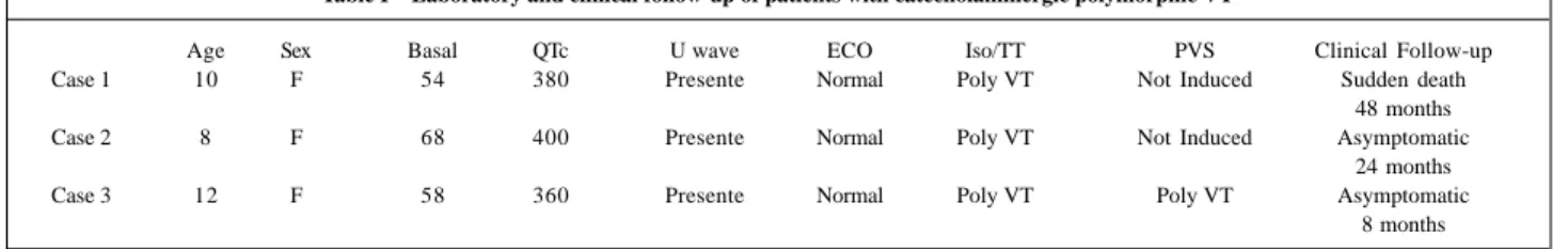
Table I – Laboratory and clinical follow-up of patients with catecholaminergic polymorphic VT
Age Sex Basal QTc U wave ECO Iso/TT PVS Clinical Follow-up
Case 1 10 F 54 380 Presente Normal Poly VT Not Induced Sudden death
48 months
Case 2 8 F 68 400 Presente Normal Poly VT Not Induced Asymptomatic
24 months
Case 3 12 F 58 360 Presente Normal Poly VT Poly VT Asymptomatic
8 months
Echo- echocardiogram; Iso- isoproterenol infusion; TT- treadmill test; Poly VT- polymorphic VT; PVS- programmed ventricular stimulation.
morphic VT on Holter monitoring, probably related to her increasing age, which should lead to an increase in proprano-lol doses. She died suddenly in the fourth year of follow-up after having discontinued her outpatient visits for 8 months.
Case 2 - An eight-year-old female patient was referred
to us with a history of frequent near-syncope and three syncope episodes in an 18-month period, which were al-ways related to exercise or emotion. She had been followed-up in another hospital with a diagnosis of vagal hyper-reactivity, and was off drug therapy. The clinical investiga-tion showed inducinvestiga-tion of polymorphic VT when sinus ta-chycardia achieved 120 beats/min during treadmill testing and isoproterenol infusion. Short runs of supraventricular tachycardia between polymorphic VT were also observed (figure 2). The tilt-table test was negative.
She has been treated with nadolol, and no further epi-sodes of syncope have occurred. Serial exercise tests were performed, and no arrhythmia was induced.
Case 3 - A 12-year-old female patient was referred to
our institution because of several episodes of syncope over the previous five years. She had suffered four episodes in the prior 6 months. A familial history revealed sudden
cardiac death of her brother at the age of 6. She had been treated for epilepsy with carbamazepine. Her physical exami-nation was unremarkable. The tilt-table test was negative. Polymorphic VT was induced during treadmill testing (Bru-ce protocol), Holter monitoring, and isoproterenol infusion, and a diagnosis of catecholaminergic polymorphic VT was made. The electrophysiologic study induced polymorphic VT (figure 3) by programmed ventricular stimulation, asso-ciated with loss of consciousness and need of electric defi-brillation, which converted the arrhythmia to polymorphic VT of lower frequency followed by spontaneous termi-nation (Figure 4). Treatment with nadolol was successful, and VT episodes were abolished in Holter monitoring, treadmill testing, and isoproterenol infusion. After 8 mon-ths of follow-up, polymorphic VT was induced only by iso-proterenol infusion. However, the child has remained asymptomatic during clinical follow-up without VT either on Holter monitoring or treadmill testing.
Discussion
Despite of the rare occurrence, catecholaminergic po-lymorphic VT is an important cause of syncope in children with no identified heart disease and normal QT interval.
Although this arrhythmia has been clearly defined for over two decades 3, only 60 cases have been reported in the
lite-rature. Syncope was the most common symptom described. Therefore, physicians must remember the possibility of ca-techolaminergic polymorphic VT whenever syncope asso-ciated with physical effort or emotion occurs in young pati-ents, especially when the QT interval is normal.
Many cases such as those reported in the present stu-dy, in the absence of heart disease but with a normal QT interval, may be misdiagnosed as epilepsy. In a series of 21 cases, Leenhardt et al 6 reported that 50% of patients had
been previously treated as epileptics. In that study, a cor-rect diagnosis was made 24 months after the onset of symp-toms. The present study also demonstrated a significant delay before the correct diagnosis (mean time of 38 months) was made. Due to its potential lethal outcome, suspicion of catecholaminergic polymorphic VT in children with physi-cal and emotional syncope is mandatory.
Although the genetic and molecular substrate of cate-cholaminergic polymorphic VT has not yet been defined, the genetic origin of this arrhythmia is confirmed by familial history of syncope or sudden death in 40% of cases 6. In our
study, one of the 3 children had a familial history of sudden
Fig. 3 - Programmed ventricular stimulation (S1-S1=500ms, S1-S2=250ms), inducing polymorphic VT during electrophysiological study with hemodynamic collapse. Leads D1, D2, V1, V6, and right ventricle.
cardiac death. Due to the low penetrance of congenital long QT syndrome 7, it is possible that catecholaminergic
poly-morphic VT may be a variant of this entity. Further research using new technologies is necessary to confirm this hypo-thesis.
The features in 12-lead ECG are nonspecific. Relative sinus bradycardia and the presence of U waves are the only characteristic findings on ECG 8.
The mechanism and electrophysiological substrate for catecholaminergic polymorphic VT has not been clearly identified. Leenhardt et al 6 suggested that triggered activity
may be the involved mechanism. Later, Nakagima et al 9 using
monophasic action potential recordings before and during pharmacological intervention (isoproterenol and beta-blo-ckers) in one patient with catecholaminergic polymorphic VT, have related that triggered activity due to delay after de-polarization was probably the electrophysiological mecha-nism responsible for this arrhythmia.
In our study, programmed ventricular stimulation in-duced polymorphic VT in one patient. Horowitz et al 10
distur-bances or reversible causes of polymorphic VT were ruled out in that study. Although induction of tachyarrhythmias by programmed ventricular stimulation was not frequently reported in catecholaminergic polymorphic VT, it might be compatible with the suggested triggered mechanism.
Beta-blockers are the treatment of choice for catecho-laminergic polymorphic VT. Although no differences exist among various agents, we and other authors have preferred nadolol because of its prolonged half-life. Doses must be ti-trated to carefully prevent the heart rate from exceeding 130 beats per minute 6. Because in any situation patients are
usually growing up, doses must be adjusted by kg/weight. Other antiarrhythmic drugs such as amiodarone and class I drugs have proved to be ineffective 6.
Classically, the success of treatment can be assessed by the disappearance of symptoms and the absence of VT on Holter recordings and exercise tests 4-6. Isoproterenol
infusion was also used in our patients to reproduce increa-sed adrenergic discharge. However, the value of this me-thod for evaluation of therapeutic efficacy is not completely defined. In patients who achieved clinical therapy efficacy, it was usual to observe PVC or even periods of nonsustai-ned VT on clinical follow-up.
Sudden death can occur in 50% of the cases before the age of 20 in patients not treated with beta-blockers 6.
Four deaths have been reported among patients treated with beta-blockers. In one case, the patient forgot to take the drug on the day of the event. In another, the death oc-curred in the context of viral myocarditis, and in two others under nonspecific circumstances. Our patient was taking propranolol regularly, but we could not confirm whether she took the medication on the day of death.
Because beta-blockers are highly effective in patients with catecholaminergic polymorphic VT, nonmedical treat-ment such as implantable cardioverter defibrillation is not mandatory.
Conclusion
Although vagal hyperreactivity is a major cause of syncope in the pediatric population, catecholaminergic po-lymorphic VT must be individualized in children without heart disease but with a normal QT interval, especially when episodes of syncope are related to physical effort or emo-tion. Delay in diagnosis and inadequate therapy can lead to sudden cardiac death.
1. Ruckman RN. Cardiac causes of syncope. Pediatr Rev 1987; 9: 101-8. 2. Pratt JL, Fleisher GR. Syncope in children and adolescents. Pediatr Emerg Care
1989; 5: 80-2.
3. Reid DS, Tynan M, Braidwood L, Fitzgerald GR. Bidirectional tachycardia in a child. A study using His bundle electrography. Br Heart J 1975; 37: 339-44. 4. De Paola AA, Horowitz LN, Marques FB, et al. Control of multiform ventricular
tachycardia by propranolol in a child with no identifiable cardiac disease and sudden death. Am Heart J 1990; 119: 1429-32.
5. Eisenberg SJ, Scheinman MM, Dullet NK, et al. Sudden cardiac death and poly-morphous ventricular tachycardia in patients with normal QT intervals and normal systolic cardiac function. Am J Cardiol 1995; 75: 687-92.
6. Leenhardt A, Lucet V, Denjoy I, Grau F, Ngoc DD, Coumel P. Catecholaminergic polymorphic ventricular tachycardia in children. A 7-year follow-up of 21 patients. Circulation 1995; 91: 1512-9.
References
7. Priori SG, Napolitano C, Schwartz PJ. Low penetrance in the long-QT syndrome: clinical impact. Circulation 1999; 99: 529-33.
8. Noh CI, Gillette PC, Case CL, Zeigler VL. Clinical and electrophysiological cha-racteristics of ventricular tachycardia in children with normal hearts. Am Heart J 1990; 120: 1326-33.
9. Nakajima T, Kaneko Y, Taniguchi Y, Hayashi K, Takizawa T, Suzuki T, Nagai R. The mechanism of catecholaminergic polymorphic ventricular tachycardia may be triggered activity due to delayed afterdepolarization. Eur Heart J 1997; 18: 530-1.