S H O R T R E P O R T
Open Access
Two novel CMY-2-type
β-lactamases encountered
in clinical Escherichia coli isolates
Vera Manageiro
1,2, Eugénia Ferreira
1, Margarida Pinto
3,6, Fernando Fonseca
4,7, Mónica Ferreira
1,
Richard Bonnet
5and Manuela Caniça
1*Abstract
Background: Chromosomally encoded AmpCβ-lactamases may be acquired by transmissible plasmids which consequently can disseminate into bacteria lacking or poorly expressing a chromosomal blaAmpCgene. Nowadays,
these plasmid-mediated AmpCβ-lactamases are found in different bacterial species, namely Enterobacteriaceae, which typically do not express these types ofβ-lactamase such as Klebsiella spp. or Escherichia coli. This study was performed to characterize two E. coli isolates collected in two different Portuguese hospitals, both carrying a novel CMY-2-typeβ-lactamase-encoding gene.
Findings: Both isolates, INSRA1169 and INSRA3413, and their respective transformants, were non-susceptible to amoxicillin, amoxicillin plus clavulanic acid, cephalothin, cefoxitin, ceftazidime and cefotaxime, but susceptible to cefepime and imipenem, and presented evidence of synergy between cloxacilin and cefoxitin and/or ceftazidime. The genetic characterization of both isolates revealed the presence of blaCMY-46and blaCMY-50genes, respectively,
and the following three resistance-encoding regions: a Citrobacter freundii chromosome-type structure encompassing a blc-sugE-blaCMY-2-type-ampR platform; a sul1-type class 1 integron with two antibiotic resistance gene cassettes (dfrA1 and
aadA1); and a truncated mercury resistance operon.
Conclusions: This study describes two new blaCMY-2-typegenes in E. coli isolates, located within a C. freundii-derived
fragment, which may suggest their mobilization through mobile genetic elements. The presence of the three different resistance regions in these isolates, with diverse genetic determinants of resistance and mobile elements, may further contribute to the emergence and spread of these genes, both at a chromosomal or/and plasmid level.
Keywords:β-lactamase, Resistance regions, Genetic environment, Escherichia coli Background
AmpCβ-lactamases, along with Class A β-lactamases, are
a major group of clinically important enzymes [1,2]. They belong to class C according to the Ambler classification and to group 1 following the functional classification of Bush-Jacoby [3,4], whose prevalence is increasing world-wide [1]; theseβ-lactamases are associated with infections caused by pathogenic Gram-negative bacteria, particularly Escherichia coliand Klebsiella pneumoniae. The identifica-tion of isolates containing plasmid-mediated AmpC-β-lactamase (PMAβ) is epidemiologically and clinically rele-vant due to the limitations of treatment options [5].
AmpC enzymes hydrolyse amino- and ureidopenicillins, and cephamycins (cefoxitin and cefotetan) and, at a low level, oxyiminocephalosporins (ceftazidime, cefotaxime, and ceftriaxone) and aztreonam: they are not inhibited by β-lactamase inhibitors such as clavulanic acid [1]. AmpC-producing isolates are susceptible to carbapenems and to zwitterionic cephalosporins (cefepime and cefpirome).
In this study, we performed the phenotypic and molecu-lar characterization of two new CMY-2-types (designated CMY-46 and CMY-50), both encoded by probably chromo-somal inducible ampC genes, produced by two clinical E. coli isolates. The genetic environment of blaCMY-46 and
blaCMY-50was also investigated.
* Correspondence:manuela.canica@insa.min-saude.pt
1Department of Infectious Diseases, National Reference Laboratory of Antibiotic Resistances and Healthcare Associated Infections, National Institute of Health Dr. Ricardo Jorge, Av. Padre Cruz, 1649-016 Lisbon, Portugal Full list of author information is available at the end of the article
© 2015 Manageiro et al.; licensee BioMed Central. This is an Open Access article distributed under the terms of the Creative Commons Attribution License (http://creativecommons.org/licenses/by/4.0), which permits unrestricted use, distribution, and reproduction in any medium, provided the original work is properly credited. The Creative Commons Public Domain Dedication waiver (http://creativecommons.org/publicdomain/zero/1.0/) applies to the data made available in this article, unless otherwise stated.
Methods
Bacterial isolate collection
Two clinical E. coli strains (INSRA1169 and INSRA3413) were isolated, in 1999, from urine samples of two patients of 77 years and 7 months old, in two different hospitals in Portugal. E. coli DH5α (pBK-CMY-2) strain was used as control for antimicrobial susceptibility tests.
Antimicrobial susceptibility tests
Minimal inhibitory concentrations were determined by a microdilution method according to guidelines of the French Society of Microbiology (SFM 2013, http://www. sfm-microbiologie.org/) against seven β-lactams, alone or in combination with clavulanic acid, and against ciprofloxa-cin, gentamicin and trimethoprim. Isolates non-susceptible to one third-generation cephalosporin, cefoxitin and/or exhibiting synergy with boronic acid and/or cloxacillin, were considered as presumptive AmpC producers. Imipi-nem and clavulanic acid were used in order to identify in-duction effect of AmpC [1,6]. Disks of inducing agents (imipenem 10μg and amoxicillin plus clavulanic acid 25 + 10 μg) and disks of cephalosporins (cefotaxime 30 μg and
ceftazidime 30 μg) were placed on Mueller–Hinton agar
plates, 20 mm apart. Positive induction was demonstrated by the antagonism effect surrounding the cephalosporin disks adjacent to the inducers.
Isoelectric point determination
β-Lactamases were characterized by isoelectric focusing of ultrasonicated bacterial extracts with the control strains expressing pI 5.2, 5.6, 7.6, 9.0, 9.2, as previously described [7].
Molecular characterization of ampC and ESBL-encoding genes
The presence of acquired ampC (blaCMY, blaMOX, blaFOX,
blaLAT, blaACT, blaMIR, blaDHA, blaMOR, blaACC) and
blaESBLgenes (blaTEM, blaSHV, blaOXA-1-type, blaCTX-M) was
investigated by multiplex PCR assays with primers and conditions as described elsewhere [7-10], and those from Table 1. Controls were included in all assays.
Gene transfer experiments
Transferability of the blaCMYgenes was attempted by both
broth mating-out assays and electroporation. Conjugation experiments were performed at 37°C, using recipient strains E. coli C600 RifR, StrR and E. coli J53 NaN3R,
according to the antibiotic susceptibilities of the clinical isolates used as donor. Transconjugants were selected on
MacConkey agar plates containing 250 μg/ml of
rifampi-cin, 160 μg/ml of streptomycin or 160 μg/ml of sodium
azide plus 10 μg/ml of cefoxitin. Plasmid DNA was
extracted from clinical strains, using the Wizard Plus Midipreps DNA Purification kit (Promega), and used to
transform electrocompetent E. coli DH5α ΔampC by elec-troporation, as previously described [7]. Transformants
were selected on Luria broth medium containing 10 μg/
ml of cefoxitin.
Cloning experiments
The blaCMY-2, blaCMY-46and blaCMY-50genes were
amp-lified with iProofTM High-Fidelity DNA Polymerase
(Bio-Rad, Hercules, CA), using primers from Table 1. Amplicons (1169 bp) were ligated in the SmaI site of the phagemid pBK-CMV (Stratagene) downstream of its in-ducible lacZ promoter and transformed into electrocom-petent E. coli DH5α ΔampC cells. A gene Pulser II apparatus (Bio-Rad, Hercules, CA) was used for standard electroporation techniques, as previously described [7]. Recombinant bacteria were selected on LB agar plates containing 10μg/ml of cefoxitin.
Genetic background characterization
The presence of class 1 integrons was determined in both isolates through PCR amplification of the integrase-specific intI1gene with the same specific primers and conditions as reported previously [11] (Table 1). PCR-mapping and
se-quencing of the genetic environment of blaCMY-46 and
blaCMY-50 was performed using primers targeting genes
known for promoting antibiotic resistance and integrons (Table 1). Sequence alignments and generation of resistance cassette contigs were performed using Bionumerics (Ap-plied Maths). Gene identity was confirmed at the NCBI website (http://www.ncbi.nlm.nih.gov/).
Findings
The two clinical E. coli isolates INSRA1169 and INSRA3413 were resistant to amoxicillin, amoxicillin plus clavulanic acid, cephalothin, cefoxitin, ceftazidime, cefo-taxime, gentamicin and trimethoprim, but susceptible to cefepime and imipenem (Table 2). INSRA1169 was also nonsusceptible to ciprofloxacin. Synergy between cloxacil-lin and cefoxitin plus cefotaxime and boronic acid, along with the absence of synergy between extended-spectrum cephalosporins and clavulanic acid, suggest that the resist-ance to extended-spectrum cephalosporins was mediated
by the overproduction of AmpCβ-lactamases. The
resist-ance phenotype was not transferable, neither in conjuga-tion assays with E. coli C600 as a recipient, or in transformation assays by electroporation of plasmid-DNA preparations into E. coli DH5α. This might suggest a chromosomal location of AmpC-encoding genes.
Transformants, obtained after cloning of DNA ampli-cons of INSRA1169 and INSRA3413 in pBK-CMV plas-mid vector (Figure 1a), showed a resistance phenotype similar to that of clinical isolates. However, they were susceptible to ciprofloxacin, gentamicin and trimetho-prim, like the control strain E. coli DH5α (pBK-CMY-2)
(Table 2). Both the clinical strains and the transformants produced β-lactamases exhibiting an alkaline isoelectric
point (pI 9.2) compatible with AmpC-typeβ-lactamases.
Indeed, PCR revealed the absence of blaESBL genes plus
the presence of chromosomal E. coli ampC-type gene; the sequence of cloned DNA fragments identified two new CMY-2-like genes, which were not related to chromosome-mediated E. coli AmpC gene. The chromo-somal location of such genes has only been observed in Salmonellaspp. and Proteus mirabilis isolates [12-14].
The deduced amino acid sequences confirmed that
the new genes encoding the β-lactamases CMY-46 (in
INSRA1169) and CMY-50 (in INSRA3413), which were new variants of CMY-2, differed by 9 and 13 amino acid substitutions, respectively (Table 3). Two of these muta-tions (Q193K plus P208A for CMY-46 and N194S plus
D198N for CMY-50) are in the Ω loop (between amino
acids 178 and 226), which interacts by hydrogen bond-ing with helix H-2 close to the active Ser64. Substitu-tions in this region have been linked to the extension of the hydrolysis spectrum [15]. However, CMY-46 and
CMY-50β-lactamases did not confer resistance to
cefe-pime and conferred low level of resistance to ceftazi-dime and cefotaxime, which suggests that, in contrast to extended-spectrum AmpCs, they have moderate or no extended-spectrum activity (Table 2) [15-18].
The study of sequences surrounding blaCMY-46 and
blaCMY-50 revealed the presence of the blc gene (encoding
an outer membrane lipoprotein) and the sugE gene (encod-ing a small MDR protein responsible for resistance to qua-ternary ammonium compounds) downstream of their open reading frames (Figure 1a). Upstream, an ampR gene encod-ing the usual transcriptional regulator of ampC genes was observed in an opposite direction of transcription (Figure 1a). The presence of an intact ampC-ampR segment in both new blaCMYgenetic regions implied that the production of
CMY-46 and CMY-50 is inducible, which was corroborated by the used phenotypic induction method. This
ampC-ampR region was identical to the sequence flanking the
blaAmpCgene in the C. freundii chromosome [6], except for
AmpRCMY-46(that had 4 amino acid substitutions), but none
were located in the helix-turn-helix region or in other
Table 2 MICs of antibiotics for CMY-46- and CMY-50-producingE. coli isolates and E. coli transformants and recipientsa
E. coli strain MIC (μg/ml)b
AMX AMCc CF CAZ CCAZc CTX FEP FOX IMP CIP GEN TMP
DH5α ΔampC 8 8 8 0.25 0.125 0.06 0.03 4 0.25 ≤0.125 ≤0.125 ≤0.25
DH5α (pBK-CMY-2) >2048 >2048 1024 32 16 8 0.25 64 0.5 ≤0.125 1 ≤0.25 INSRA1169 (CMY-46 + TEM-1) >2048 >2048 1024 16 4 4 0.032 64 2 4 >128 >128 DH5α (pBK-CMY-46) >2048 >2048 1024 32 4 8 0.25 64 0.5 ≤0.125 2 ≤0.25
INSRA3413 (CMY-50) >2048 >2048 1024 16 8 4 0.25 64 2 ≤0.125 64 64
DH5α (pBK-CMY-50) >2048 >2048 1024 16 8 4 0.25 64 0.5 ≤0.125 0.5 ≤0.25
a
E. coli EcDH5α (pBK-CMY-2) was control strain; E. coli DH5α (pBK-CMY-46) and E. coli DH5α (pBK-CMY-50) were transformants of E. coli INSRA1169 (harboring CMY-46 and TEM-1 enzymes) and E. coli INSRA3413 (harboring CMY-50 enzyme), respectively; E. coli EcDH5α was the recipient strain.
b
AMX, amoxicillin; AMC, amoxicillin-clavulanic acid; CF, cephalothin; CAZ, ceftazidime; CCAZ, ceftazidime-clavulanic acid; CTX, cefotaxime; FEP, cefepime; FOX, cefoxitin; IMP, imipenem; CIP, ciprofloxacin; GEN, gentamicin and TMP, trimethoprim.
c
Clavulanic acid, at fixed concentration of 2μg/ml.
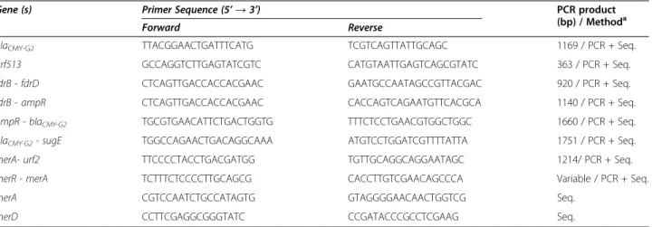
Table 1 Primers, drawn in this study, used for PCR amplification and sequencing of PMAβ genes and for PCR mapping ofblaCMY-46andblaCMY-50
Gene (s) Primer Sequence (5’ → 3’) PCR product
(bp) / Methoda
Forward Reverse
blaCMY-G2 TTACGGAACTGATTTCATG TCGTCAGTTATTGCAGC 1169 / PCR + Seq.
orf513 GCCAGGTCTTGAGTATCGTC CATGTAATTGAGTCAGCGTATC 363 / PCR + Seq.
fdrB - fdrD CTCAGTTGACCACCACGAAC GAATGCCAATAGCCGTTACGAC 920 / PCR + Seq. fdrB - ampR CTCAGTTGACCACCACGAAC CACCAGTCAGAATGTTCACGCA 1140 / PCR + Seq. ampR - blaCMY-G2 TGCGTGAACATTCTGACTGGTG TTTCTCCTGAACGTGGCTGGC 1660 / PCR + Seq.
blaCMY-G2- sugE TGGCCAGAACTGACAGGCAAA ATGTCCTGGATCGTTTTATTA 1751 / PCR + Seq.
merA- urf2 TTCCCCTACCTGACGATGG TGTTGCAGGCAGGAATAGC 1214/ PCR + Seq.
merR - merA TCTTTCTCCCCTTGCAGCG CACCTTGTCGAACAGCCCA Variable / PCR + Seq.
merA CGTCCAATCTGCCATAGTG GTAGGGGAACAACTGGTCG Seq.
merD CCTTCGAGGCGGGTATC CCGATACCCGCCTCGAAG Seq.
a
positions related to AmpR function [20,21]. The promoter regions of our blaCMY-2-type and ampR genes harbored no
sequence element associated with increased strength of the promoter [20,21]. In addition, the frdD, frdC, and frdB genes that are usually adjacent to ampC-ampR in the C. freundii chromosome were not identified in the sequences flanking blaCMY-46or blaCMY-50.
Class 1 integrons, also detected in INSRA1169 and INSRA3413 (Figure 1b), comprised the integrase-encoding gene intI1, two gene cassettes, aacA1 and dfrA1, and qacEΔ1 plus sul1, which were probably responsible for the observed resistances to trimethoprim and aminoglycosides.
We also found a truncated mercury resistance operon (Figure 1c), which was previously reported as belonging to a“kan region” that included a kanamycin resistance gene [22]. This finding is of concern since mercury resistance may help to promote antibiotic resistance through indirect selection [23].
In summary, this study describes two new blaCMY-2-type
genes located within a C. freundii-derived fragment.
Considering that CMY-type β-lactamases, detected in E.
coli, are derived from the C. freundii chromosomal AmpC [1] and that chromosome-derived genes are usu-ally mobilized by MGE [24,25], the presence of three
urf2 merE merD merA
truncated mer operon, 852bp
C
intl1 dfrA1 aadA1 qacE 1 sul1
Class 1 integron, 3,191bp
B
A
E. coli: genetic environment of blaCMY-typegenes, 2954bp blaCMY-type
ampR blc sugE
C. freundii :chromosomal region (JH414884), 34,816bp
574,378bp 588,947bp 592,040bp 609,195bp
Figure 1 Schematic representation of the same three structures found within clinical isolates expressing blaCMY-46and blaCMY-50. The
directions of transcription of the corresponding genes are depicted by arrows. A: sequence, including the genetic environment of blaCMY-type
genes, compared with C. freundii chromosomal region (GenBank JH414884); B: class 1 integron, with attI1 site (grey circle) and the two attc regions (open circles); C: truncated mercury resistance operon.
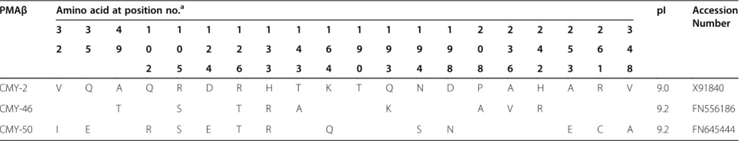
Table 3 Comparison of amino acid substitutions of two new CMY-typeβ-lactamases
PMAβ Amino acid at position no.a pI Accession
Number 3 3 4 1 1 1 1 1 1 1 1 1 1 1 2 2 2 2 2 3 2 5 9 0 0 2 2 3 4 6 9 9 9 9 0 3 4 5 6 4 2 5 4 6 3 3 4 0 3 4 8 8 6 2 3 1 8 CMY-2 V Q A Q R D R H T K T Q N D P A H A R V 9.0 X91840 CMY-46 T S T R A K A V R 9.2 FN556186 CMY-50 I E R S E T R Q S N E C A 9.2 FN645444 a
resistance regions with diverse resistance determinants and MGE in this study, suggests the dynamics of bacteria in the transference of antibiotic resistance. In addition, they might also trigger the future emergence and spread of these resist-ant determinresist-ants both at a chromosomal or/and plasmid level.
Availability of supporting data
The data set supporting the results of this article is in-cluded within the article.
Abbreviations
PMAβ:Plasmid-mediated AmpC-β-lactamase; SFM: French Society of Microbiology; MDR: Multidrug-resistance; MGE: Mobile genetic elements; ESAC: Extended-spectrum AmpC cephalosporinases; ESBL: Extended-spectrum β-lactamase.
Competing interests
The authors declare that they have no competing interests.
Authors’ contributions
VM performed the experiments, data interpretation and drafted the manuscript; EF and MF took part in the experiments; MP and FF worked on microbiology and clinical data; RB participated in data interpretation and reviewing of the manuscript; and MC conceived the study, and contributed to data interpretation and reviewing of the manuscript. All authors read and approved the final manuscript.
Acknowledgements
This study was supported by a grant from the Department of Infectious Diseases, National Institute of Health Dr. Ricardo Jorge (in 2012). V. Manageiro was supported by grant SFRH/BPD/77486/2011 from the Fundação para a Ciência e a Tecnologia, Lisbon, Portugal. This work was funded by“Strategic Plan for Environmental and Natural Sciences, Project UI/ 211–2011–2012” referenced as “Pest-OE/AGR/UI0211/2011” from CECA-ICETA. The GenBank accession numbers for the new AmpC-encoding genes are FN556186 for blaCMY-46and FN645444 for blaCMY-50.
Author details
1Department of Infectious Diseases, National Reference Laboratory of Antibiotic Resistances and Healthcare Associated Infections, National Institute of Health Dr. Ricardo Jorge, Av. Padre Cruz, 1649-016 Lisbon, Portugal. 2
Centre for the Study of Animal Sciences (ICETA), University of Oporto, Oporto, Portugal.3Laboratory of Microbiology, Hospital Garcia de Orta, EPE, Almada, Portugal.4Laboratory of Clinical Pathology, Hospital de Santa Luzia, Viana do Castelo, Portugal.5CHU Clermont-Ferrand, Laboratoire de Bactériologie, Clermont-Ferrand, France.6Present address: Laboratory of Microbiology, Centro Hospitalar de Lisboa Central, EPE, Lisbon, Portugal.7Present address: Laboratory of Clinical Pathology, Centro Hospitalar de Póvoa de Varzim-Vila do Conde, EPE, Póvoa de Varzim, Portugal.
Received: 1 October 2014 Accepted: 24 February 2015
References
1. Jacoby GA. AmpCβ-lactamases. Clin Microbiol Rev. 2009;22:161–82. 2. Rice LB, Bonomo RA.β-Lactamases: which ones are clinically important?
Drug Resist Updat. 2000;3:178–89.
3. Ambler RP, Coulson AF, Frere JM, Ghuysen JM, Joris B, Forsman M, et al. A standard numbering scheme for the class Aβ-lactamases. Biochem J. 1991;276:269–70.
4. Bush K, Jacoby GA. Updated functional classification ofβ-lactamases. Antimicrob Agents Chemother. 2010;54:969–76.
5. Pai H, Kang CI, Byeon JH, Lee KD, Park WB, Kim HB, et al. Epidemiology and clinical features of bloodstream infections caused by AmpC-type- β-lactamase-producing Klebsiella pneumoniae. Antimicrob Agents Chemother. 2004;48:3720–8.
6. Lindquist S, Lindberg F, Normark S. Binding of the Citrobacter freundii AmpR regulator to a single DNA site provides both autoregulation and activation of the inducible ampCβ-lactamase gene. J Bacteriol. 1989;171:3746–53.
7. Mendonça N, Leitão J, Manageiro V, Ferreira E, Antimicrobial Resistance Surveillance Program in Portugal (ARSIP), Caniça M. Spread of extended-spectrum β-lactamase CTX-M-producing Escherichia coli clinical isolates in community and nosocomial environments in Portugal. Antimicrob Agents Chemother. 2007;54:1946–55.
8. Pérez-Pérez FJ, Hanson ND. Detection of plasmid-mediated AmpC β-lactamase genes in clinical isolates by using multiplex PCR. J Clin Microbiol. 2002;40:2153–62.
9. Jones-Dias D, Manageiro V, Francisco AP, Martins AP, Domingues G, Louro D, et al. Assessing the molecular basis of transferable quinolone resistance in Escherichia coli and Salmonella spp. from food-producing animals and food products. Vet Microbiol. 2013;167:523–31.
10. Manageiro V, Ferreira E, Caniça M, Manaia CM. GES-5 among theβ-lactamases detected in ubiquitous bacteria isolated from aquatic environment samples. FEMS Microbiol Lett. 2014;351:64–9.
11. Leverstein-Van Hall MA, Paauw A, Box AT, Blok HE, Verhoef J, Fluit AC. Presence of integron-associated resistance in the community is widespread and contributes to multidrug resistance in the hospital. J Clin Microbiol. 2002;40:3038–40.
12. Shahada F, Sekizuka T, Kuroda M, Kusumoto M, Ohishi D, Matsumoto A, et al. Characterization of Salmonella enterica serovar Typhimurium isolates harboring a chromosomally encoded CMY-2β-lactamase gene located on a multidrug resistance genomic island. Antimicrob Agents Chemother. 2011;55:4114–21.
13. Zioga A, Whichard JM, Joyce KJ, Tzelepi E, Tzouvelekis LS, Miriagou V. Evidence for chromosomal and plasmid location of CMY-2 cephalosporinase gene in Salmonella serotype Typhimurium. J Antimicrob Chemother. 2008;61:1389–90.
14. D’Andrea MM, Literacka E, Zioga A, Giani T, Baraniak A, Fiett J, et al. Evolution and spread of a multidrug-resistant Proteus mirabilis clone with chromosomal AmpC-type cephalosporinases in Europe. Antimicrob Agents Chemother. 2011;55:2735–42.
15. Nordmann P, Mammeri H. Extended-spectrum cephalosporinases: structure, detection and epidemiology. Future Microbiol. 2007;2:297–307.
16. Rodríguez-Martínez JM, Poirel L, Nordmann P. Extended-spectrum cephalosporinases in Pseudomonas aeruginosa. Antimicrob Agents Chemother. 2009;53:1766–71.
17. Rodríguez-Martínez JM, Nordmann P, Ronco E, Poirel L. Extended-spectrum cephalosporinase in Acinetobacter baumannii. Antimicrob Agents Chemother. 2010;54:3484–8.
18. Rodríguez-Martínez JM, Fernández-Echauri P, Fernández-Cuenca F, Diaz-de-Alba P, Briales A, Pascual A. Genetic characterization of an extended-spectrum AmpC cephalosporinase with hydrolysing activity against fourth-generation cephalosporins in a clinical isolate of Enterobacter aerogenes selected in vivo. J Antimicrob Chemother. 2012;67:64–8.
19. Bauernfeind A, Stemplinger I, Jungwirth R, Giamarellou H. Characterization of the plasmidicβ-lactamase CMY-2, which is responsible for cephamycin resistance. Antimicrob Agents Chemother. 1996;40:221–4.
20. Bartowsky E, Normark S. Interactions of wild-type and mutant AmpR of Citrobacter freundii with target DNA. Mol Microbiol. 1993;10:555–65. 21. Hanson ND, Sanders CC. Regulation of inducible AmpCβ-lactamase expression
among Enterobacteriaceae. Curr Pharm Des. 1999;5:881–94.
22. Call DR, Singer RS, Meng D, Broschat SL, Orfe LH, Anderson JM, et al. bla CMY-2-positive IncA/C plasmids from Escherichia coli and Salmonella enterica are
a distinct component of a larger lineage of plasmids. Antimicrob Agents Chemother. 2010;54:590–6.
23. Baker-Austin C, Wright MS, Stepanauskas R, McArthur JV. Co-selection of antibiotic and metal resistance. Trends Microbiol. 2006;14:176–82. 24. Frost LS, Leplae R, Summers AO, Toussaint A. Mobile genetic elements: the
agents of open source evolution. Nat Rev Microbiol. 2005;3:722–32. 25. Norman A, Hansen LH, Sørensen SJ. Conjugative plasmids: vessels of the