Identification of the Multi-Resistance Gene
cfr
in
Escherichia coli
Isolates of Animal Origin
Hui Deng., Jian Sun., Jun Ma, Liang Li, Liang-Xing Fang, Qijing Zhang, Ya-Hong Liu, Xiao-Ping Liao*
College of Veterinary Medicine, National Reference Laboratory of Veterinary Drug Residues (SCAU), South China Agricultural University, Guangzhou, China
Abstract
Previous study indicated that the multi-resistance gene cfr was mainly found in gram-positive bacteria, such as StaphylococcusandEnterococcus, and was sporadically detected inEscherichia coli. Little is known about the prevalence and transmission mechanism ofcfrinE. coli. In this study, the presence ofcfrinE. coliisolates collected during 2010–2012 from food-producing animals in Guangdong Province of China was investigated, and the cfr-positive E. coli isolates were characterized by PFGE, plasmid profiling, and genetic environment analysis. Of the 839E. coliisolates, 10 isolates from pig werecfrpositive. All thecfr-positive isolates presented a multi-resistance phenotype and were genetically divergent as determined by PFGE. In 8 out of the 10 strains, thecfrgene was located on plasmids of,30 kb. Restriction digestion of the
plasmids withEcoRI and sequence hybridization with acfr-specific probe revealed that thecfr-harboring fragments ranged from 6 to 23 kb and a,18 kbcfr-carrying fragment was common for the plasmids that were,30 kb. Four different genetic
environments of cfr were detected, in which cfr is flanked by two identical copies of IS26, which may loop out the intervening sequence through homologous recombination. Among the 8 plasmids of,30 kb, 7 plasmids shared the same
genetic environment. These results demonstrate plasmid-carried cfr in E. coli and suggest that transposition and homologous recombination mediated by IS26might have played a rule in the transfer of thecfrgene inE. coli.
Citation:Deng H, Sun J, Ma J, Li L, Fang L-X, et al. (2014) Identification of the Multi-Resistance GenecfrinEscherichia coliIsolates of Animal Origin. PLoS ONE 9(7): e102378. doi:10.1371/journal.pone.0102378
Editor:Alvaro Galli, CNR, Italy
ReceivedApril 3, 2014;AcceptedJune 17, 2014;PublishedJuly 18, 2014
Copyright:ß2014 Deng et al. This is an open-access article distributed under the terms of the Creative Commons Attribution License, which permits unrestricted use, distribution, and reproduction in any medium, provided the original author and source are credited.
Data Availability:The authors confirm that all data underlying the findings are fully available without restriction. The sequences identified in the study (see Fig. 4) have been deposited in GenBank under accession numbers KJ453116 (Fig. 4a), KJ453117 (Fig. 4b), KJ453114 (Fig. 4c) and KJ453115 (Fig. 4d) respectively. Funding:This work was supported by the National Science Fund for Distinguished Young Scholars (Grant No. 31125026), the National Natural Science Foundation (Grant No. 31272609), Program for Changjiang Scholars and Innovative Research Team in University of Ministry of Education of China (Grant No. IRT13063) and Science and Technology Program of Guangzhou, China (Grant No. 2011J2200054). The funders had no role in study design, data collection and analysis, decision to publish, or preparation of the manuscript.
Competing Interests:The authors have declared that no competing interests exist. * Email: [email protected]
.These authors contributed equally to this work.
Introduction
Encoding a methyltransferase that modifies 23S rRNA at A2503, thecfrgene confers resistance to five chemically distinct classes of antimicrobials, including Phenicols, Lincosamides, Oxazolidinones, Pleuromutilins and Streptogramin A [1], and also reduces the susceptibility to selected 16-member-ring macro-lides such as josamycin and spiramycin [2]. Since its first identification on plasmid pSCFS1 fromStaphylococcus sciuri[3], the cfr gene has been identified on plasmid or chromosome in other staphylococcal species [4,5], and subsequently in other genera of gram-positive bacteria such asBacillus [6–8], Entero-coccus[9–12],Streptococcus[13],MacrococcusandJeotgalicoccus [14]. In those bacteria, plasmids and various insertion sequences have played an important role in the dissemination of thecfrgene between species and genera [15]. In gram-negative bacteria, the cfr gene has been sporadically detected in Escherichia coliand Proteus vulgaris[16–18], and little is known about the prevalence and transmission mechanism of the cfr gene in these bacteria. Here, we present the first study on the prevalence of thecfrgene in E. coliisolated from food animals in China. In addition, the main transmission mechanism of the cfr gene in E. coli was characterized by plasmid and genetic environment analysis.
Materials and Methods
Ethics statement
This study protocol was reviewed and approved by the South China Agriculture University Animal ethics committee. The owners of the farm animals from which faecal swabs were taken gave permission for their animals to be used in this study.
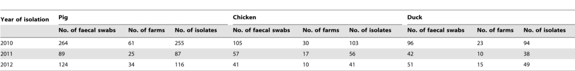
Bacterial strains and Antimicrobial susceptibility testing A total of 839E. coliisolates were isolated from faecal swabs of diseased food-producing animals submitted to the Veterinary Research Institute, Guangdong Academy of Agricultural Sciences, in Guangdong Province of China during 2010 and 2012 (Table 1). Between three and five herds were sampled from each farm, and all the samples were from 225 farms all over Guangdong province. Bacterial DNA was extracted by a DNA extraction kit (Omega, USA) following the manufacturer’s instructions. The presence of thecfrgene inE. coliwas determined by PCR amplification and sequence analysis with the primers described in a previous study [4]. The susceptibilities of cfr-positive isolates to ceftiofur, ampicillin, cefotaxime, streptomycin, kanamycin, gentamicin, amikacin, florfenicol, chloramphenicol, tetracycline, ciprofloxacin, enrofloxacin, and trimethoprim-sulfamethoxazole were tested by
Year of isolation Pig Chicken Duck
No. of faecal swabs No. of farms No. of isolates No. of faecal swabs No. of farms No. of isolates No. of faecal swabs No. of farms No. of isolates
2010 264 61 255 105 30 103 96 23 94
2011 89 25 87 57 17 56 42 10 38
2012 124 34 116 41 10 41 51 15 49
doi:10.1371/journal.pone.0102378.t001
Table 2.Characteristics of thecfr-positiveE. colistrains and the correspondingcfr-carrying plasmids.
Strain
Year of
isolation Resistance profilea Phylogroup cfr-carrying plasmid
transformation approximate size (kb) EcoRI fragment(kb) genetic environment ofcfrb
FS-P54 2010 CEF,AMP,CTX,STR,KAN,GEN,FFC,CHL,TET,CIP,ENR,SXT B + 30 18 A 1ZF13D 2011 AMP,STR,KAN,GEN,FFC,CHL,TET,CIP,ENR,SXT D + 30 18 A 2ZX7S 2011 AMP,STR,KAN,GEN,AMK,FFC,CHL,TET,CIP,ENR,SXT A + 45 23 B
3ZX12D 2011 AMP,STR,KAN,GEN,FFC,CHL,TET,CIP,ENR,SXT F + 30 18 A 8ZG1D 2011 AMP,STR,KAN,GEN,AMK,FFC,CHL,TET,CIP,ENR,SXT G + 9,75 9,23 C 8ZB6D 2011 AMP,CTX,STR,KAN,GEN,FFC,CHL,TET,CIP,ENR,SXT A + 30 18 A
8ZG6D 2011 AMP,STR,KAN,GEN,FFC,CHL,TET,CIP,ENR,SXT F + 30 18 A 8ZG8D 2011 CEF,AMP,CTX,STR,KAN,GEN,FFC,CHL,TET,CIP,ENR,SXT E + 30 18 A 8ZG12D 2011 CEF,AMP,CTX,STR,KAN,GEN,FFC,CHL,TET,CIP,ENR,SXT C + 30 21 D
FS13Z3C 2012 CEF,AMP,CTX,STR,KAN,GEN,FFC,CHL,TET,CIP,ENR,SXT H - 30 18 A
aCEF, ceftiofur; AMP, ampicillin; CTX, cefotaxime; STR, streptomycin; KAN, kanamycin; GEN, gentamicin; AMK, amikacin; FFC, florfenicol; CHL, chloramphenicol; TET, tetracycline; CIP, ciprofloxacin; ENR, enrofloxacin; SXT,
Trimethoprim-sulfamethoxazole.
bA, Figure 4a; B, Figure 4b; C, Figure 4c; D, Figure 4d.
doi:10.1371/journal.pone.0102378.t002
The
Multi-Resistance
Gene
cfr
in
Escherichia
coli
ONE
|
www.ploson
e.org
2
July
2014
|
Volume
9
|
Issue
7
|
agar dilution method according to the guidelines of the Clinical and Laboratory Standards Institute (CLSI) [19].Escherichia coli ATCC 25922 was used as the control strain.
PFGE
Pulsed field gel electrophoresis analysis of XbaI-digested genomic DNA of allcfr-positive strains was performed using the CHEF-MAPPER System (Bio-Rad Laboratories) as described previously [20]. The PFGE patterns were analyzed with BioNumerics software (Applied Maths, Sint-Martens-Latem, Belgium) using the Dice similarity coefficient with a cut-off at 80% of the similarity values to indicate identical PFGE types.
Assay ofcfrtransfer
Mating experiments were performed as previously described [21], using azide-resistantE. coliJ53 or streptomycin-resistantE. coli C600 as recipient strain. Transconjugants were selected on tryptic soy agar plates containing florfenicol (10 mg/L) and azide (100 mg/L) or streptomycin (512 mg/L). Plasmid DNA of cfr-positive strains was extracted by QIAGEN Plasmids Midi Kit, and was then transformed by electroporation into the recipient strain E. coli DH10B. Putative transformants were selected on brain heart infusion agar plates containing florfenicol (10 mg/L).
Plasmid characterization
The size ofcfr-carrying plasmid in every parental strain and their transformants was determined using S1-treated genomic DNA followed by PFGE and southern hybridization with probe specific forcfrgene. All plasmids from transformants were further analysed by restriction fragment length polymorphism (RFLP) using EcoRI (TaKaRa Biotechnology, Dalian, China) restriction enzyme followed by hybridization performed on RFLP gels. Because of the failure in electrotransformation, the cfr-carrying plasmid in strain FS13Z3C was analyzed with plasmid extracted from host cell. To investigate the genetic environment of thecfr gene, 3 pairs of primers used in previous studies [4,17,22] were used for PCR mapping, inverse PCR and sequencing based on the known structure in earlier studies [16,17].
Results
Bacterial strains and Antimicrobial susceptibility testing Among the 839 E. coli isolates, 10 isolates from pig were positive for thecfrgene as determined by PCR, which was further confirmed by sequencing the PCR product. Sampling information showed that all thecfr-carrying isolates were from different farms except strains 8ZB6D and 8ZG6D which were isolated from different animals of the same farm. Susceptibility testing showed that all thecfr-positiveE. colistrains presented a multiresistance phenotype, including resistance to chloramphenicols, quinolones,
ampicillin, kanamycin, gentamicin, tetracycline and trimethoprim-sulfamethoxazole (Table 2).
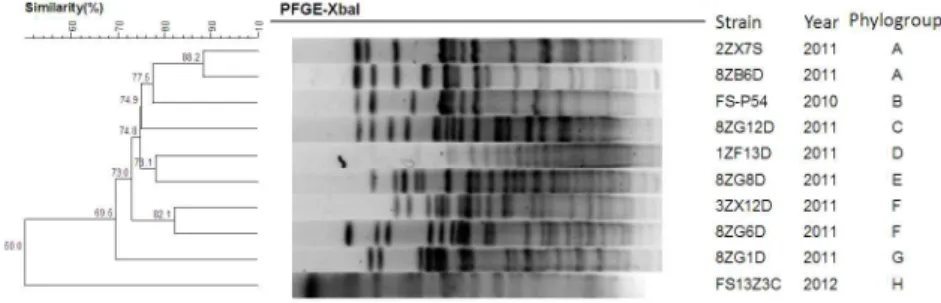
PFGE
The dendrogram of the cfr-positive strains generated from cluster analysis of the PFGE profiles is shown in Figure 1. The 10 strains displayed different PFGE patterns and belonged to 8 phylogenetic groups (designated A to H). Most of the phylogenetic groups comprised a single strain, while Group A and F were represented by 2 strains, respectively.
Transfer of resistance
The conjugation experiment ofcfr-positive strains usingE. coli C600 orE. coliJ53 as recipient strain failed, but electrotransfor-mation was achieved in all the strains except FS13Z3C. Susceptibility testing of the 9 transformants revealed drastically increased florfenicol MICs (range, 16 to.256 mg/L) compared with DH10B (1 mg/L). Co-transfer of resistance to at least one other antimicrobial was observed in 8 transformants except 8ZG1D-21, which was only resistant to florfenicol. Transformant 8ZG1D-21 was moderately resistant to florfenicol with MIC of 16 mg/L and sensitive to chloramphenicol with MIC of 8 mg/L.
Plasmid characterization
The result of S1-PFGE revealed that the tencfr-positive strains possessed multiple plasmids of varying sizes, and at least two plasmids were electroporated into the recipient strain. Southern hybridization withcfr-specific probe identified thecfrgene located on an approximately 30 kb plasmid in all cfr-positive strains except 8ZG1D and 2ZX7S (Figure 2 and Table 2). Strain 2ZX7S had acfr-carrying plasmid of,45 kb and strain 8ZG1D harbored
twocfr-positive plasmids that were ,9 kb and ,75 kb in size,
respectively. The RFLP profiles ofcfr-carrying plasmids from 9 Figure 1. UPGMA dendrogram, PFGE patterns, phylogenetic group and isolation date ofcfr-positiveE. coli.
doi:10.1371/journal.pone.0102378.g001
Figure 2. Location of thecfrgene in the 10E. colistrains and their corresponding transformants.(a) S1-PFGE of thecfr-positive strains and their transformants, (b) subsequent southern hybridization withcfr-specific probe. Lanes: M, Low Range PFG Marker; 1, 8ZG1D; 2, 8ZG1D-21; 3, 8ZG12D; 4, 8ZG12D-50; 5, 1ZF13D; 6, 1ZF13D-22; 7, 2ZX7S; 8, 2ZX7S-41; 9, 8ZG6D; 10, 8ZG6D-59; 11, 3ZX12D; 12, 3ZX12D-6; 13, 8ZG8D; 14, 8ZG8D-81; 15 FS-P54; 16,FS-P54-2; 17,8ZB6D; 18, 8ZB6D-30; and 19,FS13Z3C.
doi:10.1371/journal.pone.0102378.g002
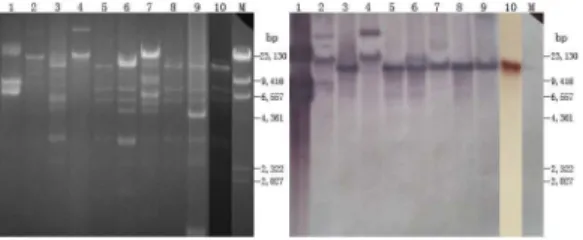
transformants and thecfr-positive strain FS13Z3C were obtained withEcoRIdigestion and are presented in Figure 3. The plasmids of 5 transformants and the cfr-positive strain FS13Z3C showed similar pattern (lanes 3, 5, 6, 8, 9 and 10 in Fig. 3). Although hybridization bands of undigested plasmids due to incomplete enzyme digestion were observed in 3 lanes (Figure 3 lanes 1, 2 and 4), southern hybridization performed on the RFLP gel revealed that the plasmids yieldedcfr-harboring fragments ranging from 9 to 23 kb, in which a fragment of,18 kb was observed in 7 out of
the 8 plasmids of,30 kb. Additionally, even though the 2
cfr-carrying plasmids in 8ZG1D-21 cannot be characterized respec-tively by RFLP, hybridization showed that the 2 cfr-positive plasmids of,9 and,75 kb yieldedcfr-carrying fragments of,9
and,23 kb separately.
Genetic environment ofcfr
The result of PCR mapping and sequencing revealed the presence of 4 different genetic environments (Figure 4a-d; GenBank accession number KJ453116, KJ453117, KJ453114 and KJ453115) with thecfrgene flanked by two copies of IS26 located in the same orientation. Among the 8 plasmids of,30 kb,
7 plasmids shared the similar genetic environment, in which the cfrgene was oriented in the opposite direction of IS26(Figure 4a). In contrast, the other 3 strains showed different environments, in which cfrwas in the same orientation with IS26(Figure 4b–d). Structural comparison of the genetic environments showed localized high homology (.98%) with plasmid pEC-01 fromE. coliLYP-C-BCTb11 and chromosomal fragment fromP. vulgaris PV-01 [16,17]. To determine the stability of these structures, inverse PCR was performed, and amplicons of approximately
1570 bp (Figure 4a), 1562 bp (Figure 4b), 2520 bp (Figure 4c) and 4412 bp (Figure 4d) were obtained. Sequence analysis of these amplicons further confirmed the structure of these genetic environments obtained by PCR mapping.
Discussion
In our study, 10cfr-positive isolates were detected from 839E. coliisolated between 2010 and 2012, and all the positive isolates were from swine farms where florfenicol is extensively used to prevent and cure diseases caused by a variety of bacterial pathogens in China [23]. PFGE analysis of the 10cfr-positiveE. colirevealed that these strains were genetically divergent. Most of the strains form a distinct phylogenetic group in the dendrogram based on genetic similarity, which suggested that the spread of the cfr gene in E. coli was not due to clonal dissemination but horizontal transfer.
S1 nuclease PFGE and hybridization showed that thecfrgene was located on an approximately 30 kb plasmid in all but two cfr-positive strains. Strains 2ZX7S and 8ZG1D harboredcfr-carrying plasmids of different sizes. The failure of the conjugation assays usingcfr-positive strains suggests that thecfrgene was carried by nonconjugative plasmids in these strains. Recently cfr was identified on a conjugative plasmid inE. coli [18], which may further accelerate the dissemination of the cfr gene among different Gram-negative bacteria. Although we were not successful in obtaining transformants with a single plasmid after several attempts, RFLP and hybridization profiles of the plasmids showed that 7cfr-carrying plasmids yielded the same-sizedcfr-harboring fragment of ,18 kb after digestion with EcoRI, and all the 7
plasmids are,30 kb in size with the same genetic environments
confirmed by PCR mapping and inverse PCR, implying that the 7 plasmids are likely identical and originated from the same source. Interestingly, compared with the 9 strains in which a single cfr-carrying plasmid was harbored, strain 8ZG1D and the transfor-mant 8ZG1D-21 have twocfr-positive plasmids. Considering the presence of IS26flanking thecfrgene, IS-mediated recombination may account for the transfer ofcfrbetween plasmids in the strain. Interestingly, the result of antimicrobial susceptibility testing showed that 8ZG1D-21 was moderately resistant to florfenicol and sensitive to chloramphenicol, suggesting that thecfrgene may confer low-level resistance to chloramphenicols inE. coli.
Four different genetic environments were detected surrounding thecfr gene, all of which have two copies of IS26of the same orientation flanking thecfrgene. Previous studies have suggested that insertion element IS26can mediate the transfer ofcfrgene Figure 3. RFLP and hybridization profiles of cfr-carrying
plasmids in the 9 transformants and strain FS13Z3C.Lanes: M,
l-HindIII marker; 1, 8ZG1D-21; 2, 8ZG12D-50; 3 1ZF13D-22; 4, 2ZX7S-41;
5, 8ZG6D-59; 6, 3ZX12D-6; 7, 8ZB6D-30; 8, FS-P54-2; 9, FS13Z3C; and 10 8ZG8D-81.
doi:10.1371/journal.pone.0102378.g003
Figure 4. Genetic environment of thecfrgene in this study, and structural comparison with plasmids pEC-01 (accession number JN982327) fromE. coliLYP-C-BCTb11 and Chromosomal fragment fromP. vulgarisPV-01 (accession number JF969273). The arrows represent the positions and transcriptional direction of the ORFs. The IS26elements are shown as light grey boxes. Regions with homology of over 98% are indicated by grey shading. Bacteria corresponding to each genetic environment are as follows: structure a (FS-P54, 1ZF13D, 3ZX12D, 8ZB6D, 8ZG6D, 8ZG8D and FS13Z3C), structure b (2ZX7S), structure c (8ZG1D), structure d (8ZG12D) (see Tables 2).
[16,17]. In our study, structural comparison of the genetic environments showed that part of the segment between IS26 shares high homology (.98%) with plasmid pEC-01 fromE. coli LYP-C-BCTb11 and chromosomal fragment from P. vulgaris PV-01 [16,17], further suggesting that IS26may have played an important role in the transfer of thecfrgene. Furthermore, inverse PCR performed on all of the cfr-positive strains can obtain an amplicon, and subsequent sequencing analysis showed that the pair of intact IS26 flanking the cfr gene can loop out the intervening sequence through homologous recombination, which can further accelerate the transfer ofcfrgene.
To conclude, we present the first study on the prevalence of the cfrgene inE. colifrom food producing animals. The identified cfr-positive E. colistrains were limited to pigs, coinciding with the extensive use of florfenicol for swine production. PFGE analysis showed that thecfr-positiveE. colistrains were genetically diverse;
however, plasmid and genetic environment analysis suggested that most of these strains harbored the samecfr-carrying plasmid of
,30 kb. Additionally, the results suggest that transposition and
homologous recombination mediated by IS26as well as transfor-mation ofcfr-carrying plasmids may be the main mechanism for horizontal spread of thecfrgene inE. coli. Further surveillance and investigation are necessary to facilitate the control of cfr spread in gram-negative bacteria.
Author Contributions
Conceived and designed the experiments: YHL XPL HD. Performed the experiments: HD JM. Analyzed the data: JS HD. Contributed reagents/ materials/analysis tools: LL LXF. Contributed to the writing of the manuscript: QJZ HD.
References
1. Long KS, Poehlsgaard J, Kehrenberg C, Schwarz S, Vester B (2006) The Cfr rRNA methyltransferase confers resistance to Phenicols, Lincosamides, Oxazo-lidinones, Pleuromutilins, and Streptogramin A antibiotics. Antimicrob Agents Chemother 50: 2500–2505.
2. Smith LK, Mankin AS (2008) Transcriptional and Translational Control of the mlrOperon, Which Confers Resistance to Seven Classes of Protein Synthesis Inhibitors. Antimicrob Agents and Chemotherapy 52: 1703–1712.
3. Schwarz S, Werckenthin C, Kehrenberg C (2000) Identification of a plasmid-borne chloramphenicol-florfenicol resistance gene in Staphylococcus sciuri. Antimicrob Agents Chemother 44: 2530–2533.
4. Kehrenberg C, Schwarz S (2006) Distribution of florfenicol resistance genesfexA andcframong chloramphenicol-resistantStaphylococcus isolates. Antimicrob Agents Chemother 50: 1156–1163.
5. Toh SM, Xiong L, Arias CA, Villegas MV, Lolans K, et al. (2007) Acquisition of a natural resistance gene renders a clinical strain of methicillin-resistant Staphylococcus aureus resistant to the synthetic antibiotic linezolid. Mol Microbiol 64: 1506–1514.
6. Dai L, Wu CM, Wang MG, Wang Y, Huang SY, et al. (2010) First report of the multidrug resistance genecfrand the phenicol resistance genefexAin aBacillus strain from swine feces. Antimicrob Agents Chemother 54: 3953–3955. 7. Wang Y, Schwarz S, Shen Z, Zhang W, Qi J, et al. (2012) Co-location of the
multiresistance genecfrand the novel streptomycin resistance geneaadYon a small plasmid in a porcineBacillusstrain. J Antimicrob Chemother 67: 1547– 1549.
8. Zhang WJ, Wu CM, Wang Y, Shen ZQ, Dai L, et al. (2011) The new genetic environment ofcfr on plasmid pBS-02 in aBacillus strain. J Antimicrob Chemother 66: 1174–1175.
9. Liu Y, Wang Y, Wu C, Shen Z, Schwarz S, et al. (2012) First report of the multidrug resistance gene cfr in Enterococcus faecalis of animal origin. Antimicrob Agents Chemother 56: 1650–1654.
10. Liu Y, Wang Y, Schwarz S, Li Y, Shen Z, et al. (2013) Transferable multiresistance plasmids carryingcfrinEnterococcusspp. from swine and farm environment. Antimicrob Agents Chemother 57: 42–48.
11. Liu Y, Wang Y, Schwarz S, Wang S, Chen L, et al. (2014) Investigation of a multiresistance gene cfr that fails to mediate resistance to phenicols and oxazolidinones inEnterococcus faecalis. J Antimicrob Chemother 69: 892–898. 12. Diaz L, Kiratisin P, Mendes RE, Panesso D, Singh KV, et al. (2012) Transferable plasmid-mediated resistance to linezolid due tocfrin a human
clinical isolate of Enterococcus faecalis. Antimicrob Agents Chemother 56: 3917–3922.
13. Wang Y, Li D, Song L, Liu Y, He T, et al. (2013) First report of the multiresistance genecfrinStreptococcus suis. Antimicrob Agents Chemother 57: 4061–4063.
14. Wang Y, Schwarz S, Shen Z, Zhou N, Lin J, et al. (2012) Detection of the staphylococcal multiresistance gene cfr in Macrococcus caseolyticus and Jeotgalicoccus pinnipedialis. J Antimicrob Chemother 67: 1824–1827. 15. Shen J, Wang Y, Schwarz S (2013) Presence and dissemination of the
multiresistance gene cfr in Gram-positive and Gram-negative bacteria. J Antimicrob Chemother 68: 1697–1706.
16. Wang Y, He T, Schwarz S, Zhou D, Shen Z, et al. (2012) Detection of the staphylococcal multiresistance genecfrinEscherichia coliof domestic-animal origin. J Antimicrob Chemother 67: 1094–1098.
17. Wang Y, Wu CM, Schwarz S, Shen Z, Zhang W, et al. (2011) Detection of the staphylococcal multiresistance genecfrinProteus vulgarisof food animal origin. J Antimicrob Chemother 66: 2521–2526.
18. Zhang WJ, Xu XR, Schwarz S, Wang XM, Dai L, et al. (2014) Characterization of the IncA/C plasmid pSCEC2 from Escherichia coliof swine origin that harbours the multiresistance genecfr. J Antimicrob Chemother 69: 385–389. 19. CLSI (2012) Performance Standards for Antimicrobial Susceptibility Testing;
Twenty-Second Informational Supplement. CLSI document M100-S22 Wayne, PA: Clinical and Laboratory Standards Institute.
20. Gautom RK (1997) Rapid pulsed-field gel electrophoresis protocol for typing of Escherichia coliO157:H7 and other gram-negative organisms in 1 day. J Clin Microbiol 35: 2977–2980.
21. Chen L, Chen ZL, Liu JH, Zeng ZL, Ma JY, et al. (2007) Emergence of RmtB methylase-producingEscherichia coliandEnterobacter cloacaeisolates from pigs in China. J Antimicrob Chemother 59: 880–885.
22. Dhanji H, Patel R, Wall R, Doumith M, Patel B, et al. (2011) Variation in the genetic environments ofbla(CTX-M-15)inEscherichia coli from the faeces of travellers returning to the United Kingdom. J Antimicrob Chemother 66: 1005– 1012.
23. Wang Y, Zhang W, Wang J, Wu C, Shen Z, et al. (2012) Distribution of the multidrug resistance genecfrinStaphylococcusspecies isolates from swine farms in China. Antimicrob Agents Chemother 56: 1485–1490.