Development of
an innovative
immunosensor
for
Leishmaniasis
screening
Sofia Sampaio Esteves
Mestrado em Bioquímica
Departamento de Química e Bioquímica 2017
Orientador
Anabela Cordeiro da Silva, Professora Associada
com Agregação, Faculdade de Farmácia da
Universidade do Porto
Coorientador
Todas as correções determinadas pelo júri, e só essas, foram efetuadas. O Presidente do Júri,
Acknowledgements
Primeiramente, gostaria de agradecer à Professora Doutora Anabela Cordeiro da Silva pelas oportunidades que me proporcionou, pelo tempo dispendido e por me ter acompanhado como orientadora ao longo deste ano de trabalho, sem a qual nada teria sido possível.
À Professora Doutora Célia Amorim, minha co-orientadora e pilar desde o início do desenvolvimento do trabalho, por me ter apoiado incansavelmente e por me relembrar todos os dias que o mais importante nesta vida é existir equilíbrio.
Gostaria de agradecer à Vitacontrol, empresa que tornou possível o desenvolvimento da tese durante esta etapa da minha vida académica.
Agradeço ao Laboratório de Sensors & Biosensors do departamento de Química Aplicada da Faculdade de Farmácia da Universidade do Porto e aos seus elementos integrantes por me terem acolhido de braços abertos, pelo ótimo ambiente e por deixarem uma marca tão positiva após este ano de trabalho.
À Professora Doutora Conceição Montenegro e ao Professor Doutor Alberto Araújo por terem sempre críticas construtivas a fazer e conselhos a dar no sentido de melhoria científica e pessoal, um imenso obrigada.
Ao Álvaro, à Ana Luísa e à Micha um enorme obrigada! Não tenho como agradecer o companheirismo, a amizade, os ensinamentos, a ajuda no desenvolvimento de trabalho científico e as palavras de apoio sempre que foram necessárias.
Gostaria de agradecer ao Laboratório de Imunologia da Faculdade de Farmácia da Universidade do Porto, com especial agradecimento à Carla Lima, que me ajudou sempre que pode.
Ao Instituto de Investigação e Inovação em Saúde (I3S), em particular ao grupo Parasite Disease e aos seus membros, especialmente ao Doutor Nuno Santarém por me ter ensinado tanto e por me ajudar a dar os primeiros passos na área das vesículas extracelulares.
Gostaria de agradecer ao grupo de investigação Plasmodium Vivax and Exosome (PVREX) do ISGlobal/IGTP, onde foi possível desenvolver parte do meu projeto, e aos seus respetivos elementos por estarem sempre prontos a ajudar.
Ao Professor Hernando A del Portillo e à Professora Assistente de Investigação Carmen Fernandez-Becerra, por me terem recebido tão bem no seu laboratório em Badalona e por todo o conhecimento científico que me transmitiram, um sincero obrigada.
Gostaria ainda de agradecer à E-COST por me proporcionar a oportunidade de realizar uma Short Term Science Mission e por ter financiado a estadia em Barcelona.
Um enorme obrigada aos amigos de longa data e aos mais recentes, que se têm cruzado na minha vida e têm enriquecido a mesma de alguma forma. Ao Quereis, por ter começado por um pequeno grupo e por se ter expandido para 21 elementos, grandes amigos, com quem partilho 5 anos de percurso académico, imensas recordações e a quem desejo um futuro brilhante!
Ao meu pai, por me mostrar que a vida é feita de tempos e contratempos mas o mais importante é manter a dedicação e perseverança. À minha mãe, por ser a minha personificação de alegria e o meu exemplo de pessoa. À Inês, por ser a pessoa que me atura a tempo inteiro e por ser a pessoa a quem recorro a toda a hora. A todos os meus avós e ao Paulinho, pelo mimo e apoio desde sempre.
Resumo
Leishmaniose é uma doença tropical negligenciada causada pelo parasita Leishmania e é transmitida a humanos e cães pela mosca da areia fêmea Phlebotominae. A forma mais severa da doença é a leishmaniose visceral e é fatal se não tratada, estimando-se até 30 000 de mortes por ano. Devido a vários fatores como alterações climáticas e migração, o número de casos de leishmaniose tem vindo a aumentar. Outro problema é a falta de especificidade dos métodos de diagnóstico atuais, que resultam em falsos resultados. Indivíduos infetados, mas sem sintomas, são potenciais propagadores desta doença. Neste contexto, existe uma necessidade emergente de diagnóstico simples e eficiente capaz de fazer triagem da doença e consequentemente controlo da sua disseminação.
Neste trabalho, pretende-se desenvolver um imunossensor imobilizando biomarcadores de soros caninos em elétrodos impressos, já que estes são descartáveis, baratos, fáceis de usar e de resposta rápida.
Técnicas eletroanalíticas, como voltametria cíclica e potenciometria, são usadas para a caracterização dos imunossensores. O protocolo usado na imobilização da proteína baseia-se na imobilização do antigénio pela cauda de histidina (atualmente a proteína recombinante CPX2) a micropartículas magnéticas funcionalizadas com ácido nitrilotriacético, sendo o mesmo protocolo usado pelo grupo para citometria de fluxo. Um dispositivo de microfluídica que contém o elétrodo impresso foi especialmente desenvolvido para este trabalho, onde um campo magnético é gerado no elétrodo de trabalho para reter as partículas magnéticas, que foram previamente imobilizadas com o respetivo antigénio e anticorpos. Para obter volumes reprodutíveis, a suspensão que continha as partículas magnéticas foi propulsionada até ao elétrodo de trabalho através de uma válvula FIA, com a ajuda de uma bomba peristáltica. Os resultados obtidos são promissores, no entanto verifica-se alteração da superfície do elétrodo devido à adsorção inespecífica de proteína. Atualmente, está-se a trabalhar no sentido de bloquear a superfície do elétrodo de forma a evitar adsorções inespecíficas e a avaliar formas de remover proteínas que possivelmente adsorvam.
Em paralelo, exossomas foram isolados de plasmas de pacientes infetados com leishmaniose (sintomáticos, assintomáticos e curados). Estas vesículas são constituídas por proteínas antigénicas envolvidas na resposta imune contra o parasita e têm potencial para serem utilizados como novos biomarcadores da doença. Esta hipótese
foi verificada pela resposta das várias frações de material extracelular contra soros de cães e humanos infetados com leishmaniose.
Palavras-chave: Leishmania infantum, Leishmaniose visceral, Reservatórios de
doença, Cães, Casos assintomáticos, Imunossensores, Partículas micromagnéticas, Elétrodos impressos, Resposta potenciométrica, Vesículas extracelulares.
Abstract
Leishmaniasis is a tropical neglected disease caused by the parasite Leishmania and it is transmitted to humans and dogs by the female Phlebotominae sandflies. The most severe form of the disease is visceral leishmaniasis that is fatal if left untreated. It is estimated that every year up to 30 000 people die. Due to several issues such as climatic changes and migration, the number of leishmaniasis cases has been rising. Another concerning issue is the lack of specificity in the current diagnosis methods, that lead to false results and unidentified infected dogs (asymptomatic cases) that can also contribute to the spreading of the disease. There is an urge to have an efficient and simple device to diagnose and screen dog serums, as they are natural disease reservoirs and therefore we want to control the silent spreading.
In this project, we intend to create an immunosensor based in a well-known canine leishmaniasis biomarker by using screen-printed electrodes, as they are disposable, cheap, they respond quickly and they are easy to use.
Electroanalytical techniques, as cyclic voltammetry and potentiometry, are currently in use to characterize the immunosensor. The protocol used for the protein immobilization is based in the immobilization of the antigen through its histidine tail (currently the recombinant protein - CPX2) to magnetic microbeads functionalized with nitrilotriacetic acid, the same strategy used by the group in flow cytometry protocol. A flow device containing the screen-printed electrode was specifically developed for this work, where a magnetic field is generated in the working electrode for fixing the magnetic beads with the respective immobilization of the antigen and antibody. To allow reproducible volumes, the dispersion containing the magnetic beads is propelled to the working electrode by using flow injection analysis, with the help of a peristaltic bomb. When the samples go through the flow system, a magnet is activated which assures the magnetic microbeads fixation to the screen-printed electrode. The obtained results are promising; however, an alteration of the electrode surface is verified due to unspecific protein adsorption. We are currently working to block the electrode surface to avoid unspecific adsorption and to evaluate the best approach to remove proteins that might adsorb. In parallel, exosomes (EXos) were isolated from plasma of infected patients leishmaniasis (symptomatic, asymptomatic and cured). These vesicles are constituted by antigenic proteins involved in the immune response against the parasite and might have potential as new disease diagnostic markers. This hypothesis was verified by the
response of several extracellular material fractions against leishmaniasis infected human and canine serums.
Key words: Leishmania infantum; Visceral Leishmaniasis; Reservoirs; Dogs;
Asymptomatic cases; Immunosensors; Micromagnetic beads; Screen-printed Electrodes; Potentiometric response; Extracellular vesicles.
Index
Acknowledgements ... i
Resumo ... iii
Abstract ... v
List of Figures ... x
List of Tables ... xiv
Abbreviations ... xv
Introduction ... 1
1. Leishmaniasis - a neglected tropical disease ... 1
1.1 Pathological forms of the disease ... 1
1.1.1 Cutaneous Leishmaniasis ... 2
1.1.2 Mucocutaneous Leishmaniasis ... 2
1.1.3 Visceral Leishmaniasis ... 2
1.2 Epidemiology ... 3
1.3 Transmission ... 4
1.3.1 Leishmania life cycle ... 4
1.3.2 Transmission Pattern ... 5
1.3.2.1 Zoonotic disease ... 5
1.3.2.1.1 Dogs as main reservoirs ... 5
2. Prevention and treatment ... 7
3. Screening, detection and surveillance... 7
4. Diagnosis methodologies ... 8
4.1 Parasitological diagnosis ... 8
4.2 Serological diagnosis ... 9
4.2.1 Direct Agglutination Test ... 10
4.2.2 Enzyme-Linked Immunosorbent Assay (ELISA) ... 11
4.2.3 Indirect Fluorescent Antibody Technique (IFAT) ... 11
4.2.4 Immunochromatographic tests ... 12
4.3 Detection of parasite DNA ... 13
5. Recombinant proteins and novel biomarkers ... 14
6. Biosensors ... 16
6.1 Immunosensors ... 17
6.2 Electro analytical biosensors ... 19
6.2.1 Amperometric biosensors ... 20 6.2.2 Impedance biosensors ... 21 6.2.3 Potentiometric biosensors ... 22 6.2.4 Conductometric biosensors ... 23 7. Nanotechnology ... 23 Methods ... 25
1. Reagents & Materials ... 25
2. Immunosensor development ... 25
2.1 Electrogeneration of a Poly(pyrrole)-NTA Chelator Film ... 25
2.2 Development of an Electrochemical Immunosensor based on magnetic microbeads ... 26
2.3 Preparation of antigen-coated magnetic microbeads ... 26
2.4 Sample incubation ... 26
2.5 Microfluidic platform incorporating magnetic field controller ... 27
2.6 Measurements ... 28
2.7 Protein quantification ... 29
3. Identification of new promising markers from human plasma exosomes to detect leishmaniasis asymptomatic cases ... 30
3.1 Recovery of extracellular material from L. infantum ... 30
3.2 Western blot analysis ... 31
3.3 ELISA - Enzyme-Linked Immunosorbent Assay ... 31
3.4 Sample collection... 31
3.5 Plasma-EVs Isolation by SEC ... 32
3.6 Exosome-characterization: Bead-based assay for FACS analysis ... 32
Results ... 34
1. Electrogeneration of a Poly(pyrrole)-NTA for Histidine-Tagged Proteins Immobilization ... 34
2. Immobilization of antigen in magnetic microbeads ... 35
3. Identification of new promising markers from human plasma exosomes to detect Leishmaniasis asymptomatic cases ... 48
3.1. Isolation and characterization of exosomes from negative controls ... 53
3.2. Isolation and characterization of exosomes from Active-Infected Patients .... 56
3.3. Isolation and characterization of extracellular vesicles from Supernatant from L. infantum culture ... 59
3.4. Isolation and characterization of exosomes from Asymptomatic – Infected Patients ... 60
3.5. Isolation and characterization of exosomes from Healed patients ... 61
Discussion ... 62
References ... 64
List of Figures
Figure 1 - Pathological forms of leishmaniasis – visceral, mucocutaneous and cutaneous107. ... 2
Figure 2 - Distribution of Endemic Visceral Leishmaniasis worldwide 2015. World Health Organization15. ... 3
Figure 3 - Leishmania spp. life cycle. Ref 168 © (2001) American Society for Microbiology
4. ... 4
Figure 4 - Schematic diagram of biosensor elements: target analyte; bioreceptor; transducer. Image adapted from “The role of biosensors in the detection of emerging infectious diseases”, 2006 33 ... 17
Figure 5 - Representative voltammogram resulting of a cyclic voltammetry ... 21 Figure 6 - EIS Nyquist plot (Zimag against Zreal). Image adapted from “Electrochemical
biosensors and nanobiosensors”, 2016, Portland Press Limited 85 ... 21
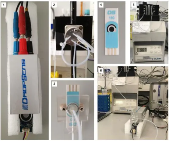
Figure 7 - Magnetic field controller ... 27 Figure 8 - 1. Dropsens switch box and electrical connections; 2. FIA valve; 3. Microfluidic Platform; 4. Screen-printed carbon electrode with silver pseudoreference electrode; 5. Peristaltic pump; 6. Assembled system ... 28 Figure 9 - Experimental procedure scheme. Incubation of supermagnetic silica microspheres coated with nitrilotriacetic acid and nickel ions with the histidine tail of CPX2. Posterior incubation of antigen-coated microbeads with canine serums for 30 minutes; injection in the microfluidic platform with the magnetic field on and reading of samples in the SPCE resorting to cyclic voltammetry. ... 29 Figure 10 - Experimental procedure to recover extracellular material of Leishmania infantum. ... 30 Figure 11- Schematic Representation of the Reversible Immobilization of Histidine-Tagged Biomolecules to an Electrogenerated Poly(pyrrole)-NTA Film97. ... 34
Figure 12 - Representation of the supermagnetic silica microspheres with Fe3O4 core
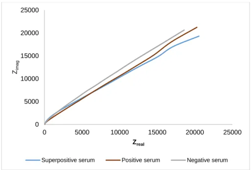
covered by hydroxyl groups coated with nitrilotriacetic acid and nickel ions and respective chelation with histidine residues of proteins of interest. ... 36 Figure 13 - Graphical representation of a negative, positive and superpositive serums measured by Electrochemical Impedance Spectroscopy (EIS). Zimag represents the
imaginary part of impedance and Zreal represents the real part of impedance. ... 37
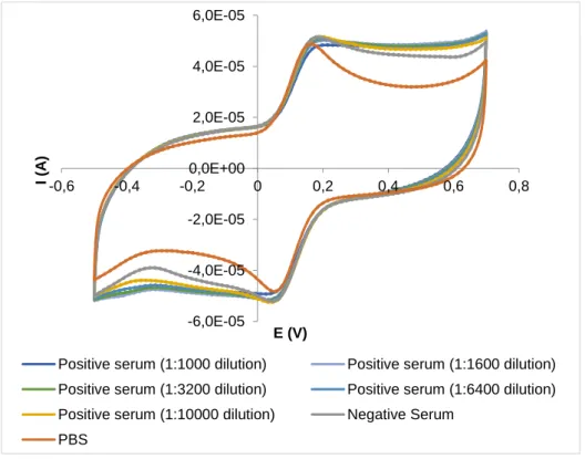
Figure 14 - Graphical representation of a negative, positive and superpositive serums measured by cyclic voltammetry. I (A) represents current measured in amperes and E (V) represents potential measured in volts. ... 38
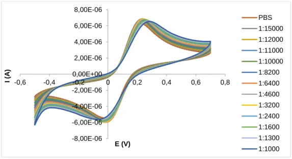
Figure 15 - Graphical representation of several dilutions of a positive serum obtained by Square Wave Voltammetry (SWV). I represents current measured in amperes (A) and E represents potential measured in volts (V). ... 38 Figure 16 - Graphical representation of several dilutions of a positive serum obtained by cyclic voltammetry (CV). I represents current measured in amperes (A) and E represents potential measured in volts (V). ... 40 Figure 17 - Voltammograms obtained with several dilutions of a positive serum resorting to cyclic voltammetry. I represents current measured in amperes (A) and E represents potential in volts (V). ... 41 Figure 21 - Three calibration curves obtained with several dilutions of a positive canine serum at a scan rate of 50 mV. They were obtained with Nernst equation, in which potential (E) in volts (V) is dependent of the logarithm of concentration (Log (C)). ... 44 Figure 22 - Calibration curves obtained with several dilutions of a positive canine serum after optimizing some parameters. They were obtained with Nernst equation, in which potential (E) in volts (V) is dependent of the logarithm of concentration (Log (C)). ... 45 Figure 23 - Two consecutive calibration curves with the same sample dilutions of a positive canine serum. They were obtained with Nernst equation, in which potential (E) in volts (V) is dependent of the logarithm of concentration (Log (C)). ... 46 Figure 24 - Voltammograms corresponding to the results of a cyclic voltammetry of the same sample – beads. I represents current measured in amperes (A) and E represents potential in volts (V). ... 47 Figure 25 - Voltammograms corresponding to several readings of the same sample - microbeads immobilized with CPX2. I represents current measured in amperes (A) and E represents potential in volts (V). ... 47 Figure 26 - Averages of the absorvance using ELISA technique of different exoproteome fractions (Total exoproteome - EXO; Extracellular vesicles - EVs; Vesicle depleted exoproteome - VDE) and Soluble Promastigote Leishmania antigens (SPLA) against positive canine serum (+) and negative canine serum (-) for Leishmaniasis. The second column of each fraction was diluted 10 x and the third column was diluted 100 x. ... 49 Figure 27 - Ratio of the absorvance between the positive (+) and the negative (-) canine serum responses for different exoproteome fractions (Total exoproteome - EXO; Extracellular vesicles - EVs; Vesicle depleted exoproteome - VDE) and against Soluble Promastigote Leishmania antigens (SPLA). The second column of each fraction was diluted 10 x and the third column was diluted 100 x. ... 50 Figure 28 - Results of the Western Blot with different volumes (50 μL; 5 μL and 0,5 μL) of EVs and VDE fractions against different serums - positive canine serum (left image) and negative canine serum (right image) ... 51
Figure 29 - Western blot after stripping the membrane and doing incubation with different serums for the wells corresponding to EVs and VDE fractions (5 μL and 0,5 μL of extracellular material). ... 51 Figure 30 - Ratio between response against positive canine serum (+) and negative canine serum (-) with different fractions of exproteome of Leishmania infantum (Total exoproteome - EXO; EVs - extracellular vesicles; VDE - vesicle depleted exoproteome) for different volumes of extracellular material. ... 52 Figure 31 - Results of an ELISA for exoproteome fractions (EVs - extracellular vesicles; VDE - vesicle depleted exoproteome) and SPLA - soluble promastigotes Leishmania antigens against human samples from an infected and a non-infected patient. ... 53 Figure 32 - Protein quantification (●) by nanodrop (Abs 280 nm); Bead-based assay for FACS analysis (●) for fractions 1-10 obtained from SEC for sample 1AT (negative control) and respective controls. Fr5 and Fr6 represent fractions 5 and 6 with a concentration of proteins of 0,109 and 0,375 μg/μL, respectively. C1 (-) - exosomes + beads + isotype (1:5000) + secondary antibody; C2 (-) - beads + Isotype (1:5000) + secondary antibody ... 54 Figure 33- Protein quantification (●) by nanodrop (Abs 280 nm); Bead-based assay for FACS analysis (●) for fractions 1-10 obtained from SEC for sample 2CA (negative control) and respective controls. Fr5 and Fr6 represent fractions 5 and 6 ... 54 Figure 34 - Protein quantification (●) by nanodrop (Abs 280 nm); Bead-based assay for FACS analysis (●) for fractions 1-10 obtained from SEC for sample 3AF (negative control) and respective controls. Fr5 and Fr6 represent fractions 5 and 6 with a concentration of proteins of 0,082 and 0,266 μg/μL, respectively. C1 (-) - exosomes + beads + isotype (1:5000) + secondary antibody; C2 (-) - beads + Isotype (1:5000) + secondary antibody ... 54 Figure 35 - Nanosight profile for the negative controls (1AT, 2CA, 3AF). Size and concentration information for the fraction with more EVs. ... 55 Figure 36- Protein quantification (●) by nanodrop (Abs 280 nm); Bead-based assay for FACS analysis (●) for fractions 1-10 obtained from SEC for sample 5878 (active) and respective controls. Fr5 and Fr6 represent fractions 5 and 6 with a concentration of proteins of 0,056 and 0,307 μg/μL, respectively. C1 (-) - exosomes + beads + isotype (1:5000) + secondary antibody; C2 (-) - beads + Isotype (1:5000) + secondary antibody ... 56 Figure 37 - Protein quantification (●) by nanodrop (Abs 280 nm); Bead-based assay for FACS analysis (●) for fractions 1-10 obtained from SEC for sample 3935 (active) and respective controls. Fr5 and Fr6 represent fractions 5 and 6 with a concentration of proteins of 0,283 and 1,034 μg/μL, respectively. C1 (-) - exosomes + beads + isotype
(1:5000) + secondary antibody; C2 (-) - beads + Isotype (1:5000) + secondary antibody ... 57 Figure 38 - Protein quantification (●) by nanodrop (Abs 280 nm); Bead-based assay for FACS analysis (●) for fractions 1-10 obtained from SEC for sample 4544 (active) and respective controls. Fr4, Fr5 and Fr6 represent fractions 4, 5 and 6 with a concentration of proteins of 0,052; 0,178 and 0,472 μg/μL, respectively. C1 (-) - exosomes + beads + isotype (1:5000) + secondary antibody; C2 (-) - beads + Isotype (1:5000) + secondary antibody ... 57 Figure 39 - Nanosight profile for the active Leishmaniasis patients (3935 FR6; 4544 FR5; 61962 FR5 & FR6). Size and concentration information for the fractions with more EXos. ... 58 Figure 40 - Results of nanodrop (Abs 280 nm) for fractions 1-8 obtained from SEC for samples 5892 and 61962 (active). Fractions 5 and 6 from sample 5892 contained 0,0346 and 0,288 μg/μL. Fractions 5 and 6 from sample 61962 contained 0,128 and 0,379 μg/μL ... 59 Figure 41 - Results of nanodrop (●) (Abs 280 nm), bead-based assay for FACS analysis (●)and respective controls (fluorescence) for fractions 1-12 obtained from 10 mL column SEC for supernatant sample from Leishmania infantum culture. Fr10 represents fraction 10 with 0,01 μg/μL. C1 (-) - exosomes + beads + isotype (1:5000) + secondary antibody; C2 (-) - beads + Isotype (1:5000) + secondary antibody ... 59 Figure 42 - Results of nanodrop (●) (Abs 280 nm), bead-based assay for FACS analysis (●) and respective controls (fluorescence) for fractions 1-12 obtained from 1 mL column SEC for supernatant sample from Leishmania infantum culture. Fr5 represents fraction 5 with 0,01 μg/μL. C1 (-) - exosomes + beads + isotype (1:5000) + secondary antibody; C2 (-) - beads + Isotype (1:5000) + secondary antibody ... 60 Figure 43 - Results of nanodrop (Abs 280 nm) for fractions 1-8 obtained from SEC for samples 608, 8844 and 621 (asymptomatic). ... 60 Figure 44- Results of nanodrop (Abs 280 nm) for fractions 1-8 obtained from SEC for samples 3081, 64047 and 3092 (healed). ... 61
List of Tables
Table 1 - Plasma samples from negative controls; asymptomatic; symptomatic and healed visceral leishmaniasis patients. ... 32 Table 2 - Size (mode) and concentration (particles/mL or particles/frame) of the fractions (FR) enriched in EXos for negative controls. ... 56 Table 3- Size (mode) and concentration (particles/mL or particles/frame) of the fractions (FR) enriched in EXos for active Leishmaniasis plasmas ... 58 Table 4 - Results of nanodrop for samples 608, 8844 and 621 (asymptomatic) in μg/μL. ... 61 Table 5 - Results of nanodrop for samples 3081, 64074 and 3092 (healed) in μg/μL. . 61
Abbreviations
CL Cutaneous Leishmaniasis
CSA Crude Soluble Antigens
CV Cyclic Voltammetry
CVL Canine Visceral Leishmaniasis
DAT Direct Agglutination Test
EIS Electrochemical Impedance Spectroscopy
ELISA Enzyme-Linked Immunosorbent Assay EVs Extracellular Vesicles
EXO Total Exoproteome EXos Exosomes
FC Flow Cytometry
FIA Flow Injection Analysis
HIV Human Immunodeficiency Virus
IC Immunochromatografic
IFAT Indirect Fluorescent Antibody Technique
IgG Immunoglobulin G
IMAC Immobilized Metal-Ion Affinity Chromatography
MCL Mucocutaneous Leishmaniasis NTA Nitrilotriacetic Acid
PCR Polymerase Chain Reaction PTFE Polytetrafluoroethylene
PVC Polyvinyl chloride
qrtPCR Real-time quantitative PCR
SAM Self-Assembled Monolayer SLA Soluble Leishmania Antigen
SPLA Soluble Promastigote Leishmania Antigens SPE Screen-Printed Electrodes
spp. Species
SWV Square Wave Voltammetry
VDE Vesicle Depleted Exoproteome VL Visceral Leishmaniasis
Introduction
1. Leishmaniasis - a neglected tropical disease
Infectious diseases are the second cause of mortality around the world, according to the World Health Organization (WHO)1. Among the parasitic diseases, Leishmaniasis is the
second cause of death after Malaria. This neglected tropical disease exhibits high morbidity and mortality putting at risk people from 98 different countries worldwide2. It is
endemic in 88 countries and 1.8 million of new cases are estimated to occur each year3.
Leishmaniasis is a vector-borne disease caused by the parasites Leishmania species and transmitted by the bite of infected female phlebotomine sandflies. Combining 30 species of phlebotomine sandflies to more than 20 species of Leishmania species capable of causing disease, makes this opportunistic infection more complex to understand.4
Several factors such as climate changes and changes in animal migratory movements have been contributing to the spread of this infectious disease5.
Organism prevalence differs by geographical distribution, being an endemic disease in the tropics, subtropics and the Mediterranean basin6. The protozoan parasites of the
genus Leishmania take advantage of the mammalian host to survive and affect predominantly least developed and emerging countries with limited resources and individuals with deficient immune system (for example, individuals infected with HIV – Human Immunodeficiency Virus)5,7.
1.1 Pathological forms of the disease
More than 20 Leishmania species are known to cause disease and the outcome of the infection will depend not only on the parasite species but also on the immune response of the host and the environment8. Different parasite species can cause identical
pathology9.
Leishmania species (spp.) lead to specific clinic-pathological categories: cutaneous
Leishmaniasis, mucocutaneous Leishmaniasis and visceral Leishmaniasis, also known as kala-azar (Figure 1). Symptoms range from skin lesions to affecting cutaneous and mucosal tissues or even
to vital visceral organs damage
4,8.1.1.1 Cutaneous Leishmaniasis
Cutaneous Leishmaniasis (CL) is the most studied form of the disease. It is caused by
L. tropica and develops as a nodule in the site of inoculation, consequence of the
infection of macrophages in the dermis that may develop to a dermal granuloma, an ulcer that heals spontaneously or several inflamed ulcers that take longer to heal (a few months to years). Histologically it is characterized by a lymphoid and monocytic infiltrate with granuloma formation. L. mexicana and L. aethiopica are responsible for a particular kind of CL in which the nodules resemble to lepromatous leprosy8,9.
1.1.2 Mucocutaneous Leishmaniasis
This clinical presentation of Leishmaniasis is caused by L. braziliensis. Mucocutaneous Leishmaniasis (MCL) on the contrary of CL, which is confined to small areas of the skin, spreads to the oral and nasal mucosa and is characterized by disfigurative ulcers that take long time to heal. These progressively destructive ulcers, unlike cutaneous Leishmaniasis, are not self-healing and appear months or years after the first episode of CL4,10.
1.1.3 Visceral Leishmaniasis
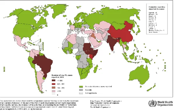
Visceral Leishmaniasis (VL), also known as kala-azar, is the most severe form of Leishmaniasis and is fatal if left untreated6. Each year there are 500 000 new cases of
VL, mainly in endemic areas (90% of these in India, Bangladesh, Brazil, Nepal and Sudan)11 (Figure 2). This disease strikes the most in poorest countries and can have a
disastrous impact when it strikes a non-immune population.
VL is caused by the Leishmania donovani complex, which includes L. donovani, L
chagasi similar to L. infantum12.
An incubation period (from 1 month to 2 years) is followed by clinical manifestations such as fever, splenomegaly, hepatomegaly, weight loss, progressive anemia, pancytopenia. These typical symptoms of this systemic infection are characteristics of the dissemination
Cutaneous
Mucocutaneous
Visceral
of parasites throughout the blood and reticuloendothelial system which lead to enlarged lymph nodes, spleen and liver8.
Post kala-azar dermal Leishmaniasis is considered a sequel of VL in which skin is the focus of infection and appears like a nodular rash. This condition is frequent in Sudan and in the Indian subcontinent13.
As an opportunistic infection, the parasite prevails in people whose immune system has been compromised. Thus, coinfection is common among HIV patients in Mediterranean countries, Brazil, east Africa and the Indian subcontinent14.
Figure 2 - Distribution of Endemic Visceral Leishmaniasis worldwide 2015. World Health Organization15.
1.2 Epidemiology
Leishmania spp. capable of causing disease are divided in two main groups: Old world
species (L. major, L. tropica, L. aethiopica, L. donovani, L. infantum) and New World species (L. mexicana, L. amazonensis, L. brasiliensis, L. guyanensis, L. chagasi).8
Visceral Leishmaniasis (VL) is caused by L. donovani (East Africa and the Indian subcontinent) and L. infantum (Europe, North Africa) in the Old World and L. chagasi in the New World (Brazil). While L. donovani infects all age groups, L. infantum infects mostly children and immunosuppressed individuals. Among other regions, L. infantum is responsible for VL in children in the Mediterranean basin. However, due to increasing prevalence of human immunodeficiency virus (HIV) infection in this region, HIV-VL coinfection in the adult population is being reported frequently. L. chagasi similar to L.
infantum causes VL in children in Latin America, where lymphadenopathy is a dominant
clinical feature16.
Due to urbanization and HIV pandemic, the incidence of VL is rising. More than 350 million people live in active parasite transmission areas, putting their health at risk17.
Furthermore, each year there are up to 30 000 deaths due to this severe form of the disease9.
This disease affects mostly least developed countries and poorest regions, where malnutrition, displacement, poor housing, weak immune system and lack of resources are common issues. Although not fully recognized, socioeconomics subjects are tightly connected to poverty and endemicity of this disease9.
1.3 Transmission
1.3.1 Leishmania life cycle
Sandflies used to be limited to their natural distribution areas but, as previously mentioned, several causes (migration, climate changes, economic development, etc.) lead to the spread of sandflies as well as Leishmania reservoirs and consequently lead to the disease incidence.
There are more than 800 known species of phlebotomine sandflies but only certain species of sandfly of the genus Phlebotomus (Old World) or Lutzomyia (New World) transmit the Leishmania spp. parasites11.
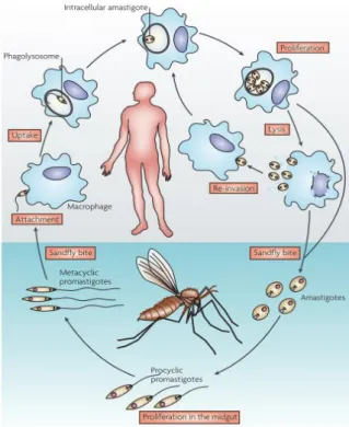
The parasites have a digenetic life cycle that includes an extracellular developmental stage in the female phlebotomine sandfly and another one in the vertebrate host, mostly intracellular. The parasite development in sandflies occurs in the alimentary tract, where promastigotes (the motile, flagellated, extracellular form of the parasites) are formed. Maturation of the parasite occurs in the midgut, where it develops to the infectious development form of the parasite: metacyclic promastigotes.
The transmission of the parasites to the mammalian host occurs during insect blood feeding. Usually, an inoculum contains around 100–1000 metacyclic promastigotes that are rapidly engulfed by mono and polymorphonuclear cells. Within these cells, the parasites go through other morphological changes, differentiating to an ovoid shape with short flagellum known as amastigote. Once they take this form, they multiply and the cycle becomes complete when the sandfly takes another blood meal with the amastigotes18,19 (Figure 3).
1.3.2 Transmission Pattern
There are two types of VL transmission: zoonotic, when the disease can be transmitted from animal to vector to human, and anthroponotic is transmitted from human to vector to human. Transmission characteristics also differ according to geographical regions, being L. donovani responsible for the anthroponotic transmission and L. infantum for the zoonotic transmission. Anthroponotic transmission occurs in the Indian subcontinent and the zoonotic type is mainly transmitted in the Mediterranean basin and South America regions.
1.3.2.1 Zoonotic disease
Zoonotic visceral Leishmaniasis is a serious public health problem that affects some mammals and results in significant mortality and morbidity where it is endemic20. Animals
such as wild canid, marsupials and rodents have been described as reservoirs of human VL21. However, zoonotic VL epidemics have been associated only in areas where canine
Leishmaniasis is endemic.
Canine visceral Leishmaniasis (CVL) is caused by Leishmania infantum in the Mediterranean area, Middle East and Asian countries and is caused by Leishmania
chagasi in Latin America. Due to their genotypic relationships, these two species are
considered identical22.
1.3.2.1.1 Dogs as main reservoirs
Although dogs are recognized as the main source of infection, the current control diagnosis methods and treatment are directed to humans23.
In fact, from an epidemiological point of view, canine visceral Leishmaniasis is more important than Leishmaniasis affecting humans since its incidence is higher and both symptomatic and asymptomatic dogs are infectious to sandflies24. Besides, CVL is a
veterinary issue because in endemic areas such as the Mediterranean Basin and Brazil, domestic dogs are the primary source of infection by the vector, allowing its life cycle perpetuation.
After an incubation period that can range from three months to several years, dogs might start presenting some clinical signs. Nevertheless, some dogs remain asymptomatic and never develop clinical signs11.
Thus, dogs can present different forms of the disease: symptomatic, oligosymptomatic and asymptomatic. It is of great relevance mentioning that all forms of the disease enable the transmission of the parasite.
Symptomatic CVL typically results in death and it is based on the presence of at least two clinical manifestations. Infected dogs present cutaneous alterations such as alopecia, onychogryposis, dermatitis, skin ulceration and visceral manifestations with splenic, renal, hepatic, and neurological disorders. Other symptoms as anorexia and weight loss are also usual25. Infected dogs can remain asymptomatic, not presenting
detectable clinical signs of disease. This is a grave problem, since these animals can contribute to the maintenance of Leishmania cycle24. Dogs that develop a few mild
symptoms are classified as oligosymptomatic.
Despite evidences showing that killing seropositive animals leads to a decrease incidence in CVL and VL in children, this control method is not well accepted26.
However, due to lack of accuracy in diagnosis methods the number of detected infected dogs cannot be elucidative of the real prevalence of infection. The number of infected dogs are estimated in few millions but it is believed that the real burden of disease is much higher23.
We would expect that seroprevalence of CVL was higher in rural areas, where the sandflies are mainly found but the fact that there are more hosts infected in urban areas leads to the increase of CVL and higher prevalence when compared to rural areas. Unlike expected, urban areas favor the transmission of parasite due to high number of dogs and gardens. It provides a good environment to sandflies proliferation27.
A sensitive and specific method to detect canine Leishmaniasis in early stages is needed to avoid false positives and false negatives that lead to unnecessary euthanasia and
disease transmission, respectively11. Detection of asymptomatic dogs is critical for
controlling the spread of the disease among dogs and between dogs and humans. The foundation to an effective control is prevention, screening and detection of the disease, since treating infected dogs is not that effective (relapses occur frequently and they can regain infectivity weeks after treatment)28. Besides, VL drugs might lead to
parasite resistance.
2. Prevention and treatment
One approach to control the infection is doing vector control. This can be achieved with insecticides, such as pyrethroids and DDT. However, sandflies may develop resistance to DDT but remain sensitive to other insecticides. Bed nets and spraying are also alternative options to minimize the contact of some species with the host. Nevertheless, consequences concerning sandflies populations resistance are still under study29,30.
Regarding dog protection, collars impregnate with deltamethrine reduce the risk of infection in dogs.
Few drugs are available for the treatment of Leishmaniasis and besides the risk of developing drug resistance, treatment efficacy depends on strains and species9.
Furthermore, there is no Leishmaniasis human vaccine available. Vaccination would be a good option to prevent infection31.
Vaccination of dogs would still be the best strategy if an efficient vaccine was developed. Being lauched in Portugal in 2011, CaniLeish® was the first vaccine for canine
Leishmaniasis in the European Union. In Brazil, LeishTec® vaccine was also registered
but only about 40% of protection against infection was found. Despite the efforts,
Leishmania vaccinology still has a lot to improve till an effective and universal vaccine is
developed32.
3. Screening, detection and surveillance
Monitoring and controlling the spread of emerging diseases is a topic that should concern all individuals.
Disease surveillance is a crucial component to eliminate or at least to minimize the spreading of this infectious disease before the burden is devastating33.
The first step to treat a condition or to take preventive measures is to diagnose. Taking appropriate actions will lead to improved health outcome. However, to achieve proper and accurate diagnosis a robust, rapid diagnosis test is needed5. Once we have a
diagnosis test with these characteristics efficient clinical and epidemiological management of infections can be made34.
There is an urge to develop a tool both sensitive and specific that allows an easy screening in endemic regions, capable of identifying asymptomatic cases35. This
diagnosis method should also be accessible, providing the possibility of doing periodic monitorization.
The process to diagnose should be automated, requiring inexpensive reagents and minimal operator intervention without diminishing the fidelity of the results. Considering that this disease affects mostly less developed countries the tests should be cheap and easy to perform in difficult field conditions5,36.
4. Diagnosis methodologies
Accurate diagnosis still remains a problem to disease control since leishmaniasis presents a wide range of manifestations37.Clinical manifestations can also be confused
with other illnesses often common in Leishmania endemic areas such as malaria, toxoplamosis and tuberculosis38.
Many Leishmaniasis noninvasive diagnosis are available but none has both specificity and sensitivity to diagnose in endemic areas. We must have into account that age, medical history and host immune system response are crucial parameters in diagnosis. According to WHO, diagnosis of Leishmaniasis can be confirmed with more conventional laboratorial techniques such as visualization of parasite in tissues by microscopic examination of the stained specimen or in vitro culture of the parasite from biopsies or aspirates from lesions, lymph nodes, spleen and bone marrow and other diagnosis methods that include molecular detection of parasite DNA in tissue samples and serological tests that detect anti-Leishmania antibodies9.
4.1 Parasitological diagnosis
Parasitological diagnosis of Leishmaniasis is specific and remains the gold standard to diagnose this infectious disease39.
One of the reference standards for diagnosis is the demonstration of parasites in tissue samples from bone marrow, skin lesions, liver, lymph nodes and spleen. Diagnostic is obtained by observation of the Leishmania amastigote forms in stained microscopic preparations with Giemsa6.
The best results are obtained with spleen aspirates (93%-98% of sensitivity). When it comes to bone marrow aspirates, the sensitivity decreases to 60%-85% and worse results are obtained with lymph nodes aspirates (sensitivity ranges between 52% to 58%)4.
Besides this method requiring trained personnel and its sensitivity being variable, it involves invasive sampling being a risky procedure that can lead to fatal hemorrhage so it must be carried out in settings with access to surgical facilities. Lymph node and bone marrow aspirates are safer but the material obtained is more diluted and therefore less sensitive, raising the risk of diagnosing false negatives40.
The culture of parasite from infected tissues is another classical confirmatory test for VL. A major problem with this technique is that different species of Leishmania have different growth factors and contaminations are recurrent. Despite being more sensitive than microscopic examination, it is time consuming and expensive, so it is rarely used for clinical diagnosis12.
Identification of amastigotes by direct examination of aspirates must be done by experts because the results are dependent on the observer9. It can also harvest false negative
results because of the low number of parasites in some samples, particularly in asymptomatic cases, where the parasite charge is lower.
4.2 Serological diagnosis
Serological tests are based on the screening of antigens or antibodies.
The first approach is an excellent method to diagnose an infection since is more specific that antibody-based immunodiagnostic tests. Thus, antigen levels are expected to correlate with parasite charge, and this method might be useful when antibody prediction is deficient. Besides, this approach should avoid cross-reactivity and should distinguish active from past infections.6 However, this technique is still unreliable at the moment
(lack of specificity and variable sensitivity), so efforts are being made to improve this tool, as it stands as a promising approach.
Currently, most clinical and surveillance laboratories of the developing world use serological techniques to detect pathogen-specific antibodies, since direct methods are either invasive and potentially fatal or expensive.
VL infection is characterized by the presence of humoral response that leads to the production of antibodies specifically against to Leishmania spp.. Thus, serological methods to detect anti-Leishmania antibodies are useful as alternative diagnosis tests
for both human and canine Leishmania infections27. They have the advantage of being
easily applied to a large amount of samples with specificity and sensitivity, mostly used in seroepidemiological studies41.
The presence of anti-Leishmania antibodies in both asymptomatic and symptomatic infected dogs has allowed the development of agglutination tests, immunofluorescent serologic tests, such as Western blotting, immunochromatographic tests, and enzyme-linked immunosorbent assays (ELISAs).
The sensitivity depends on the methodology but the specificity will always depend on the antigen used. In most serological tests, the sensitivity and specificity data are compared against the standard methodology 16.
Serological tests are the elected to diagnose CVL. Nevertheless, when dogs present low antibody titers these tests lack sensitivity and specificity. Is not unusual to occur cross-reactivity too42. Moreover, after a successful treatment, it takes a while till antibody levels
decrease so it can mask a relapse, making impossible to diagnose it. 4.2.1 Direct Agglutination Test
As an attempt to substitute the risky procedure of splenic aspirates, a noninvasive serological test was developed. Direct agglutination test (DAT) was the first antibody detection test used for VL diagnosis and has been used for more than 25 years.
DAT is a simple semi-quantitative diagnostic tool used in many developing countries with a high sensitivity (91-100%), specificity (72-100%), accuracy, reliability and inexpensiveness4,43.
DAT detects parasite antibodies in the blood or serum of those infected through direct agglutination. Whole Coomassie-stained promastigotes, either in a suspension or in freeze-dried form are incubated with serial dilutions of serum. If the result is negative (absence of anti-Leishmania antibodies) DAT antigen accumulates at the bottom of the plate. If there are anti-Leishmania antibodies present, the antigens form a film over the well (positive result). In a positive reaction, agglutination can take till 18 hours of incubation to occur9,41.
The disadvantages of DAT are requiring moderate technical expertise (the interpretation of the results depends on the person analyzing the results, creating inter-observer discrepancy)44, serial dilutions must be done (requires a considerable volume of antigen)
DAT results remain positive long after the patient is cured (anti-Leishmania antibodies can persist for years as a result of a VL infection), so this test is not appropriate to detect relapses6.
4.2.2 Enzyme-Linked Immunosorbent Assay (ELISA)
ELISA is an important serodiagnosis technique used in almost all infectious diseases, including VL.
These ELISA assays are based on the detection of antibodies present in the blood or serums. An antigen/recombinant protein is used to coat the plate and then the samples containing antibodies are incubated. To see the development of signal, a secondary antibody conjugated to an enzyme recognizes and binds to the primary antibody. After adding a substrate, a colorful product is developed and the results can be measured by optical density techniques.
This tool is frequently used to detect anti-Leishmania antibodies due to its high sensitive and specificity as well as good reproducibility and high throughput screening of large number of samples at affordable expenses. However, the specificity of this technique depends on the antigen used.
The sensitivity of crude soluble antigens (CSA) in an ELISA is high. Nevertheless, cross-reactions with serum from patients with trypanosomiasis, toxoplasmosis and tuberculosis can occur. VL diagnosed by ELISAs is based on crude soluble Leishmania antigens (SLAs). Though its high sensitivity, the specificity is low due to the antigens related with
Leishmania and other pathogenic protozoa. It also has low competence to detect
seropositivity in asymptomatic dogs27,45,46.
ELISA’s major disadvantage is its inadaptability for field conditions in resource-poor settings and its requirement for specialized operators.
4.2.3 Indirect Fluorescent Antibody Technique (IFAT)
Fluorescent antibody techniques are extremely valuable tools that allow evaluation of anti-Leishmania antibody titers produced by infected individuals. It is a quite useful method in epidemiological studies and in clinical practice47.
IFAT is based on the use of fluorophores to detect antibodies present in the sample. It involves the use of a primary antibody to bind to the antigen and allow the formation of antigen-antibody complex and posteriorly the binding of a fluorophore-conjugated secondary antibody that results in an amplified signal that can be examined by fluorescence microscopy.
IFAT is the serological gold standard for the diagnosis of CVL in most countries around the Mediterranean basin. This methodology is different from ELISA and it resorts to the whole body parasite as antigen47. Although high specificity and sensitivity (100% and
90%, respectively), IFAT sensitivity is lower for asymptomatic infections when compared to ELISA. Also, the result interpretation depends on operator’s expertise46,48.
4.2.4 Immunochromatographic tests
Immunochromatografic (IC) tests are a great option to be used in large-scale surveys, in which antibody titers are not required.The results are always evaluated considering the epidemiological context of the area and the aim of the investigation49.
IC test principal is similar to the one used in ELISA but the main difference is that the reaction occurs in chromatographic paper by capillary action. Immunochromatographic tests are based in the formation of an antigen-antibody complex. Then, the sample migrates in the membrane and the labelled antibody gets in contact with the immobilized antibody in the membrane and it results in the formation of color. The appearance of color is indicator of the antigen presence in the sample.
IC tests are inexpensive, practical, rapid and suitable for field use. However, in resemblance to DAT, a significant portion of healthy individuals is diagnosed as positives in endemic regions after long cure period (false positive cases). So, patients with suspect of relapse are not candidates for diagnosis49.
4.2.5 Flow cytometry
Flow cytometry (FC) is considered an emerging technology for the diagnostic of several infectious diseases. It usually refers to the measurement of cells but the approach of making optical measurements in a flowing sample stream is a general analytical approach50.
Flow cytometry is a technique based on the analysis and sorting of cells or particle suspensions in a controlled fluid stream through the measurement of fluorescence and scatter induced illumination. Using specific fluorescent markers, flow cytometry can be used to the quantification of structural and functional parameters on a single-particle basis.
Flow cytometry technique can quantify specifically the antibodies against Leishmania surface antigens, restraining potential cross-reactivity against more conserved intracellular structures. All the approaches to date used live or fixed promastigotes as targets to detect specific antibodies51.
Among others, flow cytometry has high throughput capacity, possibility of quantification, high reproducibility and sensitivity and potential for multiplexing52.
Recently, alternative methodologies were proposed to improve the serological approaches. One of them is immunoglobulin G (IgG) flow cytometry to detect
anti-Leishmania braziliensis antibodies in sera of active cutaneous anti-Leishmaniasis patients 38.
This method has shown to be reliable, achieving high levels of sensitivity and specificity51. Studies have shown the FC potential to monitor postchemotherapy cure of
VL, in order to evaluate the success of treatment. The antibodies which are detected in FC are the anti-membrane-specific antibodies and are only present during active disease which makes it a perfect technique to evaluate the effectiveness of the treatment53.
4.3 Detection of parasite DNA
Polymerase chain reaction (PCR) technology has become an indispensable tool for the diagnosis of many parasitic diseases, including Leishmaniasis. Amplification-based methods include the conventional PCR (polymerase chain reaction) and qPCR (quantitative polymerase chain reaction)54.
PCR is based on the amplification of specific parasitic DNA sequences. The sensitivity of a PCR assay depends upon three factors: the physicochemical conditions of the reaction, the concentration of the DNA target and the selected PCR primers20,55,56. It has
higher sensitivity for asymptomatic animals and early-stage infections when compared to serological methods. In patients with compromised immune system, PCR is also more sensitive than the classical parasitological methods.
Some PCR drawbacks are collection of samples, storage conditions and difficult access to sophisticated equipment57. Lack of standardization in the selection of target Leishmania DNA sequences and experimental protocols used worldwide also makes it
harder to compare the sensitivity and specificity of these tests.
Real-time quantitative PCR (qrtPCR) is an innovative technology that has revolutionized molecular diagnostics by adding sensitivity, speed, broad dynamic range of target DNA quantification and reduced contamination. When compared with conventional PCR, it has the major disadvantage of costing three times more55,58.
As we have seen above, every technique has its downsides.
Parasite demonstration in tissue smears and culture provide definitive diagnosis of VL, but generally has lower sensitivity than serologic methods. Particularly, splenic aspirate has the highest sensitivity of available tissue sampling techniques, but it has a risk
associated. Moreover, microscopy techniques lack sensitivity, whereas culture requires long time to obtain a result and is vulnerable to contamination59.
Molecular diagnostic tools like PCR and real-time PCR are quite sensitive and specific but are difficult to perform and have a high cost.6 So, its use remains largely restricted
to some hospitals and research centers37.
Serological tests are often not sensitive enough to detect asymptomatic individuals so they have to be combined with classical methods of diagnosis to confirm. ELISA, IFAT, DAT and rK39 immunochromatographic strip test (ICT) are highly sensitive and specific when analyzing active VL in immunocompetent individuals. This doesn’t happen when titers decline and parasite charge is lower. Due to these reasons, tests give false-negative results frequently in immunocompromised patients and for asymptomatic
Leishmania infections60.
For those reasons, it is crucial to identify and produce new proteins capable of detecting asymptomatic Leishmaniasis and individuals in early stages of infection in VL endemic regions.
5. Recombinant proteins and novel biomarkers
Every diagnosis platform has its detection limits and sensitivity but these factors are also dependent on the specificity of proteins for producing high confidence results with lower detection limits. There is an urge to develop a marker for the active diseases6.
Antigens related to the disease are discovered and validated through genomics and proteomics research. The development of new technology to diagnose can enable rapid introduction of these new antigens into clinical practice36. This tool must provide a signal
of presence or absence of a particular antibody but also provide quantitative information36.
The main problem with Leishmaniasis diagnosis is identification of markers that can detect the presence of disease since early stages until recovery after treatment. Seroconversion does not happen right after infection so serological tests may lead to false-negative results. New recombinant antigens are needed to contribute for a more accurate Leishmaniasis diagnosis and hard work has to be put in the development of a more sensitive and specific recombinant protein-based immunoassay capable of detecting asymptomatic cases in large screening studies61,56.
Recently, several Leishmania spp. proteins were cloned, purified, and characterized to improve the diagnosis of Leishmaniasis62,63. The major advantages of using recombinant
proteins for the diagnosis of Leishmaniasis infections are: knowing the precise antigenic composition applied to the serological tests; good reproducibility and easy method standardization23,64,51,63.
Using recombinant polypeptides containing specific L. donovani / L. chagasi epitopes that elicits an immune response in the majority of dogs and humans with VL can overcome some cross-reactivity problems11. Moreover, the variability in the humoral
response concerning different parasite antigens observed in infected dogs suggests that a combination of recombinant proteins can improve the diagnosis efficiency65.
Different families of proteins like kinesin-related proteins, heat shock proteins, nuclear proteins, ribosomal proteins, enzymes and other antigens were evaluated regarding their performance (sensitivity and specificity) of immunological diagnosis40.
Recombinant protein rK39 (39-amino acid repeat of a kinesin-related protein), highly conserved in Leishmania spp., is a great tool to the diagnosis of VL in HIV patients and a prognostic indicator for monitoring patients undergoing drug treatment64,59,61. An
important aspect of anti-rK39 antibody is that the titer correlates directly with the disease activity, indicating its potential for use in predicting response to therapy16. Among the
recombinant antigens, this one showed promising diagnosis and has been extensively tested in the last 5 years with IC tests in several Leishmaniasis endemic areas4,6, with
several commercial applications (Appendix1). rk39 has also been tested for ELISA and FC assays showing high sensitivity and specificity in detecting clinical forms of CVL52.
However, rK39 doesn’t have the same accuracy in asymptomatic cases with proven infection. Several proteins such as Leishmania infantum cytosolic tryparedoxin peroxidase (LicTXNPx, also known as CPX), a member of enzymatic Leishmania cascades for detoxification of peroxides expressed in all stages of parasite development, and rK28 (a synthetic gene generated by fusing multiple tandem repeats of haspb1 and k39 genes) were already described as valuable tools for the detection of infection6,23.
LicTXNPx antigen has a highly immunogenic probe during both human and canine infections. Previous studies indicated that anti-LicTXNPx antibodies were present in both symptomatic and asymptomatic experimental canine infections, making this antigen a good candidate marker and a prognostic indicator for monitoring the response to VL treatment64.
When testing rk39 and LicTXNPx in ELISA, rK39 demonstrated a better performance in the symptomatic group23 whereas LicTXNPx showed a better performance in the
detection of infections in asymptomatic infected dogs. An ELISA with both LicTXNPx and rk39 antigens (LAM-ELISA) was performed and both symptomatic and
asymptomatic dogs had higher specificity (96.3%). LAM-ELISA is a simple and sensitive (87.1% sensitivity) test that associated with DAT may be a valuable tool for screening CVL23,27.
Attention to extracellular vesicles (EVs) has been rising in the last years as they are believed to be potential biomarkers to use in the diagnosis of diseases66. Our group has
been working with EVs as we are interested in the proteic material contained in exosomes and its great clinical impact potential in nanomedicine67. This type of vesicles
is naturally released from cells and its context of infection is not still very clear68.
Particularly, the exoproteome of L. infantum is composed by two main fractions: the vesicle fraction and the free protein fraction. It is believed that extracellular proteins derived from the parasite have an active role in host-parasite interaction69. This is an
indicator that EVs can be used as future markers for infectious diseases.
6. Biosensors
A major obstacle in defining prevalence and incidence of infection is the lack of an easy and accurate tool for the detection of infected individuals35.
Since many infectious diseases spread quickly before any symptoms are identified, the diagnosis device must be a sensitive analytical tool that can easily go down to very low detection levels of antibodies without losing selectivity. Biosensors meet these field requirements as they are simple, cheap, robust, accurate, rapid and provide high-throughput33. Even more, they offer the possibility of real-time monitoring, and the
distribution of these diagnosis tools in the field provide a rapid infection detection33 of the
disease that has a significant effect on the success of disease spreading, control, or eradication5.
A biosensor is a sensing device containing a bio-recognition element that interacts with a biomarker and a transducer that converts the changes in its physicochemical properties (optical, thermal, electrical, and thermodynamic properties) into an electrical signal (Figure 4). The biosensors can be classified based on the type of bio-recognition element or the transducing method used5. Based on the bio-recognition element, the biosensors
can be classified as enzyme sensors, immunosensors, nucleic acid probe (DNA and RNA) sensors, or cell-, tissue-, or organelle-based sensors70. Based on the transducing
method, biosensors can be classified as piezoelectric, optical, or electrochemical biosensors5.
Figure 4 - Schematic diagram of biosensor elements: target analyte; bioreceptor; transducer. Image adapted from “The role of biosensors in the detection of emerging infectious diseases”, 2006 33
There are several factors that affect the efficacy of the biosensor. Thus, all characteristics that influence the accuracy of these biosensors (biomarker/bio-recognition element ligation, surface preparation/immobilization conditions, incubation time, temperature, etc.) have to be optimized during the development process.
One of the major problems with biosensor are matrix interference and adsorption of blood components onto the sensor surface. Furthermore, biosensors have to be robust and work outside laboratories to be applied to real-time detection of infectious diseases in biological samples (i.e., blood)33.
As we intend to develop a biosensor to detect an infectious disease and the host immune response to the parasite involves a humoral response and the development of antibodies against it, we decided to focus our attention on immunosensors in this revision.
6.1 Immunosensors
Antibody-based sensors, also known as immunosensors, are well accepted due to the specificity of antibody-antigen reaction to produce a change in the transducer signal5.
They are considered more versatile than enzyme-based biosensors, because the antigens can be produced specifically to any biomolecule of interest71. These biosensors
are ideal as they rely on the high affinity between the antibodies and the antigens (recognition element), resulting in very low detection limits72,73, with less level of
interferences, non-destructive approach to sample, stability, good precision and high sensitivity74. These devices should be compact and allow simple operation75.
The disadvantage of these immunosensors is that the antigen is not easily released from the antibody after the measurement has been made. Thus, several strategies have been applied to design inexpensive biosensors71. Another option involves the use of a flow cell
configuration in which the immunochemicals can be partially removed from the sensor before the next measurement71.
Independently of the biosensor’s type, immobilization of the bioreceptor onto a solid support is a crucial aspect in determining the overall performance of the device33.
To obtain a diagnosis device with the characteristics mentioned above, we have to overcome some drawbacks regarding sensitivity, stability and longevity that mostly depend on the amount of the immobilized immune molecules on the surface, their conformational stability, remaining activity after the immobilization procedure and their orientation on the sensor surface74. The last is particularly important since
immunoglobulins are asymmetrical molecules and wrong immobilization can lead to hindered interaction with the biomarker. Highly controlled orientation immobilizations are preferable to maximize their antigen-binding efficiency and achieve the best immunoassay sensitivity and selectivity. Oriented binding may require many chemical modifications that decrease the control over the nanostructures76.
There are three main methods of immobilization: adsorption, entrapment and covalent binding.
Adsorption is a rapid and simple procedure, especially for disposable biosensors. This physical type of immobilization is based on Van der Waals attraction between biomolecule and the solid support surface. The most relevant drawbacks of this technique are that bonding forces between biomolecule and support are weak and an even monolayer cannot be guaranteed. As a consequence, the biological component can be leached during the assay, depending on experimental conditions such as pH, ionic strength, temperature and solvent71.
Electropolymerization is an easy and attractive example of the entrapment technique. A biological molecule is homogenized in a monomer matrix and this is then deposited by electropolymerization71.
Immobilization cross-linking, or covalent immobilization, is used to coat electrode surfaces with specific biotransducer molecules. Covalent immobilization of the biorecognition component (enzyme, antibody, cell, etc.) can be achieved by use of the self-assembled monolayer (SAM) procedure71. The method is based on the formation of
three-dimensional links between the biological material and multifunctional reagents. Antibodies can be directly immobilized on the surface of the transducer by covalent bonding through amino, carboxyl, or aldehyde groups5. This technique has several
advantages, such as stability and reusability of immunosensors due to covalent bonds74.
Several immunosensor technologies advances have been made in the last years, with distinct notability to transducer approaches and their clinical potential such as for the detection of Salmonella pullorum77, hepatitis B77,78 and dengue79.