Development of a label-free immunosensor based on surface plasmon
resonance technique for the detection of anti-
Leishmania infantum
antibodies in canine serum
Dˆenio E.P. Souto
a,b,d, Jussara V. Silva
a, Helen R. Martins
b, Alexandre B. Reis
c,
Rita C.S. Luz
a,d, Lauro T. Kubota
d, Fla´vio S. Damos
a,d,naInstituto de Ciˆencia e Tecnologia, Universidade Federal dos Vales do Jequitinhonha e Mucuri, 39100-000 Diamantina, MG, Brazil bDepartamento de Farma´cia, Universidade Federal dos Vales do Jequitinhonha e Mucuri, 39100-000 Diamantina, MG, Brazil cDepartamento de Ana´lises Clı´nicas, Universidade Federal de Ouro Preto, 35.400-000 Ouro Preto, MG, Brazil
dInstituto Nacional de Ciˆencia e Tecnologia em Bioanalı´tica, Unicamp, 13083-970 Campinas, SP, Brazil
a r t i c l e
i n f o
Article history:
Received 15 December 2012 Received in revised form 24 January 2013 Accepted 25 January 2013 Available online 20 February 2013
Keywords: Leishmania infantum
Visceral leishmaniasis Surface plasmon resonance Immunosensor
Scanning electrochemical microscopy
a b s t r a c t
In this work, a surface plasmon resonance (SPR) immunosensor was developed using an 11-mercaptoundecanoic acid (11-MUA) modified gold SPR sensor chip for the detection of anti-Leishmania infantumantibodies. The soluble antigens ofL. infantumwere securely immobilized on an SPR gold disk by an 11-MUA self-assembled monolayer. Cyclic voltammetry (CV), electrochemical impedance spectroscopy (EIS) and scanning electrochemical microscopy (SECM) techniques were employed in the characterization of the antigen immobilization. After the immunosensor construction, canine serum positive for visceral leishmaniasis was added to its surface and showed significant variation in the SPR angle, indicating excellent sensitivity of the technique for antigen–antibody interaction detection. Moreover, the addition of negative serum was accompanied by a smaller response, demonstrating that the immunosensor shows good specificity against anti-L. infantum antibodies. Therefore, this work demonstrates the successful development of an SPR sensor for anti-L. infantumantibodies detection in short time, showing a great perspective as a sensing system of visceral leishmaniasis in endemic regions.
&2013 Elsevier B.V. All rights reserved.
1. Introduction
Leishmaniasis is a widespread zoonotic disease, which represents a serious public health problem with various clinical manifestations, depending on theLeishmaniaspecies (Lukes et al., 2007). According to disease-burden estimation, leishmaniasis is the second-ranking cause of mortality and the fourth-ranking in morbidity among neglected tropical diseases (Mathers et al., 2007). An estimated 12–15 million people are infected world-wide, with approximately 1.5–2 million new cases per year. Of these, about 500.000 are visceral leishmaniasis (VL) and 1.500.000 are cutaneous leishma-niasis (CL) (World Health Organization WHO, 2010). Whereas CL has a tendency to heal spontaneously and leave resulting scars, VL is fatal when left untreated, causing an estimated 59.000 deaths per year (35.000 men and 24.000 women) (Den Boer et al., 2011; Desjeux, 2004;Srivastava et al., 2011). Consequently, VL has aroused
the concern of health authorities due to its appearance in large urban centers or its re-emergence in many cities around the world (Alves and Bevilacqua, 2004;Ashford, 2000).
VL is caused by species belonging to theLeishmania donovani complex.L. donovaniandLeishmania infantumare the etiological agents found in the Old World, while only the latter is observed in the Americas (Murray et al., 2005). Dogs are considered the most important epidemiological reservoirs ofL. infantumand serve as an abundant source of infection for the sandfly vectorLutzomyia longipalpis, due to its high skin parasitism (Reis et al., 2006). The large number of dogs infected in endemic areas carry the parasite to the insect vectors, constituting an important link in maintaining the transmission of infection to humans (Alves and Bevilacqua, 2004). Therefore, the diagnosis and euthanasia of positive dogs is essential to the success of controlling of LV (Moreno and Alvar, 2002).
The clinical aspects for diagnosing VL are a challenge, owing to nonspecific clinical signs and symptoms in oligosyntomatic and the high percentage of asymptomatic cases in humans and dogs (Teixeira-Neto et al., 2010). Thus,adefinitive diagnosis depends on the laboratory tests, including parasitological and serological tech-niques, to confirm infection. Considering the high production of Contents lists available atSciVerse ScienceDirect
journal homepage:www.elsevier.com/locate/bios
Biosensors and Bioelectronics
0956-5663/$ - see front matter&2013 Elsevier B.V. All rights reserved. http://dx.doi.org/10.1016/j.bios.2013.01.067
n
Corresponding author at: Instituto de Ciˆencia e Tecnologia, Universidade Federal dos Vales do Jequitinhonha e Mucuri, 39100-000 Diamantina, MG, Brazil. Tel.:þ55 38 3532 1283; fax:þ55 38 3532 1223.
anti-L. infantumantibodies during the course of an infection, the serological tests are currently recommended to make a VL diagnosis. The most common techniques are the enzyme-linked immunosor-bent assay (ELISA), using crude antigen, and the indirect immuno-fluorescence assay (IFA), based on IgG antibodies.
However, cross reaction with other trypanosomatides, and low sensitivity to detect the asymptomatic cases, are the main limitations of these techniques. In addition, these tools are not easily adapted to large serological surveys of dogs in endemic areas because of the time involved in implementing and conduct-ing the various steps (Teixeira-Neto et al., 2010). In order to improve canine diagnosis, simpler and faster, new approaches have been developed and validated but, currently, commercial rapid detection assay of anti-Leishmania spantibodies in humans and animals has generally shown lower sensitivity and specificity when compared with ELISA and IFA (Andrade et al., 2009).
In this sense, the immunosensors have attracted great interest in recent years for the monitoring of biological and chemical agents in a variety of applications. Several signal-transduction platforms have recently been developed, such as electrochemical impedance spectroscopy (EIS)-based immunosensors (Gamero et al., 2010;Im et al., 2010;Venugopal et al., 2011), Quartz crystal microbalance (QCM)-based immunosensors (Buchatip et al., 2010;Chen et al., 2011;Salmain et al., 2011) and surface plasmon resonance (SPR)-based immunosensors (Arya et al., 2011; Hong et al., 2011; Liu et al., 2011). Among them, SPR-based sensors have been widely recognized as a potential analytical tool due to their extreme sensitivity to small changes in the refractive index near to the sensor surface caused by the variation of the mass on the transducer surface. SPR requires neither label nor tracer, thus reducing the number of steps while allowing for the analysis of biomolecular interactions in real time. Furthermore, the SPR technique permits the investigation of kinetics and thermody-namic parameters and also the regeneration of the sensor surfaces and low cost analysis.
The procedure for the immobilization of biomolecules is essen-tial, since the species to be immobilized must keep their properties for recognition, to develop an efficient SPR-based immunosensor. Thus, the use of self-assembled monolayers (SAMs) as an initial step for preparation of biosensors has provided greater sensitivity and stability to these systems (Cancino and Machado, 2012).
A SPR based-immunosensor was developed for direct and highly sensitive detection of anti-L. infantumantibodies through theL. infantumantigens immobilized on SAM of 11-MUA on the gold substrate. SECM was used for 2-D electrochemical imaging of L. infantumantigens immobilized on SAM of 11-MUA on the gold substrate. To our best knowledge, this is the first SPR-based immunosensor for anti-L. infantumantibodies detection.
2. Materials and methods
2.1. Reagents and chemicals
11-MUA, N-hydroxysuccinimide (NHS), N-(3-Dimethylaminopro-pyl)-N-ethylcarbodiimidehydrochloride (EDC), 4-(2-hydroxyethyl) piperazine-1-ethanesulfonic acid (Hepes) and ethanolamine (EA) were purchased from Sigma-Aldrich Chemical (St. Louis, MO, USA). Ethylenediamine tetraacetic acid (EDTA), NaCl, KCl and HCl were purchased from Isofar (Rio de Janeiro, Brazil). Tween 20 (surfactant P20) and KOH were obtained from Vetec Quı´mica Fina LTDA (RJ, Brazil). Ethanol (99.5%) and monobasic sodium phosphate were purchased from DinˆamicasQuı´mica (Sao Paulo, Brazil).~
HBS-EP pH 7.4 buffer solutions were obtained by mixing solu-tions of 10.0 mmol L1Hepes pH 7.4, 3.0 mmol L1EDTA pH 8.0, 150.0 mmol L1NaCl and 0.005% surfactant P20. All solutions were
prepared with water purified in a Milli-Q system. The ethanolic solution of 11-MUA was freshly prepared with ethanol before using, and all chemicals were of analytical grade and used as received.
2.2. Apparatus
Cyclic voltammetry (CV), differential pulse voltammetRy (DPV) and electrochemical impedance spectroscopy (EIS) were carried out with an AUTOLAB PGSTAT 128N Potentiostat/Galva-nostat from Eco Chemie B.V., (the Netherlands). For these mea-surements a cell with capability of 10 mL, with an Ag/AgCl electrode (KCl 3.0 mol L1) as reference electrode, a platinum wire as counter electrode and gold film of sensor disc as working electrode (0.0314 cm2) were used.
SECM measurements were carried out using a CHI-920C scanning electrochemical microscope (CH Instrument, Austin, TX, USA) that uses a combination of stepper motors and an XYZ piezo block in order to position the UME (ultramicroelectrode). A four-electrode electrochemical cell was used, a Pt UME as first working electrode (Tip), a gold SPR disk as the second working electrode (substrate), a Pt wire as the counter electrode and an Ag/AgCl (3 mol L1KCl) as the reference electrode. All potentials are reported with respect to this reference throughout the manu-script. The SECM approach curves were obtained at 10
m
m diameter Pt tip sealed with glass. The RG value (RG is the ratio of the radius of insulating sheath (rg) to the radius of conductive disk (a), so that RG¼rg/a) of the tips used in this work were obtained at about 10.Approach curves were recorded by moving the tip toward the surface of gold SPR disk at the speed of 2.5
m
m s1, while the tip was held at a constant potential for a diffusion-limited current of solution-phase probe at the tip. For kinetic measurements, a family of approach curves, obtained at different substrate poten-tials, was fitted to theory to yield kinetic electron transfer constant (kET) values using the analytical approximation method for fitting approach curves described by Bard (Wei et al., 1995).The biomolecular interactions were investigated using an SPR system Autolab Springle fromEco Chemie B.V., (the Netherlands), in which the optical system comprises a glass prism and a planar gold SPR disk, both purchased from Metrohm-Autolab. The flow rate and temperature of the SPR system were maintained at 48
m
l/min and 23711C, respectively, during the measurements. It functions with alaser diode fixed at a wavelength of 670 nm, using a vibrating mirror to modulate the angle of incidence of the p-polarized light beam on the SPR substrate. The instrument is equipped with a cuvette and based on the Kretschmann configuration, which is also the most commonly used configuration in SPR.
2.3. Production of soluble antigens of Leishmania infantum
The present study employed soluble antigens obtained from promastigotes of L. infantum (MHOM/BR/1070/BH46). Promasti-gotes were harvested in an LIT (Liver Infusion Tryptose) medium. The parasites were washed three times by centrifugation in phosphate buffer solution (PBS) at pH 7.2, for 10 min. After, the parasites were disrupted with three 1-min cycles at 40 W ultra-sound (SonifiersCell Disruptor
—Branson Sonic Power Co., USA) in an ice bath. Sonicated culture material was then centrifuged at 37,000g for 1 h and 30 min at 41C. The supernatant was
transferred to dialysis tubes and dialyzed through PBS for 24 h at 41C. The remaining material was filtered in disposable sterile
2.4. Canine serum
The canine serum samples were obtained from dogs naturally infected with L. infantum from endemic regions, with diagnoses established by the Secretary of Health in the city of Belo Horizonte, Brazil, and confirmed in the laboratory of Immunopathology at the Federal University of Ouro Preto (UFOP), Ouro Preto, Minas Gerais, Brazil. In addition, sera from healthy mongrel dogs housed in non-endemic regions (those raised in the Animal Center of the Federal University of Ouro Preto) were used as controls. The collection of these samples was approved by the ethics committee on research/ UFOP, project number 2007/83.
2.5. The formation of 11-MUA SAM on the gold surface
The cleaning of the bare gold surface is of critical importance for self-assembled monolayer formation and should be done accomplished systematically. The gold electrode was immersed in a piranha solution (1:3 mixture of 30% H2O2–H2SO4conc.) for 3 min. The surface was then washed copiously with water. After that, the gold films were immersed in absolute ethanol solution consisting of 11-MUA in different concentrations (0.1, 1.0 and 10.0 mmol L1) during different immersion periods (0.5, 2, 8, 16, 24 and 60 h). After optimization of these parameters, the gold films were rinsed several times with ethanol and water. After that, the gold films were dried with pure N2stream. In the next step SAM was activated by introducing NHS–EDC reagent (aqu-eous solution of 150 mmol L1NHS and 100 mmol L1EDC) for approximately 15 min. The sensor disc was rinsed in HBS-EP buffer at pH 7.4, and immediately after it was attached to the prism of the SPR instrument and activated SAM on sensor disc was ready. The SPR angle was monitored until the baseline stabilization.
2.6. Antigen immobilization on the sensor
The immobilization of the solubleL. infantumantigens on the activated 11-MUA SAM was carried out in the SPR system. In this step various concentrations ofL. infantumantigen (10, 18, 30, 40 and 50
m
g mL1) were investigated, as well as different immobi-lization periods of these antigens on the sensor disc (1, 5, 10, 30 and 60 min), were studied. After the establishment of the baseline by the addition of 200m
L of HBS-EP buffer at pH 7.4, 100m
L of L. infantum antigen dissolved in HBS-EP buffer at pH 7.4 was injected in the cell. Then, a covalent crosslinking by amino reactive group of antigen with the ester terminals was achieved. After that, the immunosensor was thoroughly rinsed with HBS-EP buffer at pH 7.4 to remove the excess of unbounded material. At following, the unbound reactive ester groups were deactivated by a brief flow of 100 mmol L1ethylenediamine (EA) solution for about 5 min to prevent non-specific binding.2.7. Detection of the immunoreaction between soluble antigens of L. infantum and anti-L. infantum antibodies using the SPR technique
In order to evaluate the diagnostic capability of the developed immunosensor, 20 canine sera were employed: 10 which were negative for visceral leishmaniasis (VL) and 10 which were positive. These canine sera were used for preparing a pool of animals both positive and negative to VL. Sera were prepared with different dilutions (1:50, 1:100, 1:400, 1:800, 1:1600, 1:3200, 1:6400, 1:12800, 1:25600, 1:51200 and 1:102400) in HBS-EP buffer at pH 7.4. For each dilution factor mentioned, were injected 200
m
L on the immunosensor. The immunoreaction between immobilized antigen and antibody (the association phase) was studied for different periods (60, 300, 600, 1800 and3600 s). An optimum time period of 600 s was found to be satisfactory to get a stable SPR signal for successful interaction and binding of reaction product onto the sensor surface. Every association phase was followed by a brief flow of carrier HBS-EP buffer at pH 7.4 to wash away the unreacted/loosely bound species (the dissociation phase).
3. Results and discussion
3.1. Evaluation of L. infantum antigens immobilization
In order to immobilize theL. infantumantigens, the character-istics of 11-MUA SAM produced by the adsorption of 11-mercaptoundecanoic acid on a gold-coated glass SPR sensor disk were evaluated. In this sense, under optimized conditions, 11-MUA (1.0 mmol L1) ethanolic solution and a 24-h immersion period of gold substrate were sufficient for stable SAM formation on the gold surface. To obtain further information and control regarding the SAM characteristics, the dielectric constant (
e
) and thickness (d) of the thiol film adsorbed on the gold substrate were investigated through SPR (Bruijn et al., 1991). The values found fore
anddwere 2.17 and 13.3 ˚A, respectively, for surface coverageG
of 26.6 1012mol cm2, which are in agreement to the literature (Bruijn et al., 1991; Zhang et al., 2009) indicating a good quality in 11-MUA SAM formation.For the covalent protein immobilization, the COOH terminal groups in 11-MUA SAM were activated previously with coupling agents (EDC/NHS) followed by immersion in PBS-EP solution (pH 7.4), and the resonant angle was monitored until the baseline stabilization. As shown inFig. 1A, the injection of L. infantum antigen in the SPR cuvette containing the NHS/EDC-activated 11-MUA SAM produced an increase in the local dielectric con-stant, resulting in an increase of 180 millidegrees (m1) in the SPR angle due to the antigen immobilization. This procedure led to the formation of amide bonds between the NHS ester group on the activated surface and L. infantum antigen, which contained reactive amine groups (L ¨ofas and Whirter, 2006). Subsequently, the L. infantum antigen-modified sensor surface was cleansed with HBS-EP buffer at pH 7.4 (Fig. 1A) to wash off the nonspecific bound antigen. The change of the resonant angle was monitored and the signal was calculated from the SPR angles after the washing procedure.
After optimizing the conditions for preparing the immunosen-sor, the signal relationship with respect to theL. infantumantigen concentration is presented inFig. 1B. As shown by the inset figure, the shift in the SPR angle also increased in proportion to the concentration ofL. infantumantigen, thereby presenting a linear relationship between the concentration of antigen and the shift of SPR angle after the interaction time of about 30 min.
These results indicate the efficiency of the 11-MUA SAM produced forL. infantumantigen immobilization, since the addi-tion of antigen in the absence of SAM was not accompanied by displacement of the SPR angle (
D
y
SPR), noting that the process of solubleL. infantumantigen immobilization was highly stable on the sensor disc.3.2. Characterization of immunosensor by cyclic voltammetry (CV), electrochemical impedance spectroscopy (EIS) and scanning electrochemical microscopy (SECM)
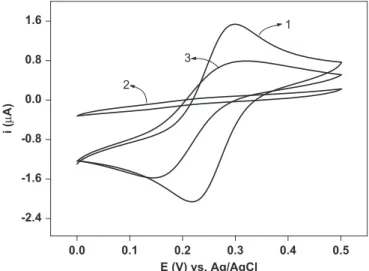
attachment of 11-MUA after an adsorption time of 24 h (curve 2), and the adsorption ofL. infantumantigens onto the activated self-assembled 11-MUA (curve 3).
As can be seen, two well-defined peaks characteristic of Fe(CN)63/Fe(CN)64 were observed on the bare gold electrode. However, the 11-MUA SAM/Au surface resulted in preventing the access of the Fe(CN)63/Fe(CN)64 redox species to the electrode. Hence, no Faradaic peaks for Fe(CN)63/Fe(CN)64 were observed between 0.0 V and þ0.5 V vs Ag/AgCl. On the other hand, an increase in the peak current of the redox couple was observed after the incubation of the 11-MUA/Au electrode surface with L. infantumantigens. EIS is a sensitive and powerful characteriza-tion tool that can provide detailed informacharacteriza-tion on charge-transfer resistance (Rct), which reflects the electron transfer kinetics of the redox probe at the electrode surface (Luz et al., 2005; Pei et al., 2001).
Fig. 3shows the Nyquist plots of the bare gold electrode (curve 1), the MUA SAM modified gold electrode (curve 2) and the L. infantumantigen/MUA SAM modified gold electrode (curve 3) in the presence of the redox probe measured at the formal potential. The measured EIS data were simulated with a modified Randles equivalent circuit as shown in inset ofFig. 3. This circuit
consists of the ohmic resistance of the electrolyte (Rs) between the gold working electrode and the Ag/AgCl reference electrode, Rct of the redox probe moving through the film, the Warburg impedance (Rw) resulting from the diffusion of ions from the bulk of the electrolyte to the interface and a constant phase element (CPE) is used instead of a pure capacitor to compensate for the non-ideal capacitive response of the interface (Ding et al., 2005; Hleli et al., 2006;Norlin et al., 2002;Randles, 1947).
The bare gold electrode exhibits an almost straight line with a small semicircle at high frequencies (Fig. 3, curve 1), indicating a diffusion-limited electron transfer process (Rct¼2.90 k
O
). After immobilization of the MUA, the value ofRctincreased to 1600 kO
(Fig. 3, curve 2), which indicated the successful formation of a MUA SAM, thus blocking the electron transfer of the redox probe to the electrode surface. After immobilization of theL. infantum antigen on MUA SAM, theRctdecayed to 43.2 kO
, confirming the0 500 1000 1500 2000 2500
0 50 100 150 200 250
Wash step Immobilization step
Injection of buffer
Injection of
L. infantum
Δθ
SPR
/m
°
Time / sec
0 400 800 1200 1600 2000
0 50 100 150 200 250
5 4 2
3 1
Δθ
SPR
/ m
°
Time / sec
R: 0.9995
Δθ
/ m
°
Conc. / μg.mL
Fig. 1.Sensograms regarding the immobilization of solubleL. infantumantigens in
HBS-EP buffer pH 7.4 onto the activated self-assembled 11-MUA. (A) Immobilization and washing steps of the antigen (50mg mL1). (B) Immobilization steps for the antigen at different concentrations: 10mg mL1; 20mg mL1; 30mg mL1; 40mg mL1and 50mg mL1, curves 1, 2, 3, 4, and 5, respectively. Inset: a linear relationship between the concentration of antigen and the SPR angle shift, after an adsorption time of about 30 min.
0.0 0.1 0.2 0.3 0.4 0.5
-2.4 -1.6 -0.8 0.0 0.8 1.6
3
2
1
i (
μ
A)
E (V) vs. Ag/AgCl
Fig. 2.Cyclic voltammograms obtained for the aqueous solution Fe(CN)63/4
(1 mmol L1) redox reaction on the bare gold electrode (curve 1), on 11-MUA (1.0 mmol L1) ethanol solution after a modification time of 24 h (curve 2) and after antigen immobilization on SAM (curve 3). The measurements were per-formed in 0.1 mol L1KCl and a scan rate of 20 mV/s.
0 10 20 30 40 50 60 70 80 90 100
0 20 40 60 80 100
3 2
1
Z
''(kohm)
Z'(kohm)
Fig. 3.(A) Nyquist plots (Z00vsZ0) of a bare gold electrode (curve 1), an MUA SAM
trend observed in cyclic voltammetry experiments. It is possible that the antigen immobilization on SAM caused defects in the structure of the monolayer, thus decreasing the electron transfer resistance at the surface. Indeed, the terminal carboxylic group of MUA is negatively charged in the pH solution (pH 7.4). The redox probe of [Fe(CN)6]3/4was also negatively charged, and thus the redox probe was difficult to approach on the electrode surface due to the strong electrostatic repulsion. After activation with EDC/NHS, the majority of terminal carboxylic groups were con-verted to NHS esters, so that repulsive interaction with the anionic probe [Fe(CN)6]3/4at the electrode interface did not occur, thus promoting the transfer of the redox ions [Fe(CN)6]3/4 to the electrode surfaces. It was observed that the monolayers became less insulating, and that an increase in current response occurred. The constant phase element values were extracted from compu-ter simulated spectra which are 7.89
m
F cm2, 0.954m
F cm2and 0.450m
F cm2 for the bare gold electrode, MUA SAM and L. infantumantigen/MUA SAM, respectively. It is well known that long-chain alkanethiols, such as MUA, form very well ordered defect-free monolayers, which constitute a barrier to the electron transfer and the penetration of ions towards the electrodic substrate(Campuzano et al., 2006). Thus, when a MUA SAM with a long
alkane chain is deposited on an electrode surface, the interfacial capacitance was 0.954
m
F cm2, value that is more than 8-fold lower than the characteristic capacitance of bare gold electrode.Scanning electrochemical microscopy (2-D imaging and approach curves) measurements were carried out in order to investigate the effects of electrostatic repulsion between the redox couple and the films, as well as the defects in the MUA SAM andL. infantumantigen/MUA SAM on the electron transfer of the redox probe to the electrode surface. Thus, the SECM experi-ments were performed for the MUA SAM modified gold electrode and theL. infantumantigen/MUA SAM modified gold electrode in the presence of hydrophilic species of [Fe(CN)6]4.
Fig. 4shows the SECM approach curves of [Fe(CN)6]3/4 on the MUA SAM modified gold electrode (Fig. 4A) and the L. infantumantigen/MUA SAM modified gold electrode (Fig. 4B). Approach curves were collected as a function of substrate poten-tial, while the tip potential was set to the value at which the diffusion-controlled oxidation of the probe [Fe(CN)6]4 could occur at tip (Etip¼0.5 V vs Ag/AgCl) and the substrate potential wasstepped to greater negative values progressively. This proce-dure was carried out in the same region on the substrate to obtain a family of effective heterogeneous electron transfer constants, keff, values as a function of substrate potential. According to the theory, the following equations can be used to extract the first orderkeff(Mirkin et al., 1992;Wei et al., 1995):
IkT¼I k
S 1
IinsT IC
T !
þIinsT ð1Þ
IkS¼
0:78377 Lð1þ1=
L
Þþ½0:68þ0:3315expð1:0672=LÞ ½1þFðL,
L
ð2Þ
whereIC
T,I
k
T,IinsT represent the normalized tip current for diffusion-controlled regeneration of the redox mediator, finite substrate kinetics, and insulating substrate, respectively, at a normalized tip-substrate separation, L¼d/a; Ik
T is the kinetically controlled substrate current; and
L
¼kETd/D, where kET is the apparent heterogeneous rate constant (cm/s), and F(L,L
)¼(11þ2.3L
)/ [L
(110–40L)]. These currents are normalized by the tip current at an infinite tip-substrate separation,iT,N. The analytical approx-imations forICTandIins
T (Mirkin et al., 1992;Wei et al., 1995)
ICT¼ 0:78377
L þ0:3315exp 1:0672
L
þ0:68 ð3Þ
IinsT ¼ 1
0:15þ1:5358=Lþ0:58expð1:14=LÞ þ0:0908exp½L6:3=1:017L ð4Þ
Effective heterogeneous electron transfer constants for the MUA SAM modified gold electrode and theL. infantum antigen/ MUA SAM modified gold electrode were obtained with 1 mmol L1 of [Fe(CN)
6]4. For blocking monolayers, the rate constants at one region of the substrate were fit to the Buttler– Volmer equation to obtain both the heterogeneous rate constant, kET1, and the electron transfer coefficient,
a
(Kiani et al., 2008)kET¼koETexp½afðEsEoÞ ð5Þ
wheref¼F/RT,Fis the Faraday constant,Ris the gas constant and Tis the temperature.
It can be seen in Fig. 4 that in the case of the L. infantum antigen/MUA SAM modified gold electrode, deviation from pure
0.4 0.8 1.2 1.6 2.0
1 2 3
4 5
Normalized tip current
L = d / a
0 1 2 3 4 5
0 1 2 3 4 5
0.4 0.8 1.2 1.6 2.0
3
4
2
1 5
Normalized tip current
L = d / a
Fig. 4.SECM approach curves obtained on a gold electrode coated by (A) MUA
negative feedback begins at lower potentials compared to that of the MUA SAM modified electrode. Indeed, thekETvalues obtained were higher forL. infantumantigen/MUA SAM compared to MUA SAM, which is consistent with cyclic voltammetry and electrochemical impedance experiments. As substrate potential varied, the rate of the redox couple oxidation through the SAM increased, and the approach curves change from negative to positive feedback. The rate constants obtained were fit to Eq. (5) and a kET1 value of 3.26105cm s1and a
a
value of 0.29 were obtained for MUA SAM (see inset inFig. 4A) and akET1value of 2.58104cm s1and aa
value of 0.42 were obtained forL. infantumantigen/MUA SAM (see inset inFig. 4B).Images of electrochemical reactivity for the probe (1.0 mmol L1 ferrocyanide in 0.1 mol L1 KCl) across the sub-strate were also obtained by SECM. Scanning in a rastered pattern was performed at a constant substrate-tip separation above the tooth sample. All the images presented in this paper relate to a 300
m
m300m
m area of the substrate. The micro-electrode and the substrate were polarized at 0.5 V and 0.0 V vs Ag/AgCl, respectively. The SECM images in Fig. 5 correspond to a 2-D representation of the local tip current after approach in substrate, consisting, respectively, of 11-MUA SAM (Fig. 5A) and of antigens immobilized on 11-MUA SAM (Fig. 5B). By comparing images (A) and (B) inFig. 5, it can be seen that the results observed in this study clearly demonstrate that after immobilization of antigens on the surface, there was an increase in the current obtained from the electrochemical redox reaction of the probe used, confirming the data found by the CV and EIS techniques, as well as the kinetic data from SECM.3.3. Immunosensor response to anti-L. infantum antibodies
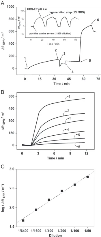
The efficiency of the SPR-based immunosensor was evaluated by adding to its surface of positive and negative canine sera for VLC.Fig. 6A shows the sensogram obtained for the steps involved in the detection of anti-L. infantum antibodies. In first step, stabilization of baseline was performed for 180 s. In second step, the immobilization of theL. infantumantigen onto the activated 11-MUA SAM is evidenced by a rise in the resonance angle (position 1,Fig. 6A). In third step, washing was performed with HBS-EP buffer at pH 7.4 (position 2,Fig. 6A) and in fourth step, to prevent non-specific binding and also for the blocking of un-reacted NHS-ester groups on sensor chip, EA was used (position 3, Fig. 6A) followed by a brief wash with HBS-EP buffer at pH 7.4 (position 4,Fig. 6A) to remove unbound species from the sensor surface. In the fifth step, the immunoreaction between the L. infantumantigen and the anti-L. infantumantibody, after injection of positive canine serum at 1:50 dilution, was monitored (position 5, Fig. 6A). An interaction time of 10 min showed a strong antigen– antibody interaction, as evidenced by a sharp rise in resonance angle shift of about 600 m1, demonstrating a very high sensitivity of the proposed label-free immunoassay. After measuring the immune response, the regeneration step was carried out using 1% (w/v) sodium dodecyl sulfate (SDS) prepared in water and a pulse of about 3 min, which was found appropriate to liberate the bound antigen– antibody, leaving the sensor surface active for the next injection. In inset ofFig. 6A, the SPR sensorgrams shows four repeated injections of positive canine serum ofL. infantum antibody and regenerated surface with 1% (w/v) SDS. The heights of the SPR sensorgrams were almost the same and we observed no reduction in the response. The relative standard deviation in the SPR angle for the four measure-ments was 4%.
Positive canine serum in different dilutions was subsequently injected onto the immunosensor surface. The sensorgrams showed in Fig. 6B involves association step due to interaction antigen– antibody. The sensitivities of the immunosensors prepared with
different amounts of anti-L. infantumantibodies were presented. It is possible to observe inFig. 6B the wide detection range and excellent sensitivity of anti-L. infantum through immunosensor construction and also note that at lower concentrations of antibodies (or larger dilutions of serum), the immunosensor sensitivity is reduced, due to the low number of interactions of antibodies on the immunosensor. Calibration plot was constructed using the logarithm of
D
y
SPRagainst dilution of positive canine sera and the figure is presented inFig. 6C. The calibration plot exhibited the linearity over the dilution range of 1:6400–1:50 with a value of 0.995 correlation coefficient.Moreover, the addition of negative serum in different dilutions (1/50–1/6400) was accompanied by a smaller response when compared with positive serum. Even more significant differences for sera with dilution factor greater than 200 were obtained. Currently the Ministry of Brazilian Health formally establishes that an animal is reagent considered when dilution factor shows equal or superior to 1:40 in ELISA and IFA, but this criterion is still controversy considering that for several authors only the dilution factor equal or higher that 1:160 should be conclusive considered. However, in this dilution factor the failure in demonstrating the infection by ELISA and IFA in asymptomatic dogs is more marked and the higher dilution can also show loss of specificity. In this sense, the results obtained by our group indicate the successful in
Fig. 5.SECM 2-D images obtained with a platinum microelectrode (r¼5mm) in
1.0 mmol L1 Fe(CN) 6
the development of an SPR-based sensor, indicating advantages when compared to conventional techniques, such as, excellent sensitivity for anti-L. infantum antibodies detection, short time analysis and real time detection. Therefore, the SPR-based
immunosensor prepared shows a great perspective as a sensing system to the diagnosis of VLC.
4. Conclusions
The development of an immunosensor for anti-L. infantum antibodies detection by the SPR technique was successfully performed. The sensor showed good response, allowing analysis of interactions in real time and without needing markers, showed a great perspective as a sensing system to VLC in endemic regions. The SPR immunosensor based on the L. infantum antigen was successfully applied to the analysis of antibodies against anti-L. infantum antibodies in canine serum up to 1: 1:6400 diluted. Area scans with SECM (2-D imaging and approach curves) clearly demonstrate that after immobilization of antigens on the surface, there was an increase in the current obtained from the electrochemical redox reaction of the probe used, confirming the data found by the CV and EIS techniques.
Acknowledgments
The authors are grateful to Conselho Nacional de Desenvolvi-mento Cientı´fico e Tecnolo´gico (CNPq), INCTBio, Fundac-ao de~
Amparoa Pesquisa do Estado de Minas Gerais (FAPEMIG) and Fundac-ao de Amparo~ a Pesquisa do Estado de S ao Paulo (FAPESP)~
for financial support. D. E. P. S is indebted to INCT-Bio for fellowships.
References
Alves, W.A., Bevilacqua, P.D., 2004. Cadernos de Sau´de Pu´blica 20, 259–265. Andrade, R.A., Arau´jo, M.S.S., Reis, A.B., Gontijo, C.M.F., Vianna, L.R., Mayrink, W.,
Martins-Filho, O.A., 2009. Veterinary Immunology and Immunopathology 128, 79–86.
Arya, S.K., Singh, A.T., Naidoo, R., Wu, P., Mcdermott, M.T., Eyoy, S., 2011. Analyst 136, 486–492.
Ashford, R.W., 2000. International Journal for Parasitology 30, 1269–1281. Bruijn, H.E., Altenburg, B.S., Kooyman, R.P.H., Greve, J., 1991. Optics
Communica-tions 82, 425–432.
Buchatip, S., Ananthanawat, C., Sithigorngul, P., Sangvanich, P., Rengpipat, S., Hoven, V.P., 2010. Sensors and Actuators B 145, 259–264.
Campuzano, S., Pedrero, M., Montemayor, C., Fata´s, E., Pingarro´n, J.M., 2006. Journal of Electroanalytical Chemistry 586, 112–121.
Cancino, J., Machado, S.A.S., 2012. Electrochimica Acta 72, 108–113. Chen, J.-C., Sadhasivam, S., Lin, F.-H., 2011. Process Biochemistry 46, 543–550. Den Boer, M., Argaw, D., Jannin, J., Alvar, J., 2011. Clinical Microbiology and
Infection 17, 1471–1477.
Desjeux, P., 2004. Clinical Microbiology and Infection 27, 305–318.
Ding, S.J., Chang, B.W., Wu, C.C., Lai, M.R., Chang, H.C., 2005. Electrochimica Acta 50, 3660–3666.
Gamero, M., Pariente, F., Lorenzo, E., Alonso, C., 2010. Biosensors and Bioelec-tronics 25, 2038–2044.
Hleli, S., Martelet, C., Abdelghani, A., Burais, N, Jaffrezic-Renault, N., 2006. Sensors and Actuators B 113, 711–717.
Hong, S.C., Chen, H., Lee, J., Hye-Kyung, P., Young, S.K., Hyun-Chul, S., Cheol-Min, K., Tae, J.P., Seok, J.L., Kwangnak, K., Hwa-Jung, K., Chulhun, L.C., Jaebeom, L., 2011. Sensors and Actuators B 156, 271–275.
Im, J.-E., Han, J.-A., Kim, B.K., Han, J.H., Park, T.S., Hwang, S., In Cho, S., Lee, W.-Y., Kim, Y.-R., 2010. Surface and Coatings Technology 205, S275–S278. Kiani, A., Alpuche-Aviles, M.A., Eggers, P.K., Jones, M., Gooding, J.J., Paddon-Row,
P.M.N., Bard, A.J., 2008. Langmuir 24, 2841–2849.
Liu, J.T., Chen, C.J., Ikoma, T., Yoshioka, T., Cross, J.S., Chang, S.-J., Tsai, J.-Z., Tanaka, J., 2011. Analytica Chimica Acta 703, 80–86.
L ¨ofas, S., Whirter, M., 2006. The art of Immobilization for SPR Sensors. In: Homola, J., Wolfbeis, O.S. (Eds), Surface Plasmon Resonance Based Sensors, Springer Verlag, New York, pp. 3–44.
Lukes, J., Mauricio, I.L., Schonian, G., Dujardin, J.-C., Soteriadou, K., Dedet, J.-P., Kuhls, K., Tintaya, K.W.Q., Jirku, M., Chocholova, E., Haralambous, C., Pratlong, F., Obornik, M., Horak, A., Ayala, F.J., Miles, M.A., 2007. Proceedings of the National Academy of Sciences 104, 9375–9380.
Luz, R.C.S, Damos, F.S., Bof, A.O., Beck, J., Kubota, L.T., 2005. Electrochimica Acta 50, 2675–2683.
Mathers, C.D., Ezzati, M., Lopez, A.D., 2007. PLoS Neglected Tropical Diseases 2007, 1–114.
0 200 400 600 800 1000
6
5
4 3 2
1
Δθ
SPR
/ m
°
Time / min
0 150 300 450 600
6 4
3
5 2
1
ΔΘ
SPR
/ m
°
Time / min
0 15 30 45 60 75
0 3 9 12
1/6400 1/1600 1/400 1/200 1/100 1/50 1.5
2.0 2.5 3.0
log (
ΔΘ
SPR
/ m
°
)
Dilution
0 20 40 60 80 -100
0 100
200 HBS-EP pH 7.4
regeneration step (1% SDS)
Δθ
SPR
/ m
°
Time / min
positive canine serum (1:800 dilution)
6
Fig. 6.(A) Sensogram showing the immobilization and immunoreaction ofL. infantum
antigen onto the activated 11-MUA modified gold surface [position 1, injection of
Mirkin, M.V., Fan, F.-R.F., Bard, A.J., 1992. Journal of Electroanalytical Chemistry 328, 47–62.
Moreno, J., Alvar, J., 2002. Trends in Parasitology 18, 399–405.
Murray, H.W, Berman, J.D., Davies, C.R., Clive, R., Saravia, N.G., 2005. The Lancet 366, 1561–1577.
Norlin, A., Pan, J., Leygraf, C., 2002. Biomolecular Engineering 19, 67–71. Pei, R., Cheng, Z., Wang, E., Yang, X., 2001. Biosensors and Bioelectronics 16,
355–361.
Randles, J.E.B, 1947. Discussions of the Faraday Society 1, 11–19.
Reis, A.B., Martins-Filho, O.A., Teixeira-Carvalho, A., Carvalho, M.G., Mayrink, W., Franc-a-Silva, J.C., Giunchetti, R.C., Genaro, O., Corrˆea-Oliveira, R., 2006.
Research in Veterinary Science 81, 68–75.
Salmain, M., Ghasemi, M., Boujday, S., Spadavecchia, J., Te´cher, C., Val, F., Le Moigne, V., Gautier, M., Briandet, R., Pradier, C.-M., 2011. Biosensors and Bioelectronics 29, 140–144.
Srivastava, P., Dayama, A., Mehrotra, S., Sundar, S., 2011. Transactions of the Royal Society of Tropical Medicine and Hygiene 105, 1–6.
Teixeira-Neto, R.G., Giunchetti, R.C., Carneiro, C.M., Vitor, R.W., Coura-Vital, W., Quaresma, P.F., Ker, H.G., De Melo, L.A., Gontijo, C.M., Reis, A.B., 2010. Veterinary Parasitology 11, 248–257.
Venugopal, M., Arya, S.K., Chornokur, G., Bhansali, S., 2011. Sensors and Actuators A: Physical 172, 154–160.
Wei, C., Bard, A.J., Mirkin, M.V., 1995. Journal of Physical Chemistry 99, 16033–16042.
World Health Organization, 2010. WHO Technical Report Series 949, pp. 1–186. Zhang, L., Zhang, J., Zhang, C.H., 2009. Biosensors and Bioelectronics 24,
![Fig. 4 shows the SECM approach curves of [Fe(CN) 6 ] 3 /4 on the MUA SAM modified gold electrode (Fig](https://thumb-eu.123doks.com/thumbv2/123dok_br/15758976.639318/5.892.454.824.101.735/fig-shows-secm-approach-curves-mua-modified-electrode.webp)