Development of a novel
3D-scaffold to promote bone
regeneration
Rafaela Maria Da Silva Presa
Dissertation for the Degree of Master of Science in
Bioengineering at Faculdade de Engenharia da
Universidade do Porto and Instituto de Ciências
Biomédicas Abel Salazar da Universidade do Porto
Development of a novel 3D-scaffold
to promote bone regeneration
Rafaela Maria Da Silva Presa
Dissertation for the Degree of Master of Science in
Bioengineering
at
Faculdade
de
Engenharia
da
Universidade do Porto and Instituto de Ciências Biomédicas
Abel Salazar da Universidade do Porto
Integrated Master in Bioengineering
Supervisor: Doctor Aureliana Sousa
Co-supervisor: Doctor Marco Araújo
“So remember to look up at the stars and not down at your feet. Try to make sense of what you see and wonder about what makes a
universe exist. Be curious. And however difficult life may seem, there is always something you can do and succeed at. It matters that you don’t just give up.”
Acknowledgments
First of all, I have to thank to my supervisor with all my heart. Thank you Filipa for your openness, your closeness, your support, your freedom, your advices, your sudden but always very accurate ideas. Thank you for being the person you are and for allowing a relationship where openness is great for sharing thoughts, ideas, moods, everything.
I could not fail to mention two other very important people who appeared with the beginning of this adventure, Marco and Filipa. Without you this way would not have been so good, full of laughter, happy moments, much help, companionship, camaraderie and friendship. Thank you for being two fantastic and wildest healthy humans there can be.
Marco, my co-supervisor, thank you for being who you are, for the good heart you are! Thank you for everything, for your help in developing my work, for all the support, for all the advices and for the good working environment that you have helped to create every day.
Filipa, thank you for being the sweet person you are. Thank you for sharing this adventure with me and always being someone with whom I could share the good and bad times. Thank you for the advices, support and all the help, but above all for the great team spirit and mutual help.
I have to thank to Manel for all the help that allowed me to develop my work and Eduardo for the support and good team spirit and work environment provided.
An acknowledgment to Susana Olhero and Paula Torres from CICECO for providing the CaP powders and the robocasting.
I have to acknowledge the support of Ricardo Vidal from the Biointerfaces and Nanotechnology i3S Scientific Platform, Maria Lázaro from the Bioimaging i3S Scientific Platform, member of the national infrastructure PPBI - Portuguese Platform of Bioimaging (PPBI-POCI-01-0145- FEDER-022122) and Daniela Silva from the Image, Microstructure and Microanalysis Unit (IMCROS) from the Materials Centre of the University of Porto (CEMUP).
All this, to end by thanking two of the most important people in my life - my parents. Thank you, mom and dad, for all understanding, support, affection, open mind and help. Without you, that wouldn’t be possible.
Bifunctional CaP-based Biomaterials for Bone Cancer Treatment and Regeneration (PTDC/CTM-CER/29940/2017
Abstract
Bone Engineering has been one of the most explored and successful fields of research on Tissue Engineering (TE). Besides trauma lesions and bone infections, also several diseases such as Osteoporosis and Bone Cancer significantly affect the functionality of the tissue thus impairing bone intrinsic regeneration ability. The appearance of large defects on bone tissue and the occurrence of fractures can result in a negative impact on quality of life of the patients, with a lot of co-morbidities associated. Considering the composition of natural bone, one of the approaches that has emerged as a successful alternative are scaffolds resulting from the combination of polymers, which act as a support matrix, with ceramic powders, which provide similar properties to the mineral phase of bone.
Such biomaterials can be explored due to their characteristics such as biodegradability profile, bioactivity and osteoconductive properties. With the rise of advanced additive manufacturing technologies such as robocasting, 3D porous scaffolds can be tailored to the specific need of each patient. With this technique is possible to produce a highly ordered and interconnected macroporous structure, which was demonstrated to be essential to provide the ideal conditions that allow the local delivery of cells, growth factors, drugs as well as providing the ideal mechanical properties for a load-bearing tissue such as bone.
In this work, we were able to test in vitro stability of a scaffold composed of chitosan and a biphasic powder of Hydroxyapatite (HA) and β-Tricalcium Phosphate (β-TCP) as well as the influence of the application of specific magnetic conditions on human Mesenchymal Stem Cells (hMSCs) behavior. The in vitro stability and also the mechanical properties were altered after sterilization process rendering scaffold with properties that were not ideal. We then developed an alternative strategy using a biphasic powder of HA and β-TCP in combination with Konjac Glucomannan (KGM) to produce a 3D-scaffold through robocasting. KGM is a natural polysaccharide which is nontoxic, biodegradable, biocompatible, has good film-forming ability and also displays interesting viscoelastic properties for the use in 3D printing systems. Furthermore, it can be chemically functionalized with specific moieties which represents an important advantage for the design of a multi-functional scaffold.
adherence and proliferation of hMSC. Although further studies are needed, these scaffolds proved to be able to allow hMSCs differentiation into the osteogenic lineage and there is some indication that may display osteoinductive properties. We were also able to functionalize KGM with dopamine as a proof-of-concept to future hyperthermia induction approaches. As an alternative strategy, polydopamine particles have also been successfully synthesized for future incorporation in the scaffolds.
Altogether, the results obtained in the scope of this thesis, suggest that this newly developed hybrid organic-inorganic scaffold is a promising candidate to be used on biomedical engineering approaches.
List of Contents
Acknowledgments………...i Abstract……….iii Abbreviation List………..viii List of Figures………..x List of Tables………..xii Introduction………..11. Bone Tissue – Organization and Composition………...1
1.1 Bone Organization………1
1.2 Bone Composition………2
1.2.1 Extracellular Matrix………2
1.2.2 Cells……….3
1.3 Bone Remodeling……….4
1.4 Bone-related therapeutic needs……….5
2. Bone healing traditional approaches………8
3. Tissue Engineering based approaches………9
3.1 Materials/Biomaterials………11 3.1.1 Calcium Phosphates……….12 3.1.2 Polymers……….13 3.1.2.1 Chitosan………13 3.1.2.2 Konjac Glucomannan………..14 3.1.3 Other Agents………..15
3.2 Fabrication techniques – General Overview………16
3.2.1 Robocasting………...17
4. Experimental Strategy Rationale………..19
5. Aim of the thesis………..21
Materials and Methods……….22
1. Materials………...22
2. Chitosan-CaP ink preparation loaded with magnetic nanoparticles (MNPs scaffolds………22
3. Preparation of the hybrid KGM-BCP printable ink (KGM scaffolds)……..23
3.1 Preparation of KGM solution………23
3.2 Preparation of the KGM-BCP ink………23
4. Robocasting of MNPs scaffolds and KGM scaffolds………...24
5. Characterization of hybrid organic-inorganic KGM scaffolds and its components………..25
5.1 Rheology………25
5.2 1H NMR………..25
5.3 Size exclusion chromatography (SEC)………..26
5.4 Attenuated total reflectance- Fourier-transform-infrared spectroscopy (ATR-FTIR) ………...………26
5.5 Powder X-ray Diffraction (XRD)………..26
5.6 Particle size distribution………27
5.7 Scanning Electron Microscopy (SEM)………27
5.8 Measurement of pore size, fiber diameter and grain size of the produced KGM scaffolds………..27
6. KGM functionalization with photothermal moieties………27
6.1 Synthesis of carboxylated KGM (KGM-SAC0) …..……….27
6.2 Functionalization of KGM-SAC0 with dopamine (KGM-SAC0-Dopamine) ……….28
6.3 Synthesis of Polydopamine particles……….28
6.4 Characterization of the obtained materials………29
7. In vitro studies………..29
7.1 Cell culture……….29
7.2 Cytotoxicity………29
7.3 hMSCs culture on 3D MNPs scaffolds and 3D KGM scaffolds……...30
7.4 Metabolic activity………...32
7.5 Biochemical Analysis………32
7.5.1 Alkaline phosphatase (ALP) activity………..……….32
7.5.2 Protein quantification……….32
7.5.3 DNA quantification……….33
7.6 Viability assay………33
7.7 Immunostainings………..33
8. Statistical analysis………...34
Results and Discussion………...35
1. Chitosan-CaP scaffolds loaded with magnetic nanoparticles (MNPs scaffolds)………..35
1.1 hMSCs viability and metabolic activity on MNPs scaffolds…………36
1.2 hMSCs morphology and collagen type I deposition on MNPs scaffolds……….40
2. Preparation and characterization of the hybrid organic-inorganic KGM scaffolds………...42
2.1 Characterization of the KGM….………..42
2.2 Characterization of the ceramic BCP powders……….45
2.3 Hybrid organic-inorganic KGM-BCP extrudable ink……….…50
2.4 Production and Characterization of the hybrid organic-inorganic KGM scaffolds…….………54
2.5 KGM scaffolds in vitro behavior assessment……….57
2.5.1 Cytotoxicity of KGM scaffolds………...………..57
2.5.2 hMSCs differentiation on KGM scaffolds………58
2.5.2.1 Metabolic cell activity of hMSCs on KGM scaffolds…..58
2.5.2.2 hMSCs morphology on KGM scaffolds………...60
2.5.2.3 ALP quantification………..63
3. KGM functionalization with photothermal moieties.………65
3.1 KGM functionalization with Dopamine………..……...65
3.1.1 Carboxylation of KGM………..………65
3.1.2 KGM-SAC0 dopamine coupling………...69
3.2 Synthesis of Polydopamine particles………72
Conclusions and Future Work………75
References………..78
Abbreviation List
3D - Three-Dimensional ALP - Alkaline Phosphatase AM - Additive ManufacturingATR-FTIR - Attenuated total reflection - Fourier-transform infrared spectroscopy BCA – Bicinchoninic acid
BCP - Biphasic Calcium Phosphate BM – Basal Medium
BTE – Bone Tissue Engineering BSA - Bovine Serum Albumin CAD - Computer Aided Design CaP - Calcium Phosphate
DAPI - 4’,6-diamidino-2-phenylindole
DMEM - Dulbecco’s Modified Eagle Medium DMSO - Dimethyl sulfoxide
DNA - Deoxyribonucleic Acid DWA – Direct Write Assembling ECM - Extracellular Matrix
EDC - 1-(3-Dimethylaminopropyl)-3-ethylcarbodiimide hydrochloride EDTA – Ethylenediamine tetraacetic acid
EM – Expansion Medium FBS - Fetal Bovine Serum GO – Graphene Oxide HA – Hydroxyapatite
hDNFs – human Dermal Neonatal Fibroblasts hMSCS – human Mesenchymal Stem Cells
1H-NMR – Proton Nuclear Magnetic Resonance
ISO - International Organization for Standardization KGM – Konjac Glucomannan
α-MEM - α-Minimum Essential Medium MES - 2-(N-morpholino) ethanesulfonic acid MNPs – Magnetic Nanoparticles
NHS - N-Hydroxysuccinimide PBS - Phosphate-Buffered Saline PFA – Paraformaldehyde
PMMA - Polymethylmethacrylate PS - Particle Size
PSD - Particle Size Distribution PVA – Polyvinyl Alcohol
P/S – Penicillin/Streptomycin RPM – Rotations per minute RT - Room Temperature SAC0 – Succinic Anhydride
SEC – Size exclusion chromatography SEM - Scanning Electron Microscopy SDS – Sodium dodecyl sulfate TCP - Tricalcium Phosphate TE – Tissue Engineering
TEM – Transmission electron microscopy UV-Vis - Ultraviolet–visible spectroscopy XRD - X-Ray Diffraction
List of Figures
Figure 1 - The chemical composition and multi-scale structure of the natural bone…..1
Figure 2 - Micrographs of bone portions………...……...3
Figure 3 - Bone remodeling cycle………...……...5
Figure 4 - Balance of bone resorption and bone formation………...6
Figure 5 - Defects on bone tissue……….7
Figure 6 -Tissue Engineering approach on Bone Tissue………...…..…10
Figure 7 - Different type of porous scaffolds used in BTE………...…11
Figure 8 - Konjac Glucomannan………14
Figure 9 - AM technique……….……….17
Figure 10 - Robocasting technique………..……….18
Figure 11 - CAD Model………24
Figure 12 - KGM scaffolds production cycle……….25
Figure 13 – Schematic Illustration of hMSCs culture on 3D KGM scaffolds……..…..31
Figure 14 - Experimental Set-Up of the MNPs scaffolds………31
Figure 15 – Live/Dead assay of hMSCs on MNPs scaffolds………..38
Figure 16 – hMSCs metabolic cell activity on the different scaffolds……….…39
Figure 17 – Cells Morphology….………40
Figure 18 - 1H NMR Spectrum of Konjac Glucomannan in D 2O…...………...…..43
Figure 19– ATR-FTIR Spectrum of Konjac Glucomannan………44
Figure 20– Konjac Glucomannan molecular weight……….…………..45
Figure 21- XRD analysis of BCP powders………46
Figure 22 – ATR-FTIR Spectra of the ceramic powders……….48
Figure 23 - Particle Size Distribution (PSD) of the powders………...49
Figure 24 - Rheological characterization of KGM solution and ink………51
Figure 25 - Shear Viscosity pattern of the Ink and KGM solution………...52
Figure 26 - SEM images of KGM scaffolds………...55
Figure 27 – Metabolic cell activity of the cytotoxicity tests……….57
Figure 28 – Metabolic cell activity of hMSCs on KGM scaffolds………58
Figure 29 – hMSCs morphology on KGM scaffolds……….61
Figure 30 – Collagen type I expression on KGM scaffolds……….61
Figure 31 – Total Protein concentration during differentiation assay………...62
Figure 33 – Synthetic route for the conjugation of dopamine to KGM………...65
Figure 34 - Incorporation of SAC0 through analysis of 1H-NMR………....66
Figure 35 – Incorporation of SAC0 through analysis of ATR-FTIR………67
Figure 36 - Incorporation of Dopamine through analysis of 1H-NMR………69
Figure 37 - Incorporation of Dopamine through analysis of ATR-FTIR ………...70
Figure 38 - TEM images of Polydopamine particles at different scales………....72
List of Tables
Table 1 – Example of commonly used polymers for BTE………...13 Table 2 – List of primary and secondary antibodies used in this study………..34 Table S1 - Statistical analysis of data from Figure 16, for the Influence of the application of an external magnetic field on the Metabolic cell activity………89 Table S2 - Statistical analysis of data from Figure 16, for the Influence of iron ions on the Metabolic cell activity………89 Table S3 - Statistical analysis of data from Figure 28, for the metabolic cell activity of hMSCs on KGM scaffolds for basal and osteogenic condition………..90
Introduction
1. Bone Tissue – Organization and Composition
The human body is composed by a multiplicity of cells that form different types of tissues [1], [2]. Bone is composed of a highly specialized form of connective tissue whose primary functions are to provide mechanical support for muscular activity (thus allowing locomotion), physical protection of organs and soft tissues, storage facility for systemic mineral homeostasis (inorganic ions such as calcium), as well as harboring the bone marrow [2].
1.1 Bone Organization
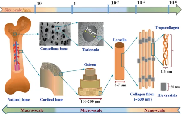
Morphologically, the bone of the mature skeleton consists of cortical (or compact) bone (80%) and cancellous (or trabecular) bone (20%). In cortical bone, densely concentric lamellae are formed by packed collagen fibril. Cancellous bone has a loosely organized, porous matrix (Figure 1) [3].
Figure 1 – The chemical composition and multi-scale structure of natural bone. Bone is constituted by
cortical bone, the densest tissue that is organized in concentric lamellae. The cancellous bone has a porous matrix. These structural differences lead to distinct function: Cortical bone provides mechanical and protective functions and cancellous bone provides metabolic functions. At a nano-scale is possible verify the two main components of the bone – Collagen – organic matrix and Hydroxyapatite – inorganic matrix. Retrieved from [4].
Structural and functional differences between cortical and cancellous bone are related to their primary functions. Cortical bone provides mechanical and protective functions and cancellous bone provides metabolic functions [3]. The distribution of the two types of bone tissue is different in the human skeleton, depending, primarily, on the principal function of the bone in question [1].
1.2 Bone Composition
1.2.1 Extracellular matrix
Bone extracellular matrix is composed of 5% of water, approximately 70% of crystals of calcium phosphate for structural reinforcement and stiffness, (inorganic phase) and 25% of proteins that together with 2% of lipids constitute the organic phase that provides flexibility and toughness to bone [5]. The inorganic phase is mainly constituted by Hydroxyapatite crystals (Ca10(PO4)6(OH)2) since calcium and phosphate ions combine in a process of
nucleation, but significant amounts of bicarbonate (HCO3-), sodium (Na+),
potassium (K+), citrate (C
6H5O73-), magnesium (Mg2+), carbonate (CO32-), fluoride
(F-), zinc (Zn2+), barium (Ba2+), and strontium (Sr2+) ions are also present.
Together with collagen, the noncollagenous matrix proteins form a scaffold for hydroxyapatite deposition. Hydroxyapatite gives to bones their hardness, resistance and strength, while the collagen fibers give them flexibility, that they are not brittle [1], [2].
The organic matrix is composed by collagenous proteins (90%), predominantly type I collagen, and noncollagenous proteins including osteocalcin, osteonectin, osteopontin, fibronectin and bone sialoprotein II, bone morphogenetic proteins (BMPs) and growth factors. There are also small leucine-rich proteoglycans [6]. In contrast with another tissues, bone tissue is poor in cells. The small number of cells is entrenched in a fibrous collagen rich matrix which forms a surface for the adherence of the inorganic salt crystals and have a crucial role in the maintenance of a healthy and functional tissue [1].
1.2.2 Cells
Although the cells represent a small volume of the bone tissue, it is constituted by four types of cells: osteoblasts, osteoclasts, osteocytes and bone lining cells [1], [2].
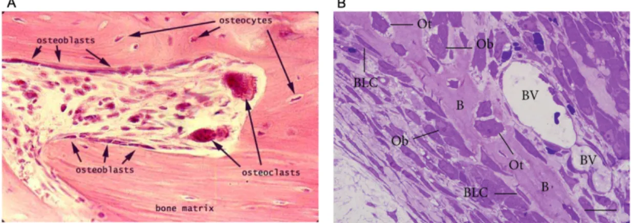
Figure 2 – Micrographs of bone portions. A – On this histological image, osteoblasts are visible on the
surface of the bone matrix, together with giant osteoclasts. Osteocytes are visible entrapped on the bone matrix. B – On this image a portion of a bony trabecula (B) is observed with Osteoblasts (Ob) and bone lining cells (BLC) present on bone surface while osteocytes (Ot) are observed entrapped in the bone matrix. Blood vessels (BV) are visible, too. Retrieved from [2], [7].
Osteoblasts are cuboidal differentiated cells found on the surface of new bone (Figure 2) responsible for the production of the extracellular matrix (ECM) of bone, and its subsequent mineralization process. Osteoblasts produce the organic matrix of bone (osteoid). Some of the osteoblasts become trapped in the lacunae within the matrix of bone and differentiate into osteocytes. Old osteoblasts become inactive, thin and elongated lining cells which lies on the bone surface. The repair process and formation of new bone requires the differentiation and the proliferation of osteoblasts [8].
The osteocyte is a mature osteoblast represent 90% of bone cells in the skeleton of an adult human [8]. They are uniform in shape and size that can be found inside the bone matrix (Figure 2) and is responsible for its maintenance, through the growth of new arms on the cell and the packing of osteoblasts and osteoclasts, they become responsible for communication between other analogous cells and bone lining cells by developing long branches, forming a wide network of intercellular communication. Osteocytes also have mechanosensitive functions since they can detect mechanical pressure allowing the adaptation of bone to daily mechanical forces. By this way, these cells seem to act as orchestrators of bone remodeling, through regulation of osteoblast and
osteoclast activities. Moreover, a chemotactic signal to osteoclastic bone resorption is the osteocyte apoptosis [2].
Bone lining cells are flat, elongated, inactive cells that cover bone surfaces that are undergoing neither bone formation nor resorption (Figure 2B). Little is known regarding the function of these cells; however, it has been speculated that bone lining cells can be precursors for osteoblasts and participate in osteoclast differentiation [2], [8].
Osteoclasts originate from hematopoietic stem cells, they are large multinucleated cells, responsible for resorbing the mineralized bone matrix and they move from different locations on the bone surface dissolving the bone (Figure 2A) [8].
1.3 Bone Remodeling
Bone is a highly dynamic tissue. Due to this particular characteristic, bone is being continuously remodeled [2]. As described above, the cells of the bone tissue interact intensively. Moreover, the osteoclasts not only are responsible for the bone resorption, but for the bone formation. Osteoclasts have the capability to communicate with osteoblasts – through the release of coupling factors - in a crosstalk that regulates the local recruitment and bone forming activity of osteoblasts [2], [9].
The bone remodeling cycle (Figure 3) is a highly complex process where there is a very important crosstalk between all the components of the tissue to ensure this process [9]. There are direct and indirect communication among the cells, as well as between the cells with the matrix through the release of several factors. This process is known by coupling process and the factors involved are designated by coupling factors [2].
Bone matrix not only acts as a support for bone cells and to the inorganic phase, but can also release several molecules that interfere in the bone cells activity and, consequently, has a participation in the bone remodeling. The molecules involved are, predominantly, adhesion molecules, such as integrins [10], [11].
of the integrins in the interaction osteocyte-bone matrix. These interactions are essential for the function of these cells, whereby signals induced by tissue deformation are generated and amplified [13].
Then, the remodeling process occurs due to the coordination actions of osteoblasts, osteoclasts, osteocytes, bone lining cells and the bone matrix [14].
Figure 3 – Bone remodeling cycle. Bone remodeling begins when osteoclasts resorb bone mineral and
matrix. Mononuclear cells prepare the resorbed surface for osteoblasts, which generate newly synthesized matrix as they differentiate. Matrix mineralization and the differentiation of some osteoblasts into osteocytes completes the remodeling cycle. Adapted from [15].
The remodeling process starts with the action of the osteoclasts, mediated by osteoclastogenic factors, that initiate bone resorption (Figure 3). During this phase, osteoclasts release some factors that inhibit the activity of the osteoblast in order to completely remove the damaged or aged bone tissue. After this phase, coupling factors and matrix-derived factors are released and with that the recruitment of osteoblasts and their differentiation is locally controlled by osteoclasts and bone matrix. At this time bone formation occurs (Figure 3).
This particular characteristic of the bone tissue is very important for the maintenance of the bone volume and bone balance [16]. Moreover, the dynamic of the bone is essential for fracture healing, skeleton adaptation to mechanical use and calcium homeostasis [2].
However, when an imbalance between bone resorption and bone formation occurs several bone diseases can appear. Thus, an equilibrium between these two process is necessary and, as previously described, it depends a lot on the action of several components and factors [14], [17].
1.4 Bone-related therapeutic needs
There are different diseases affecting the bone tissue, that leads to alterations in the bone remodeling process or are in fact a consequence of the impaired balance between these processes.
Between them, hip fracture is the most devastating type of fracture because it is associated with increased morbidity and mortality rates. Within one year of a hip fracture, 40% of patients are unable to walk independently, 60% need assistance with at least one essential activity of daily living (dressing, bathing) and 80% are unable to perform an instrumental activity of daily living, such as driving or housecleaning. In the same period of time, 14-36% of the patients die. When the fracture occur the risk of death is higher [18].
When multiple fractures occur, the patient can develop kyphosis (i.e. vertebral deformities), abdominal protrusion and a loss of >1.5 in of mature height. Respiratory problems and poor nutritional habits may occur due to thoracic and abdominal compression, respectively [18]. All of these aspects are physical and functional components that dramatically impact the quality of life of the patient. Moreover, an emotional component can be added. Depression and anxiety are typical symptoms revealed by the patients that suffer from skeletal-related events [19].
When an imbalance between the activity of osteoblasts and osteoclasts occurs, the situations that can appear and cause more impact are: bone destructions exceed bone formation or vice-versa (Figure 4).
Figure 4 - Balance of bone resorption and bone formation. If the balance between bone formation and
resorption is lost by the uncontrolled production of regulators, bone structure would be strikingly damaged and the subject would be susceptible to osteoporosis. Adapted from [20].
When bone resorption is excessive, without the corresponding amount of new formed bone different causes can be behind. Diseases, such as osteoporosis, periodontitis, Paget’s disease, bone cancer and inflammatory
These diseases have distinctive causes but, in terms of bone tissue, they lead to the same panorama: loss of bone mass and density. Then, there is a very high risk of fracture, multi-fractures can occur at the same time, impaired healing of the fracture and large defects on bone tissue, namely when the cause is a trauma or some type of cancers (Figure 5) [2], [9], [16], [18], [22].
The occurrence of fractures in a frequent manner compromises a lot the daily life of these patients. Moreover, the complications associated with the healing of the fractures increase the morbidities and, inclusively, the mortality of the patients [18], [19], [22].
Figure 5 - Defects on bone tissue. A) Osteoporosis – A progressive loss of bone mass leads to changes
in bone structure that can result in fractures. B) Bone cancer – The remotion of the tumor mass leads to the appearance of defects on bone tissue. Moreover, there are the problem of cancer cells remain on the tumor site and the recurrence of the tumor can occur. Adapted from [23], [24].
Osteopetrosis is a rare bone disease, where genetic mutations affect the resorption activity of the osteoclasts and new bone tissue starts accumulate in a disproportional manner [2].
When certain types of cancer affect a person, the skeletal system will be affected too. In the particular case of cancer, the surgical procedure of tumor’s resection leads to the appearance of big defects, that the bone by itself doesn’t have the capability of regenerate (Figure 5B) [9], [16], [25], [26]. Then, the bone tissue stays fragilized and the risk of fractures or infections highly increase. In these cases, the loss of bone mass is higher than in other pathologies and occurs more rapidly. Moreover, due to the anti-cancer therapies that are administrated to these patients, the healing process becomes more difficult [18], [22].
However, for these patients there is an extra problem. There is always the problem of the recurrence of the tumor [25]–[28]. As mentioned before, these patients are the ones who present the major bone defects. This is mainly
because, nowadays there aren’t a consensus on the clinical field about the concept of clear margins – the amount of healthy tissue in the neighborhood of the tumor that have to be removed to avoid the recurrence of the tumor. There are a lot of physicians that suggest a radical position, meaning the removal of high amounts of healthy tissue that is surrounding the tumor [29].
All the diseases that affect the bone tissue compromise its regenerative capability. In these situations, the ability of mesenchymal stem cells to differentiate into osteoblasts and form new bone is reduced [22]. As a result, there is a huge need to develop solutions in order not only to treat the background disease, but also to promote the regenerative bone process.
In the case of bone cancer, rather than advances in the promotion of bone tissue regeneration, new solutions are needed to eliminate the problem of recurrence of the tumor and thus to end the non-unanimity of medical opinions.
2. Bone healing- traditional approaches
Currently, the most used strategy is the use of bone grafts to fill space left by the fracture/tumor and to enhance healing. As first option and considered the gold standard are the autografts (bone harvest from the patient) followed by the allografts (donor bone) [9], [22], [30].
An autograft combine all the properties required in a bone graft material: it is osteoinductor (with the release of bone morphogenetic proteins and other growth factors), osteogenic (osteoprogenitor cells) and osteoconductive (scaffold) [30]. However, there are limitations associated to the use of bone grafts. Harvesting requires an additional surgical procedure that is an additional morbidity, with well-documented complications and discomfort for the patient. Furthermore, their availability is always limited [9], [22], [30]. Regarding the allograft, the problem of potential transmission of pathogens and rejections is the principal limitation [9], [22], [30]. Using the bone grafts strategy, the surgery is the cornerstone of the treatment and there are risk of secondary complications, such as infection and thromboembolism [22]. Moreover, the fractures frequently do not heal, may require multiple surgeries and frequently re-fracture the same site. Because of that, the treatment of these fractures is still a challenging, namely due
to the diminished capacity for fracture healing [22]. Therefore there is still a strong need to develop bone graft substitutes to avoid these limitations [9].
More limited are the patients that suffer of bone cancer. Currently, these patients are treated with two different therapies. These patients have to undergo to cycle(s) of chemotherapy and radiotherapy, in order to avoid the recurrence of the tumor. This therapy is a systemic therapy with a lot of side effects [25]–[27]. All of the solutions mentioned above are applied in patients to promote the regeneration of the bone. However, these patients need solutions that will have to undergo beyond the promotion of bone regeneration.
The bone graft substitutes are generating great interesting as alternatives to autologous or allogeneic bone grafts. They consist of scaffolds made of synthetic or natural biomaterials that promote the migration, proliferation and differentiation of bone cells for bone regeneration [30].
3. Tissue Engineering based approaches
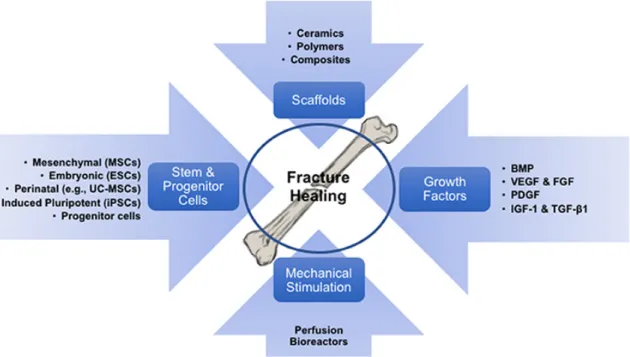
Tissue engineering (TE) is an exciting and multidisciplinary field with the objective of developing biological substitutes to restore, replace or regenerate tissue form and function. Cells, scaffolds and growth factors are generally referred to as the main components of TE (Figure 6). Scaffolds provide the three-dimensional structural support for cell attachment and subsequent tissue development, in a favorable biological environment for the growth and differentiation of cells [31].
Figure 6 - Tissue Engineering approach on Bone Tissue. Diagram illustrating the concept of skeletal tissue regeneration via scaffold-based tissue engineering strategies, involving its components (cells, biomaterials/scaffolds and growth factors), and the required exposure to mechanical environments to pre-conditioning the engineered implants. Retrieved from [32].
Approaches of TE are currently trying to achieve the gold standard solution needed for bone regeneration. The development of 3D porous scaffolds is one of the most promising biomaterial-based strategies for bone defect regeneration. The interconnected macropores are essential to supply sufficient space for cellular activity, nutrient transport and cell-cell interactions [28].
There are three essential elements required for bone tissue engineering: osteogenic cells, growth factors and a scaffold matrix that possesses all the characteristics necessary for new bone growth, i.e. (i) osteoconductivity (passive scaffold to promote vascular ingrowth and bone apposition), (ii) osteogenicity (containing osteoprogenitor cells) and (iii) osteoinductivity (provide signals to induce osteogenic differentiations of local stem cells) [33]–[36].
Because of that, the main principle of Bone Tissue Engineering (BTE) strategy is to seed cells in an artificial 3D porous matrix (Figure 7), working as a scaffold to sustain cell proliferation, differentiation and to provide guidance for tissue regeneration and growth. After implantation the natural tissue regeneration process will ideally take place, integrating the implant, leading to vascularization. The structure of the scaffold dissolves while new tissue is formed in its place [37].
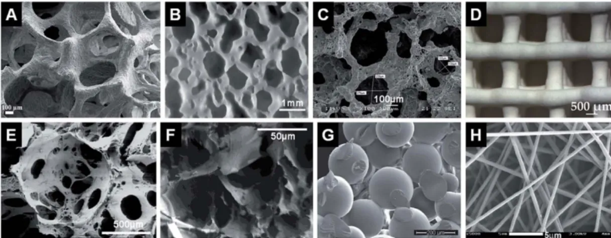
Figure 7 – Different types of porous scaffolds used in BTE. Representative internal structures of porous
scaffolds produced via (A) polymer sponge replication, (B) impregnate sintering, (C) gel-cast foaming, (D) solid free-form fabrication, (E) solvent casting and particulate leaching, (F) phase separation, (G) microsphere sintering and (H) electrospinning. Retrieved from [38].
3.1 Materials/ Biomaterials
To produce the matrix/scaffold there is a wide range of biomaterials such as collagen, hydroxyapatite (HA), β-tricalcium phosphate (β-TCP), calcium-phosphate cements and glass ceramics [30].
However, the range of materials that can be used is much higher. The materials used in BTE can be classified according two aspects: chemical composition and biological behavior. In relation to the chemical composition the materials can be subdivided in ceramics, metals, polymers, and composites, with varied degree of bioactivity. Therefore, biomaterials can be classified into four categories according to the nature of their interaction with the surrounding tissue: a) bioinert, their implantation on the body do not cause any reaction or interaction with the biological tissue, meaning that the host won´t recognize them as a strange body (ex: titanium, zirconia and alumina); b) biotolerant, they are moderately accepted by the recipient tissue which encapsulates the implant with a fibrous interface (ex: stainless steel, glass, chromium-cobalt fibers and polymethylmethacrylate (PMMA)); c) bioactive, they may cause a reaction or have an effect on the living environment, also due to their composition are capable of forming a direct link with the tissue (ex: hydroxyapatite, bioactive glasses); d) biodegradable/reabsorbable, those that suffer of slow degradation and gradual substitution of the host tissue (ex: tricalcium phosphate and bioactive glass) [39].
3.1.1 Calcium Phosphates
The materials that are used in bone regeneration should be the most similar to the natural composition of the tissue and mimic very well its structure and properties [9]. This aspect is critical on a tissue like bone, because the material that is used will highly influence the bone remodeling process [16]. Because of that, calcium phosphates (CaPs) are widely used. Their composition is similar to the mineral phase of bone and they have some key bone properties, such as biodegradability, bioactivity and osteoconductivity [9].
CaPs can be HA, β-TCP or an intimate mixture between the both, that is designated as biphasic calcium phosphate (BCP) [9]. The different types of BCPs differ one from another in the ratios of HA and β-TCP that constitute the mixture [9]. Depending on the desired effect, this difference between BCPs is crucial to the successful performance of the material. It is demonstrated that the composition of the CaP material has direct consequences on its performance, including its ability to be resorbed by osteoclasts, affecting osteoclast differentiation and activity [9], [16].
β-TCP exhibits excellent biodegradability and osteoconductivity, it can be resorbed by osteoclastic cells and subsequently replaced by new bone formation via osteoblasts [16]. On the other hand, HA has good biocompatibility but poorer resorption characteristics, meaning that HA is relatively stable in a bone defect due to slower bone remodeling kinetics [16]. Therefore, the chemical composition of CaPs affects the crosstalk between osteoclasts and osteoblasts and as a result, a scaffold containing these components should have important roles in the induction of the remodeling process. Even though this type of scaffolds should demonstrate effects on the bone regeneration process, there are some components, such as metallic ions,graphene oxide (GO) and polydopamine, that are being added to the scaffolds to enhance bone tissue regeneration. Metallic ions that are being used are gold, iron oxide (magnetite) and silver [25], [26], [28], [40], [41].
Although calcium phosphate materials have excellent characteristics, when applied to bone regeneration they need a “carrier” to act as the structural base of the scaffolds and improve their mechanical properties [42]. As a result,
the hybrid ceramic-polymer/hydrogel materials are emerging as ideal materials to be used on BTE [43].
3.1.2 Polymers
Polymers and their composites are considered as the most promising candidate materials to be used as scaffolds for bone regeneration (Table 1). Their advantageous biocompatibility and biodegradability over most metals and ceramics makes of them very attractive materials. More importantly, polymers possess highly flexible design capacity and their various properties can be easily tailored to meet specific requirements through manipulating their chemical compositions and structures. Moreover, polymers have been widely used in BTE due to their similarity with extracellular matrices [44].
Table 1 – Examples of commonly used polymers for BTE. Retrieved from [44].
3.1.2.1 Chitosan
Chitosan is one of the most well-known natural polymer and nowadays is widely used in different areas, alongside other polymers like hyaluronic acid, alginate, agar, polyvinyl alcohol (PVA) and poly (ethylene glycol) [45]. Chitosan is used because of its bioactivity, non-toxicity, biocompatibility, biodegradability
and low-allergenicity [40], [46]–[48]. Moreover, chitosan and its derivates showed to have improved physical properties such as high surface area, porosity, tensile strength and conductivity. In addition, they can be easily molded into different shapes and forms (films, fibers, sponges, beads, powder, gel and solutions) [48]. Chitosan-based biomaterials are a popular target for tissue engineering. They have certain mechanical and structural properties for providing the proper functioning of the repaired tissues. For example, Chitosan-â-tricalcium phosphate composite exhibited efficacy in enhancing osteogenesis, vascularization and repair of bone defects in combination with mesenchymal stem cells [48].
3.1.2.2 Konjac Glucomannan
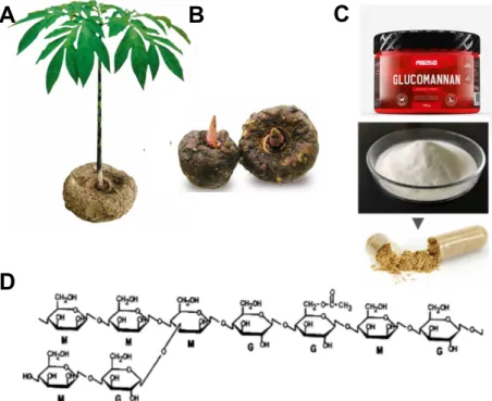
Konjac Glucomannan (KGM) is an essential natural polysaccharide and the main component of the roots and tubers of the Amorphophallus konjac plant (Figure 8A-B). KGM comprises a back-bone chain of glucose and mannose monomers in a molar ratio of 1:1.6 with ~5–10% acetyl-substituted residues at the side C-6 position. It has a highly branched chain (Figure 8D) [49].
Figure 8 - Konjac Glucomannan. A) and B) Plant and roots of the Amorphophallus konjac plant from where
the polymer is extracted; C) KGM in powder formulation used in different areas, namely food industry; D) Chemical structure of KGM. Adapted from [50], [51].
A
B
C
cholesterol reduction, the applications of this polymer have widely increased (Figure 8C). Moreover, KGM is identified as a safe material by the Food and Drug Administration [49]. Since KGM has characteristic high viscosity, good water imbibing, biocompatibility, biodegradability, non-toxic and excellent gelling film forming, it is widely used in food, chemical, medical and pharmaceutical areas. The findings of the benefits of KGM encourage more applications of KGM in biomaterial field, namely as a controlled release matrix [49], [52], [53]. The excellent gelation properties makes KGM unique and superior in relation to other natural macromolecules [53].
Having in consideration all the characteristics and potentialities of KGM, this material can be more elastic polymer than the routinely used in the fabrication of bone scaffolds, providing a matrix much more versatile that can support for a longer time the attach, migration and proliferation of cells and follow the regeneration process of big defects on bone tissue. Moreover, and thinking always on a bi/multifunctional material, this type of matrix can be an excellent reservoir of drugs/molecules to a controlled release and can be easily functionalized. KGM offers a great versatility, allowing the production of tailored multifunctional scaffolds.
In the case of bone cancer, the combination of tissue engineering methodologies and specific local tumor treatments can provide an alternative treatment option to heal large bone defects resulting from tumor resection. A potentially innovative therapeutic strategy would rely on the use of multifunctional scaffolds that have enhanced osteogenic capacity for bone regeneration combined with the ability to locally destroy residual tumor cells. In this way, with only one strategy it is possible mitigate the limitations and complications associated at each disease condition [27].
3.1.3 Other Agents
Other interesting materials can present bifunctional effects and act as anti-cancer agents. Between them, it is important to highlight. metallic ions, due to their magnetic properties, GO and polydopamine which are photosensitive agents [26], [28], [41]. Their mode of action will be provided by the application of an external magnetic field - for magnetic materials - or a laser – for photosensitive agents - which will increase the temperature - hyperthermia effect - and kill the
cancer cells. This effect is very selective (there isn’t the risk of kill the healthy cells if the temperature achieved isn’t higher than 43°C/45°C [54]–[56], destruction of the immune system and other side effects), localized and minimal invasive [28], [57].
Metallic ions naturally present in bone composition, such as strontium, magnesium, zinc, copper and even silver, have vital roles in the formation, growth, and repair of bone, and thus can be incorporated in the polymeric bases of the scaffolds. In recent years various studies demonstrated that adding these elements to CaP materials, can lead to controlled degradation, increase the mechanical strength of the materials, and positively influence the biological response, improve the performance process after implantation [58], [59].
3.2 Fabrication techniques- General Overview
In an organ, cells and their ECM are usually organized into 3D tissues. Therefore, in tissue engineering, the production of a highly porous 3D matrix is often necessary to accommodate cells and to guide their growth and tissue regeneration in three dimensions. Different techniques have been used to fabricate 3D porous ceramics scaffolds for tissue engineering and regenerative medicine [60].
Traditional techniques are relatively simple methods, including solvent casting and particulate leaching, gas foaming, fiber meshes and fiber bonding, phase separation, melt molding, emulsion freeze drying, solution casting and freeze drying [61], [62]. However, most of these methods are incapable of producing fully continuous interconnectivity and uniform pore morphology within a scaffold. In these techniques it is very difficult control the pore geometry, pore size, the spatial distribution and overall porosity of the scaffolds [63]. In addition, the use of organic solvents may cause toxic effects on cells during subsequent culture [64], [65].
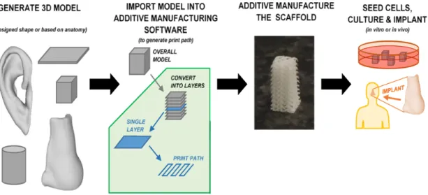
Recently, to overcome these obstacles, advanced additive manufacture (AM) techniques have emerged. AM is a diverse group of manufacturing techniques that build objects by adding material in a layer-by-layer fashion (Figure 9).
Figure 9 – AM technique.Overall procedure for additive manufacturing a tissue engineering construct, with the building of objects through the addiction of material in a layer-by-layer fashion. Through this process it is possible produce a wide variety of objects with complex shapes. Adapted from [66].
Implementation of this simple idea makes a myriad of applications possible, where complex shapes such as those in aerospace and biomedical engineering are possible of being produced with a short time of production runs. It is therefore unsurprising that additive manufacturing has received a significant amount of interest, speculation, and investment from industry and academia, and in recent years it has entered the public consciousness. This has resulted in rapid development of the technology, which has progressed well beyond sensationalized press articles and preliminary prototypes, and is already a disruptive technology in the real world.
3.2.1 Robocasting
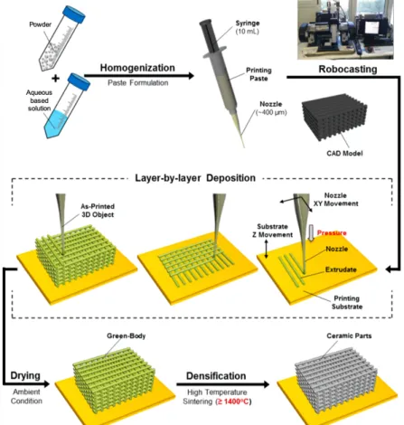
Most work on additive manufacturing has focused on metals and polymers, both of which are currently being embraced in industry. On the other hand, additive manufacturing of ceramic materials is less well developed [42], [43]. Robocasting emerged as a distinct AM and the use of ceramics starts to be developed. The use of ceramics in Robocasting is based on the following principle (Figure 10): green 3D objects are built by extruding a filament of paste (known as an “ink”) through a fine nozzle while the nozzle’s position is controlled by a computer in accordance with a Computer Aided Design (CAD) model. Objects are built up in a layer-by-layer fashion. As the object is built the extruded filament impinges on the printed part and fuses with it due to surface tension.
Thus, in robocasting the rheology of the ink is a key feature in order to print a self-supporting structure. Robocasting has shown to be a promising technique in producing a range of ceramic materials with relatively good mechanical properties. Furthermore, the flexibility of robocasting opens up a number of possibilities of new microstructures and materials that have yet to be explored [42], [43]. The most recent work developed on the robocasting technique reports the formulation of inks based on a hydrogel which acts as a carrier for a ceramic powder [43].
Figure 10 – Robocasting technique. The traditional step by step of a produced structure through
Robocasting – Ink Formulation, Layer by Layer Deposition of a structure codified by a CAD Model, Drying of the Green Body and Sinterization of the structure. Adapted from [29].
In this work scaffolds will be produced through Robocasting or Direct Write Assembling (DWA). This is an easy and fast fabrication method and considered very promising for biomedical applications. With a 3D-printing technique it is possible to produce a structure with characteristics, such as porosity, that could improve nutrition, transporting and cell ingrowth [25], [46].
4. Experimental Strategy Rationale
The scaffolds produced in this work are sintering-free scaffolds (in the production process the last step of the Figure 10 doesn’t occur). In this way, the incorporation of drugs, growth factors and other types of molecules and elements which can enhance the anti-cancer therapy is possible during the scaffold fabrication. In a traditional methodology, as a final step, the scaffolds are submitted to high temperatures to improve the mechanical properties. However, any component sensitive to the temperature will be destroyed.
Furthermore, it is not necessary to use traditional methods - soaking methods - to functionalize the material and to be subjected to its limitations. In such methodology the scaffold, after the sintering step, is immersed on a solution containing the desirable component. This usually gives limited functionalization yields. The functionalization efficiency depends on the concentration of the solution that is used, the soaking time and the interaction between the scaffold and the component. Components such as GO and polydopamine are usually deposited in thin layers through coating processes. In this way, the functionalization occurs only on the surface of the scaffold or in specific zones – if the sandwich method is applied. Thus, the surface area limits the process of functionalization.
The development of non-sintered scaffolds allows a more homogeneous distribution of the elements and a more efficient incorporation, specially when dealing with temperature-sensitive components. The increase of the amount of therapeutic component incorporated in the scaffold will provide an increase on the efficacy of the material in promoting regeneration, kill cancer cells or treat an infection. Moreover, non-sintered scaffolds are more cost-effective materials which if efficiently functionalized, can provide a decrease in the therapeutic time of the disease, as well as in the dosage of the therapeutic agent.
For example, this type of scaffolds is very attractive to be functionalized with agents such as magnetic nanoparticles, polydopamine. There are yet some works on the literature that report the use of these components on a multifunctional scaffold to promote bone regeneration and death of cancer cells. Zhang et al. demonstrated that the incorporation of iron oxide and GO into the scaffolds had an important role in the stimulation of osteogenic differentiation
of rabbit bone marrow stromal cells and the scaffolds produced have potential application in killing the residual human bone tumor cells by magnetothermal treatment [25]. Wu et al. verified that the integration of magnetic nanoparticles in the CaP scaffolds can promote the differentiation and proliferation of osteoblast cells [41]. Ma et al. reported in their work the importance of the incorporation of GO in the scaffolds to promote the regeneration of big bone defects and the potential to kill cancer cells [26]. A work performed by the same authors reveled that with the functionalization of scaffolds with polydopamine is possible induce tumor cell death in vitro, and significantly inhibited tumor growth in mice. Moreover these scaffolds could support the attachment and proliferation of rabbit bone mesenchymal stem cells, and significantly facilitated the formation of new bone tissue in rabbit bone defects [28].
Despite of the demonstrated effects of these materials, there are still some concerns related with their long-term safety. For instance, metallic nanoparticles are poorly biometabolized and have pertinent issues related to the safety of the metal itself [57]. Iron oxide nanoparticles are the most used and studied. They have excellent properties such as superparamagnetism, biocompatibility and biodegradability [40]. The carbon-based materials have been demonstrated to induce many toxic responses such as oxidative stress and pulmonary inflammation [57]. These are the problems associated with most of the current photothermal agents, promoting an increasing interest on the use of polydopamine [28].
Polydopamine is a polymer imitating melanin that occurs naturally in organs and tissues, such as hair, skin, brain medulla, iris of eyes, and brain medulla. Owing to its satisfactory non long-term toxicity during their retention in rats, biocompatibility and biodegradability, a high median lethal dose polydopamine has greater potential for biomedical application. Furthermore, it has a high photothermal conversion efficiency of 40% (much higher than previously reported photothermal agents), suggesting that dopamine is a favorable photothermal agent for tumor therapy [28], [57].
5. Aim of the thesis
The main goal of the work performed in this thesis is to produce and validate free-sintering 3D additive manufactured scaffolds, produced by robocasting to be used in bone tissue engineering. Moreover, the development of a bifunctional 3D material/scaffold: a material that at the same time can promote the regeneration of the bone and kill the cancer cells that remain on the tumor site, and that can prevent the recurrence of the tumor is a further goal.
The specific objectives included:
1. the in vitro testing of scaffolds composed by chitosan and CaP powders
that had already been developed in the scope of the project in which this thesis is inserted but were not yet validated for in vitro culture.
2. Inclusion of magnetic nanoparticles on the chitosan/ biphasic CaP and
assessment of the influence of magnetic conditions on MSCs behavior.
3. Overcome the limitations of the existent scaffolds by designing innovative
KGM printed scaffolds, in order to find an alternative to the complex handling of chitosan scaffolds and their sterilization process.
4. In vitro testing of scaffolds of KGM and BCPs for a bone regeneration
approach (cell delivery).
5. Development and optimization of functionalization of KGM with
polydopamine as a photosensitive agent to allow the induction of localized hyperthermia to induce cancer cell death while preserving healthy cells. Strategies include:
§ Production of polydopamine nanoparticles for a further incorporation on the scaffolds ink;
Materials and Methods
1. Materials
For producing KGM-BCP scaffolds: KGM was purchased from Prozis, Portugal. Biphasic Calcium Phosphate (BCP) powders – containing HA and β-TCP in their composition were kindly provided by the Department of Materials and Ceramics Engineering, CICECO - University of Aveiro, Portugal. For obtaining MNPs scaffolds: Chitosan (Low molecular weight, Sigma-Aldrich) and Calcium phosphate (CaP) powders - HA and β-TCP – were provided by Department of Materials and Ceramics Engineering, CICECO University of Aveiro, Portugal). For KGM functionalization and polydopamine synthesis: NaCl (VWR, Pennsylvania, USA), succinic anhydride – SAC0 (Sigma-Aldrich, Missouri, USA), N-Hydroxysuccinimide - NHS (Sigma-Aldrich, Missouri, USA), 1-(3-Dimethylaminopropyl)-3-ethylcarbodiimide hydrochloride - EDC (Alfa Aesar, Massachusetts, USA) and Dopamine hydrochloride (Sigma-Aldrich, Missouri, USA), hydroxylamine hydrochloride (Sigma-Aldrich, Missouri, USA), 25 % (v/v) ammonia solution (Merck, Darmstadt, Germany), 96% ethanol (Valente e Ribeiro, Belas, Portugal), MWCO 3.5 kDa dialysis membrane (Spectra/Por®, SpectrumLabs) were purchased and used as received.
2. Chitosan-CaP ink preparation loaded with magnetic
nanoparticles (MNPs scaffolds)
The prepared inks consisted in a mixture of 5 wt% chitosan solution containing 10 wt% of citric acid, HA and β-TCP powders non-doped/doped with iron, 1.5 wt% genipin as reticulating agent and 25 mg/mL magnetic nanoparticles solution (fluidMAG-Chitosan, Chemicall, Germany). The amount of ceramic added to the chitosan solution differ if the CaP powders are doped or not. If the ceramic powders are doped, the quantity of solids added is 40 wt%, while if the powders don’t have iron ions in the composition the amount decrease to 34 wt%. 1 wt% of this quantity are Magnetic Nanoparticles dried in an oven. The inks were prepared by mixing the several components using a planetary centrifugal mixer
ceramic powders were designed as BMNPs, while the ones composed by iron doped ceramic powders were named FeMNPs. This type of ink have been very well characterized as reported in previous studies [67].
3. Preparation of the hybrid KGM-BCP printable ink (KGM
scaffolds)
3.1 Preparation of KGM solution
KGM solutions with concentrations ranging from 0.2-3 wt%, (0.2 wt%, 0.5 wt%, 1 wt%, 2 wt%, 3 wt), in dH2O have been prepared by overnight stirring (1400
rpm) at room temperature (RT) using a thermomixer (eppendorf thermomixer comfort). KGM solutions with similar concentration have also been prepared in a 0.36% (w/v) Na2CO3 in order to study the influence of the KGMs’ deacetylation in
the gelation features of the polymer. The concentration of the aqueous KGM solution was optimized to achieve an adequate viscosity for printing, through the manual extrusion of the solution with syringes.
3.2 Preparation of the KGM-BCP ink
After adjusting the concentration of the KGM solution, BCP powders in 30 and 40 wt% have been added in order to obtain a printable KGM-BCP ink. For the preparation of these hybrid organic-inorganic printable inks, an adequate amount of KGM was mixed with 30 wt% or 40 wt% BCP powders and allowed to stir overnight in dH2O (1400 rpm) at room temperature using a thermomixer
(eppendorf thermomixer comfort). After this initial screening, the best KGM-BCP ink formulation (3 wt% KGM with 40 wt% BCP) was prepared using a planetary centrifugal mixer (ARE-250, Thinky Corp., Tokyo, Japan) to obtain an ink with improved homogeneity.
4. Robocasting of MNPs scaffolds and KGM scaffolds
3-D scaffolds consisting of a mesh of ceramic rods were constructed layer-by-layer via direct write assembly of the ink using a robotic deposition device (3-D Inks, Stillwater, OK). The ink was deposited through cylindrical metallic deposition nozzles (EFD Inc., East Providence, RI) with a diameter d = 410 µm, at a printing speed of 10 mm s-1 (Figure 12B and C).
The external dimensions of the MNPs scaffolds were about 9×9×3 (Figure 11A) in a total of 12 layers. Each structure consisted of 9 individual scaffolds of 3×3×3 mm (Figure 11B and D), which were produced with a pore size of 500 µm. After printing, samples were placed at 37°C overnight with controlled humidity (80%) to promote chitosan crosslinking by genipin. On the following day, the scaffolds were placed at 37°C for 24h.
Figure 11- CAD Model. The external dimensions of the scaffolds were about 15 × 15 × 3 (A) in a total of
12 layers being 9 individual scaffolds of 3 × 3 × 3 mm (B) and (D), or 6 layers and 9 individual scaffolds of 3 × 3 × 1.5 mm (C) and (D). Adapted from [36].
For the KGM scaffolds the external dimensions were about 9×9×1.5 mm (Figure 11A) in a total of 6 layers. Each structure consisted of 9 individual scaffolds of 3×3×1.5 mm (Figure 11C and D), which were produced with a pore size of 500 µm. After printing, samples were placed in an ethanol bath to promote the assembly of the structure (Figure 12D). After 1h, the scaffolds were placed at 37°C overnight with controlled humidity (80%) to promote the increase of their mechanical properties (Figure 12E).
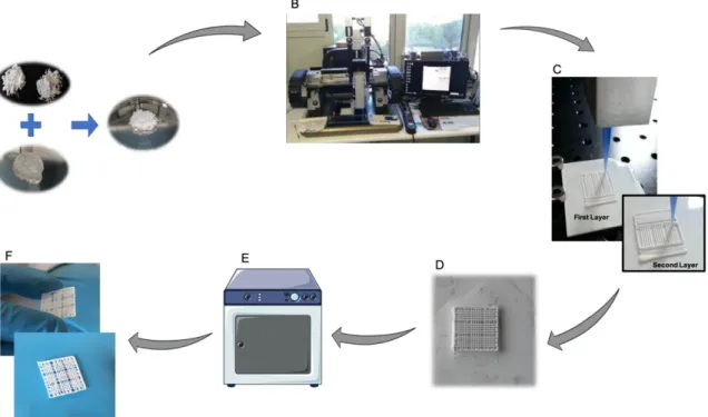
Figure 12 – KGM scaffolds production cycle. A) Production of the ink through the mixture of the ceramic
powders on the KGM solution. B) Robocasting machine where the ink is extruded through a syringe with a nozzle of 410 µm. C) Layer by Layer Deposition of the ink. D)Ethanol bath for 1h after printing of the structure to improve its’ mechanical properties. E) Scaffolds are placed 24h in an oven at 37°C with 80% of humidity. F) Final structure.
5. Characterization of hybrid organic-inorganic KGM
scaffolds and its components
5.1 Rheology
Rheological measurements of the ink and KGM solution were made using a Kinexus Pro Rheometer (Malvern, Pennsylvania, USA). The apparent viscosity of the ink, as well as of the KGM solution, was measured in viscometry mode using a cone and plate sensor system (4°/40 mm) and 150 µm gap size. Samples were placed in the bottom plate geometry and analysed in manual viscometry mode applying different shear stress (0.1, 1, 5, 10, 20, 50, 75, 100, 200, 300 and 400 Pa).
5.2 1H NMR
1H NMR spectra of KGM was recorded using a 400 MHz spectrometer
AVANCE III (Bruker). The polymer was dissolved (13.3 mg/mL) in deuterated water (D2O, Euriso-top) and transferred to NMR tubes. 3-(trimethylsilyl)
propionic-2,2,3,3-d4 acid sodium salt (TSP-d4, Euriso-top) used as internal standard (δ = 0 ppm).
5.3 Size exclusion chromatography (SEC)
Molecular weight was determined on an Agilent 1260 HPLC system equipped with quaternary pump (G1311B), injector (G1329B), column oven (G1316A), refractive index (G1362A) and dual-angle static light scattering (G7800A) detectors. Standard and samples were dissolved in the mobile phase (0.1M NaN3: 0.01M NaH2PO4, pH 6.6) at 1 g/L. Separation was carried out with
four columns (PSS, Germany): Suprema precolumn (5µm, 8x50mm), Suprema 30Å (5µm, 8x300mm), Suprema 100Å (5µm, 8x300mm) and Suprema ultrahigh (10µm, 8x300mm) at a flow rate of 1 mL/min after a 100 µL injection. Column oven and light scattering detector were kept at 30ºC, while refractive index detector was maintained at 40ºC. Detectors were calibrated with a polyethylene oxide standard (PSS, Germany) of 106 kDa (Mp) and polydispersity index 1.05. Refractive index increments (dn/dc) were calculated from the RI detector assuming accurate sample concentrations.
5.4 Attenuated total reflectance- Fourier-transform-infrared spectroscopy (ATR-FTIR)
ATR-FTIR spectra were acquired using a PerkinElmer Frontier FTIR Spectrometer (Massachusetts, USA) from 4000 to 400 cm-1 and with 4 cm -1 resolution.
5.5 Powder X-ray diffraction (XRD)
The amorphous/crystalline nature of the calcinated powders, as well as quantitative analysis of phase composition were determined using a High-Resolution X-ray Diffractometer (PANalytical X'Pert PRO) with Cu Kα radiation (k = 1.5406 Å) produced at 45 kV and 40 mA, which scanned the diffraction angles (2θ) between 5º and 70º with a step size of 0.0260º, time per step 198,645 s. A spectra-fitting software called HighScorePlus was utilized to quantify the percentages of crystalline phases for the calcined powders. This software
within a XRD spectrum. The files utilized in the evaluation of the powders were; HA (# 04-015-7245), β–TCP (#04-006-9376).
5.6 Particle Size distribution
Particle size and particle size distributions of the ceramic powders were evaluated using a particle size analyzer (COULTER LS230, UK) with Fraunhofer optical model. For that, the ceramic powders were suspended at low concentration on an electrolyte solution.
5.7 Scanning electron microscopy (SEM)
The samples were placed, with a carbon tape, in adequate supports for SEM visualization. Before the visualization the samples were coated with palladium gold. The micro and macroporosity of the scaffolds were assessed through SEM using a High resolution (Schottky) Environmental Scanning Electron Microscope with X-Ray Microanalysis and Electron Backscattered Diffraction analysis: Quanta 400 FEG ESEM / EDAX Genesis X4M.
5.8 Measurement of pore size, fiber diameter and grain size of the produced KGM scaffolds
The measurement of pore size, fiber diameter and grain size were performed on SEM images with Fiji, through the execution of a set scale on the images following the scale bar provided by the SEM equipment.
6. KGM functionalization with photothermal moieties
6.1 Synthesis of carboxylated KGM (KGM-SAC0)
A 1 wt% solution of KGM (100 mg; 0,00006 mmol) in dH2O was treated
with succinic anhydride (257 mg; 0,003 mol) added in two portions (t=0 and t=2h) at 0 ºC. The molar ratio of KGM: succinic anhydride is 1:4, calculated based on the weight of the most abundant monomer of KGM. After the addition of each portion, the pH was adjusted to 7.0 and the mixture allowed to stir for a total of 4h at room temperature. The polymer was precipitated by the addition of 99% ethanol (150 mL) and centrifuged at 4000 rpm and 20 ºC for 10 min (Eppendorf
centrifuge 5810R, rotor A-4-62). The precipitated polymer was further washed with acetone (3x 25 mL) and centrifuged in similar conditions. The white precipitate was dissolved in water, transferred to a MWCO 3500 dialysis membrane and purified through dialysis for 3 days against decreasing, changing the solution 2-3 times per day. The dialyzed solution was frozen, lyophilized, and stored at -20oC until further use. The product was obtained as a white fluffy solid
(75 mg).
6.2 Functionalization of KGM-SAC0 with dopamine (KGM-SAC0-Dopamine)
A solution of 1 wt% KGM-SAC0 (36 mg; 0,138 mmol) in MES buffer (0.1M MES, 0.3M NaCl, pH 6.5), was treated with EDC.HCl (29 mg; 0,152 mmol, 1.1 eq) and NHS (17,5 mg; 0,152 mmol; 1.1 eq), following the addition of dopamine hydrochloride (31,5 mg; 0,166 mmol; 1.2 eq) at 4 ºC under argon. The mixture was allowed to stir overnight at room temperature and protected from light. The reaction was quenched with NH2OH.HCl (10,58 mg; 0,152 mmol, 1.1 eq) and the
resultant solution was transferred to a MWCO 3500 dialysis membrane and dialysed for 3 days against decreasing NaCl concentrations, changing the solution 2-3 times per day. The resultant solution was frozen, lyophilized, and stored at -20 oC until further use. The product was obtained as a yellowish fluffy
solid (38 mg).
6.3 Synthesis of Polydopamine particles
The reaction medium was prepared by mixing 4 mL of 25% (v/v) ammonia solution, 80 mL of 96% vol. ethanol and 180 mL of deionized water under mild stirring at 30 °C. After 30 min, a solution of dopamine hydrochloride (1g; 0,005 mol) in dH2O (20 mL) was added and the reaction allowed to proceed for 24 h at
30 ºC. The color of this solution immediately turned to pale yellow and gradually changed to dark brown, indicating polymerization of dopamine [68]. The resultant suspension was centrifuged at 25 000 rpm and the pellet washed with water (3x). The precipitate was resuspended in water and freeze-dried, giving a black

![Table 1 – Examples of commonly used polymers for BTE. Retrieved from [44].](https://thumb-eu.123doks.com/thumbv2/123dok_br/15709031.1068640/32.892.126.744.538.981/table-examples-commonly-used-polymers-bte-retrieved.webp)