Vol.59: e16150613, January-December 2016 http://dx.doi.org/10.1590/1678-4324-2016150613
ISSN 1678-4324 Online Edition
BRAZILIAN ARCHIVES OF BIOLOGY AND TECHNOLOGY
A N I N T E R N A T I O N A L J O U R N A L
Rat dental pulp stem cells: isolation and phenotypic
characterization method aiming bone tissue bioengineering
Bruno Machado Bertassoli
1, Emanuela Silva Costa
1, Cristiane Aparecida Sousa
1, Juliano
Douglas Silva Albergaria
1, Kátia L. Melo Maltos
2, Alfredo Miranda Goes
3, Thais Maria da
Mata Matins
3, Gerluza Aparecida Borges Silva
1, Erika Cristina Jorge
1*1Departamento de Morfologia, Instituto de Ciências Biológicas, Universidade Federal de Minas Gerais. Av.
Antonio Carlos, 6627, Pampulha, Belo Horizonte- MG. 2Departamento de Odontologia Restauradora, Faculdade de Odontologia, Universidade Federal de Minas Gerais. Av. Antonio Carlos, 6627, Pampulha, Belo Horizonte- MG. 3Departamento de Bioquímica e Imunologia, Instituto de Ciências Biológicas, Universidade Federal de Minas
Gerais. Av. Antonio Carlos, 6627, Pampulha, Belo Horizonte- MG.
ABSTRACT
Dental pulp stem cells (DPSC) have been showing a considerable potential for regenerative medicine. Pulps were collected from lower incisors (n=2) through direct access of the tooth pulp chamber. The isolated cells were cultured in alfa-MEM 10% FBS, in standard culture conditions. At the third passage, DPSC were characterized by flow cytometry (MHCI, CD54, CD73, CD90, CD45, CD11 and CD34); RT-PCR for Nanog gene; and their differentiation capacity in osteogenic, adipogenic and chondrogenic cell lines. Isolated cells exhibited adhesion capacity to plastic; fusiform morphology, and 80% confluence reached in approximately 3 days. These cells have also revealed positive expression for CD54, CD73 and CD90 markers; and negative expression for CD11, CD34 and CD45. Nanog expression was detected by RT-PCR, expected for a mesenchymal stem cell profile. DPSC chondrogenic differentiation was confirmed by positive staining in Alcian Blue; lipidic droplets stained with oil red confirmed their capacity to differentiate in adipogenic fate; while mineralized beads, stained with alizarin red, confirmed their differentiation in osteogenic phenotype. These results indicate the viability of the isolation and expansion of rat DPSC following this method, and osteogenic differentiation potential opens new perspectives for in vivo studies and the use of these cells in cellular therapies and tissue bioengineering, aiming bone repair.
Key words: mesenchymal stem cells, dental pulp, murine, phenotypic characterization, cellular differentiation.
INTRODUCTION
Cellular therapies and the use of mesenchymal stem cells in tissue bioengineering techniques represent a promise for tissue replacement in various pathologies, including for the treatment of neurodegenerative, cardiac, pulmonary and bone diseases (Iyer et al. 2009; Arvidson et al. 2011; Tang et al. 2012; Dawson et al. 2014). Some clinical studies, especially in the context of coronary heart disease, have boosted the credibility, safety and feasibility of stem cells for human use (Orlicet al. 2001; Strauer et al. 2002; Perinet al. 2003; Santos et al. 2004; Motaet al. 2005).
Cellular therapy protocols indicate that selected cells can be administrated by both (1) intravenous infusion (Bydlowski et al. 2009); or (2) after previous culture in matrices or three-dimensional scaffolds, both as undifferentiated or partially
induced cell lines, transplanted for in loco repair
(Savitz et al. 2002; Atari et al. 2011; Atari et al. 2012). The association of chitosan and embryonic stem cells (Weng-Ning et al. 2009); PGA-PLLA and umbilical cord stem cells (Wu et al. 2004); fibrin scaffold and mesenchymalstem cells
(Bensaid et al. 2003); poly (D,L-lactic-co-glycolic
acid) hydroxyapatite and embryonic stem cells (Kim et al. 2008); phosphate ceramic biphasic calcium and mesenchymal stem cells (Arinzeh et al. 2007); are all examples of stem cells seeded onto biomaterials for tissue reconstruction.
Mesenchymal stem cells (MSC) have been
indicated as the first choice in tissue
bioengineering studies, by presenting advantages
as high differentiation potential,
immunosuppressed effects, and viability after expansion in culture (Bensaid et al. 2003; Li et al. 2005; Uematsu et al. 2005; Arinzeh et al. 2007; Lyra et al. 2007; Bertassoli et al. 2013). MSC were originally isolated from bone marrow (Arinzeh et al. 2007). After that, these cells were found in various organs and tissues, including in the dental pulp (Bensaid et al. 2003; Li et al. 2005; Uematsu et al. 2005; Arinzehet al. 2007; Lyra et al. 2007; Bertassoli et al. 2013).
Gronthos and co-workers (2000) published the first report about the isolation and identification of an adult dental pulp progenitor population. Since then, DPSC have been described as a clonogenic cellular population, highly proliferative, capable of self-renew and differentiation in adiponegic, condrogenic, and osteogenic cell lines (Gronthos
et al. 2000; Gronthos et al. 2002). Besides the dental pulp from permanent teeth, deciduous and supernumerary teeth have also been used as sources for stem cells (Miura et al. 2003; Kerkiset al. 2006; Jo et al. 2007; Kerkis et al. 2012). However, the stem cells derived from these different teeth differ in the expression pattern of the main markers, and present variations in their differentiation potentials (Jo et al. 2007).
DPSC have been showing positive results for bone repair (Gronthos et al. 2000; Gronthos et al. 2002). These cells can differentiate in osteoblasts and
originate bone tissue both in vitro and in vivo
(Lainoet al. 2005, 2006; D’Aquino et al. 2007;
Graziano et al. 2008). Some in vivo studies have
also been showing the potential of the DPSC to improve the ventricular function, to induce angiogenesis and to reduce the infarct size in rats (Gandia et al. 2007); and to differentiate in neural-crest derived melanocytes (Stevens et al. 2008), opening perspectives of their use in regenerative medicine. DPSC also present the classic genetic/antigenic profile, similar to bone marrow mesenchymal stem cells. These cells express both the embryonic stem (ES) cells transcriptional factors, Oct4 and Nanog, as well as MSC surface markers, such as CD105, CD73, and CD13. However, these cells do not express CD45, CD34, CD14, CD43, and HLA-DR (Tang et al. 2012). An important advantage of the DPSC, compared to other MSC, is that they can be isolated from patients with no major ethical and moral
limitations (Prenticeand Tarne, 2007).
Additionally, autologous cells do not activate immune rejection, and respond to growth factors inherent to the host. In adults, DPSC isolation is performed after a simple surgical procedure of teeth exodonty, mostly using molars or incisors (Tatullo et al. 2014). DPSC canal so maintain their immnunophenotypic properties and differentiation potential, even after a month of cryopreservation of the whole tooth (Tatullo et al. 2014). Thus, extracted human teeth, usually discarded as biological waste, could become an interesting source of cells for regenerative therapies, aiming not just the development of new teeth, but also other tissues as bones, muscles, and nerves (Telles et al. 2011; Sprio et al. 2012).
population. Understanding the mechanisms
involved in the DPSC in vitro manipulation and
the possible genetic or functional alterations should be considered before using these cells as a therapeutic tool. The present study aimed to isolate and characterize a rat dental pulp stem cell sub-population, collected using a simplified method of isolation. This characterized cell line will be the source for future studies aiming bone tissue bioengineering.
MATERIALS AND METHODS
Animals
Mesenchymal stem cells were collected from dental pulps of lowers incisors of adults Wistar rats (n=2 pulps in each extraction), using animals weighting approximately 250 g. Animals were obtained from the UFMG vivarium (CEBIO). All experiments were performed in accordance with the guidelines established by CEUA/UFMG (Ethics Committee on Animal Use, protocol 288/2013).
Isolation and culture of DPSC
Animals were sacrificed by decapitation after intraperitoneal anaesthesia overdose. After the dissection of the skull, upper and lower jaws were separated and released from the soft tissue. Through a midline incision between the lower incisors, the workpiece was separated into two hemi-jaws. Pieces were immersed in 1% PVP-I and taken to the laminar flow hood for pulp tissue
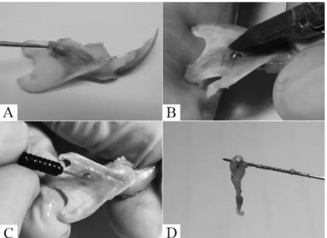
manipulation. With the aid of a scalpel, a small window was opened at the apical foramen, for the pulp tissue exposure (Fig.1A-C). Using a sterile nerve extirpated (size 21 mm, Nº 30 to 40, depending on the foramen diameter), the pulp was released from the root wall, rotating the instrument against the dentinal walls. The pulp tissue was removed without effort or tissue breakdown (Fig. 1C-D).
On a cooled glass plate, the pulp tissue was fragmented and the apical portion, known as apical button (Ohshima et al. 2005) was discarded. The pulp fragments were transferred to a falcon tube for enzymatic digestion with 1,5 mg/mL Collagenase I (Gibco), 40 min at 37°C and 5%
CO2. Digestion was interrupted with growth
medium [GM: Minimum Essential Medium Eagle, Alfa Modification (Sigma-Aldric), supplemented with 10% fetal bovine serum (FBS, Gibco), and
1% penicillin/streptomycin/amphotericin B
solution (Gibco)]. After centrifugation at 360x g, for 10 minutes at 4°C, the supernatant was discarded and the pellet containing the cells was resuspended in GM. Cells were transferred to 25
cm2 flasks and incubated at 37°C and 5% CO
2.
GM was replaced every 2 to 3 days. After reaching 70% confluence, cells were trypsinized (0.25% Trypsin in EDTA 1mM, Gibco), and transferred to
new flasks. Cells were collected for
immunophenotypic characterization and
Figure 1. Dental pulp isolation from pulp canal of Wistarrats. In (A), the hemi mandible extracted and dissected. Instrument indicates the end of the apical foramen. In (B), the opening of the apical foramen, used to extract the pulp. (C) Nerve extirpated inside the apical foramen; and in (D), extracted dental pulp.
Flow Cytometry
Antigens typically expressed in MSC (CD54, CD90 and CD73) were used as positive markers; while markers for hematopoietic stem cells (CD45, CD11b/c and CD34) were selected as negative
controls. The DPSC immune phenotypic
characterization was performed in the third
passage. Briefly, 5x10⁵ cells were incubated with
monoclonal primary antibody for 30 minutes at 4°C. After washing with PBS, cells were incubated with Alexa Fluor 488 goat anti-mouse (Molecular Probes, Life Tecnologies), diluted 1:500 in PBS, for 30 minutes at 4°C. Background fluorescence was determined in DPSC samples labeled with secondary antibody only. The following antibodies were used: CD11a/b, CD90, CD45, CD54, CD73-phycoerythrin (all from BD Biosciences, USA), CD34 (Santa Cruz Biotechnology, USA), HLA-ABC-FITC (Abcam, USA). Stained cells were next analyzed using the FACS Calibur (BD Biosciences), in 10,000 events, and the data was analyzed using WinMDI 2.8 software. Graphics were generated using data from labeled cells size versus granularity.
Total RNA isolation and reverse-transcription polymerase chain reaction (RT-PCR)
Total RNA was extracted from the first, second and third passage for the DPSC by using TRIzol Reagent (Thermo Fisher Scientific), according to
the manufacturer’s instructions. One microgram of total RNA was reverse transcribed in first-strand
complementary DNA, following the
manufacturer’s instructions of the Revert Aid TM H Minus First Strand cDNA Synthesis Kit (Thermo Fisher Scientific). The cDNA was
amplified by using TaqDNA PolymeraseMaster
mix (Thermo Fisher Scientific) and 0.4uM of each specific primer, in a final volume of 25 uL. The
sequences of the Rattus novergicus Nanog primers
Differentiation potential (chemical induction)
DPSCs were induced to differentiate toward adipogenic, osteogenic, and chondrogenic cell fates for 21 days.
For adipogenic differentiation, 1x10³ cells/cm2
were cultured in a 6-well plate in adipogenic medium (GM, supplemented with 0.5 mM isobutyl-methylxanthine, 1 mM dexamethasone, 10 mM insulin, 200 mM indomethacin), with three weekly medium changes. Oil Red O staining (Thermo Fisher Scientific) was performed
following manufacturer’s instructions, as an
indicator of intracellular lipid accumulation. Cells were washed with PBS and fixed in 10% formalin for 1 h. After washing with 60% isopropanol, cells were stained with Oil-Red O solution in 60% isopropanol for 5 min, rinsed with deionized water, and counterstained with hematoxylin for 1 min.
Osteogenic differentiation was induced by
culturing 2.5x10⁴ cells/cm2 in a 24-well plate in
osteogenic medium [GM, supplemented with 50
mM ascorbic acid (Sigma-Aldrich) and 10 mM β
-glycerophosphate (Sigma-Aldrich)], with three weekly medium changes. Cell differentiation was assessed by Alizarin Red staining, as an indicator of extracellular matrix calcification. For Alizarin Red staining, cultures were fixed in 70% ethanol, incubated with 2% Alizarin Red (Sigma-Aldrich) for 15 min, and rised with deionized water.
Chondrogenic differentiation was induced by
culturing 2.5x10⁴ cells/cm2 in a 24-well plate in
chondrogenic medium [GM, supplemented with 1 mM dexamethasone, 100 mM pyruvate, 100 U/mL
Insulin, 5 ug/mL Transferring, 50 mg/mL ascorbic acid (all chemicals from Sigma-Aldrich) and 10
ng/mL TGF-β1], with three weekly medium
changes. To evaluate chondrogenic differentiation, plates were fixed in 4% paraformaldehyde and proteoglycan deposition was detected by 1% Alcian blue staining (Sigma-Aldrich).
For all three protocols, control experiments were performed in cells plated on the same conditions and maintained in GM for 21 days.
Imaging analysis
All images were capture dusing the Motic AE31 inverted phasemicroscope withan Moticam 2300 camera and the Motic Image plus 2.0 software.
RESULTS
Cell morphology in expansion phase
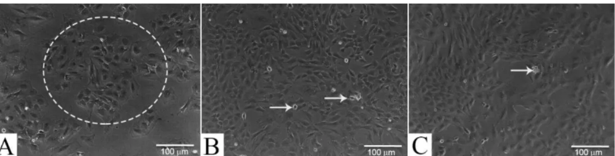
Right after seeding, cells in culture presented a heterogenic morphology, with some of them showing a fibroblast-like aspect, and others, oval shape (Fig. 2A). After the first passage, however, cells shape became predominantly fibroblastoid, presenting an elongated appearance (Fig. 2B). The fibroblast-like morphological aspect persisted until the conclusion of the experiments (Fig. 2C). Enrichment for DPSC was performed based on their adherent capacity to the plastic. DPSC colony organization was observed in all passages (Fig. 2A). These DPSC reached confluence of 80% in approximately three days. After the third passage, however, a longer time was need to DPSC reach confluence in culture.
Figure 2 - DPSC cell morphology during culture. In (A), morphology found after the first passage of the cells. A heterogeneous cell shape was found, with cells presenting both fibroblastoid and round cell shapes. Cell clusters were also observed, suggesting the colony formingunit– dottedcircle. In (B), morphologic aspect observed at the second passage; and in (C) at the third passage. Cells were predominantly fibroblastoid, presenting an elongated appearance. Arrow indicates non-adherent round shape cells insuspension, discarded during medium replecement.
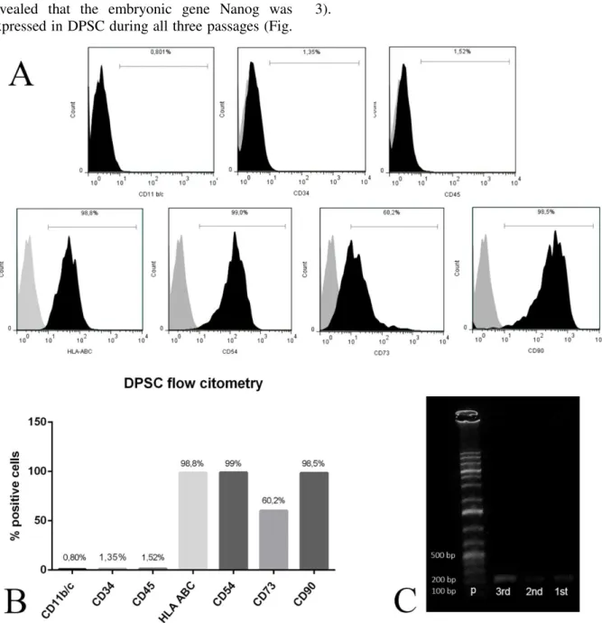
Phenotypic characterization DPSC
The immunophenotypic characterization revealed that the DPSC were positive for surface markers of nucleated cells such as HLA-ABC; and also for
revealed that the embryonic gene Nanog was expressed in DPSC during all three passages (Fig.
3).
Figure 3.DPSC characterization. In (A) and (B),the percentage of DPSC staining indicated after cytometric flow for HLA-ABC, CD54, CD73, CD90, CD11b/c, CD34,and CD45markers.DPSC staining were positive for HLA-ABC, CD54, CD73 and CD90 markers; while negative for the CD11b/c, CD34 and CD45 markers. In (C), RT-PCR result indicating the expression of the embryonic gene Nanog during all passages of DPSC culture. P - 1kb plus molecular weightladder; 3rd – DPSC in the third passage; 2nd – in the second passage; and 1st – in the first passage.
Cell plasticity analysis
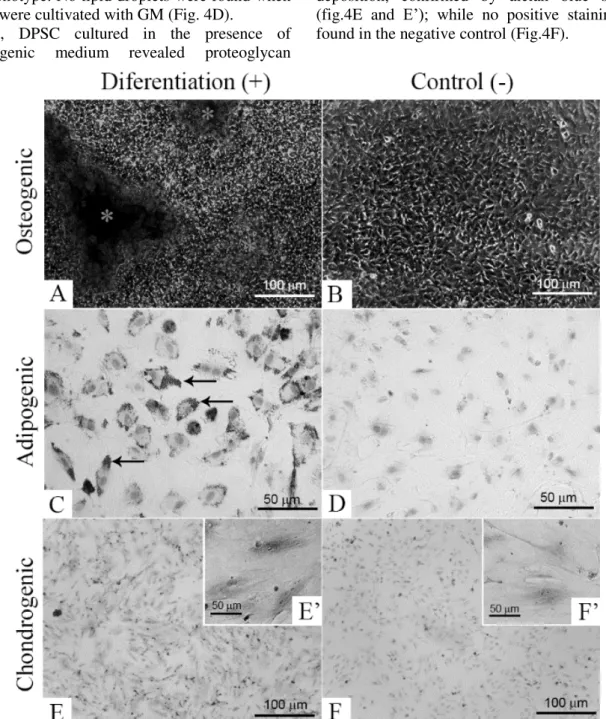
After three weeks of culture, the DPSC isolated using the proposed method from this work could successfully differentiate in all three cell fates: condogenic, osteogenic and adipogenic.
The DPSC osteogenic differentiation was
confirmed by the alizarin red staining. The results revealed the deposition of mineralized nodules in the extracellular matrix of these cells, after 21 days of culture (Fig. 4A), which is characteristic
ofosteblasts. DPSC cultivated in
non-supplemented GM, were negative for this staining (Figure 4B).
the phenotype. No lipid droplets were found when DPSC were cultivated with GM (Fig. 4D).
Finally, DPSC cultured in the presence of
condrogenic medium revealed proteoglycan
deposition, confirmed by alcian blue staining
(fig.4E and E’); while no positive staining was
found in the negative control (Fig.4F).
Figure 4. DPSC differentiation in osteogenic, adipogenic and chondrogenic cell fates. In (A), the presence of mineralized nodules stained with alizarin red (*) confirmed the DPSC differentiation in osteoblasts; and in (B), the negative control. In (C), lipid droplets stained with oil red (arrows) confirmed the positive adipogenic differentiation of the DPSC; and in (D), negative control. In (E), the positive staining of extracelular matrix protegly can by alcian blue, confirming the chondrogenic differentiation of the DPSC isolated in this work; in (E’) zoom of (E); In (F), the negative control and in (F’) zoom of (F).
DISCUSSION
The term “stem cell” can be applied to a diverse
group of cells that share two properties: (1) a capacity for prolonged or unlimited self-renewal under controlled conditions; and (2) the potential to differentiate into a variety of specialized cell
types (Dawson et al. 2014). The term
“mesenchymal stem cell” was originally used for a hypothetical common progenitor, of a wide range of “mesenchymal” (hematopoietic, non-epithelial, and mesodermal) tissues (Caplan1991). The presence of MSC in a broad range of postnatal
vitro assays and immunophenotyping (Dominici et
al. 2006). MSC were originally named “bone
marrow stem cells” by Friendenstein and
co-workers (1966), grouping those stem cells that werenon-hematopoietic, adherent to polystyrene plates, and with a fibroblast-like morphology. In this work, mesenchymal stem cells were isolated from dental pulps, using a particular method that allowed removing the pulp tissue directly from the apical foramen of the tooth. Right after seeding, the DPSC isolated using this method were found to be adherent to the polystyrene plastic. However, adhesion properties are not sufficient to classify these purified cell population as MSC (Phinney et al. 1999). In fact, previous studies have been reported that fibroblasts, macrophages, and some hematopoietic cell lines can all adhere to the plastic, and also present MSC morphological characteristics after growth (Schrepfer et al. 2007; Bydloswki et al. 2009). Cell density can also be a factor that influences the capacity of MSC to adhere to the plastic: MSC plated in low-density conditions adhere faster to the plastic, compared to high-density conditions (Deans and Moseley 2000). Additionally, DPSC from this work were not homogeneously found in the fibroblastoid-shape in the first passage. A homegeneous aspect of the DPSC, with the majority of the cells presenting a spindle/fibroblastoid morphology arranged in colony forming units, was observed in culture only after the third passage. Dental pulp connective tissue is a well known heterogenic cell population, what could be an explanation to our initial observations. This characteristic of finding fibroblast-like morphology of these cells only after a few passages was also reported in stem cells derived from bone marrow and deciduous teeth (Friendenstein et al. 1966; Gronthos et al. 2000; Gronthos et al. 2002; Jesus et al. 2011), suggesting this as a characteristic of MSC.
Another characteristic of the DPSC isolated in this work was that they initially reached confluence of 80% in approximately three days. After the third passage, however, a longer time was needed to reach confluence in culture. This observation suggested that successive passages could be inducing a decrease of the proliferation rate or even cell death in these cells. This characteristic was also described for MSC after several passages (Fehrer and Lepperdinger 2005).
Different methods of MSC isolation and expansion
were reported thus far. The “Mesenchymal and
Tissue Stem Cell Committee", of the International Society for Cellular Therapy, proposed the minimal criteria to define human MSC (Dominici et al. 2006). Following this criteria, MSC can be characterized by: (1) the capacity to adhere to the plastic; (2) the pattern of expression of surface
markers known as “Cluster of Differentiation”,
being positive for CD105 (Endoglin/SH2), CD73 (SH-3) and CD90 (Thy-1), and negative for CD45, CD34, CD14 or CD11b, CD79alpha or CD19 and HLA-DR surface molecules; and (3) the potential to in vitro differentiate in osteoblasts, adipocytes, and chondroblasts (Dominici et al. 2006; Kolf et al. 2007; Iacono et al. 2015). Although designed for human stem cells, the same designation has been referenced and used to characterize stem cells obtained from animal tissues.
The phenotypic characterization of the stem cells before the differentiation in specialized cell types can reveal the purity of the culture, as hematopoietic cells and fibroblasts could exhibit
similar morphological characteristics in vitro
(Bianco et al. 2001; Bobis et al. 2006; Ishii et al. 2005). Hematopoietic cells, for example, express the surface marker CD45 (Bobis et al. 2006; Pittenger et al. 1999), which can also be found in fibroblasts (Ishiiet al., 2005). The CD73 surface marker can either be expressed in fibroblasts or in MSC (Ishii et al., 2005). CD90 and CD54 were both found to be MSC exclusive (Covas et al. 2003).
In this study, CD90 was expressed in over 90% of the cells. This result strongly suggested that the cell culture was in fact a MSC culture. However, only 60.2% of the isolated cells were positive for CD37. According to Domicini and co-workers (2006) criteria, CD73 must be found in more than 90% of human MSC. Low expression of this particular surface marker was also observed in DPSC from humans third molar (Atari et al. 2012), suggesting that this could be a particular characteristic of DPSC. Curiously, the same authors have described a low expression of the CD90 marker in DPSC, what differs from our results. These discrepancies in surface marker expression patternmay be dueto:(1) the source of tooth/animal model; (2) the method of stem cell
isolation;(3) the cell maintenance in vitro; and (4)
proliferation characteristics of hematopoietic stem
cells and endothelial progenitor cells
(Gangenahalli et al. 2005). The DPSC from this work did not express this particular marker. The HLA-ABC antigens, also known as Major Histocompatibility Complex class I (MHC), are constitutively expressed in nucleated cells. The immunophenotypic characterization revealed that 98.8% of the DPSC isolated in this work were MHCI positive. Overall, the proposed method for stem cell isolation from dental pulp tissues resulted in a cell population expressing CD54, CD73, CD90, and MHCI markers; but not expressing the markers CD45, CD11 and CD34. Therefore, the immunophenotypic characterization of the DPSC cells isolated from rat incisors confirmed the purity of over 90% of the culture. Following the multipotency criteria, DPSC were chemically induced to differentiate, after 21 days, into osteoblasts, chondroblasts and adipocytes. The differentiation of stem cells in osteoblasts may represent a breakthrough for tissue bioengineering. Studies have been demonstrating the DPSC potential to induce regeneration of critical-sized calvarial defects in mice (Moshaverinia et al. 2014; Menicanin et al. 2014); in canine and swine models, local implantation of DPSC, associated
with β-TCP and HA/TCP scaffolds, successfully
induced bone regeneration in critical-sized orofacial bone defects (Zheng et al. 2009; Kim et al. 2009).
CONCLUSION
Stem cells can be easily isolated from dental pulp using a simplified method, which give access to the pulp via the apical foramen. DPSC isolated using this method expressed MSC surface markers, but not the hematopoietic ones. DPSC multipotentiality profile was confirmed by the differentiation of these cells into osteoblasts, adipocytes and chondrocytes. According to the minimum criteria established by ISCT, DPSC isolated from rats incisors can be considered MSC. Given their plasticity, especially for osteogenic cell fate, this study opens perspectives for the evaluation of DPSC for bone tissue bioengineering and cellular therapy, aiming the replacement or regeneration of bone tissue.
REFERENCES
Arinzeh TL, Tran T, McAlary J, DaculsiGA. Comparative study of biphasic calcium phosphate ceramics for human mesenchymal stem-cell-induced bone formation. Biomaterials.2007; 26(17): 3631– 3638.
Arvidson K, Abdallah BM, Applegate LA, BaldiniN, Cenni E, Barrena EG, et al. Bone regeneration and stem cells. J Cell Mol Med.2011;15(4): 718-746. Atari M, Barajas M, Hernandez-Alfaro F, Gil C,
Fabregat M, Ferre´SPadro E, et al. Isolation of pluripotent stem cells from human third molar dental pulp. HistolHistopathol. 2011; 26(8):1057-1070.
Atari M, Serrano JC, Recio CG, Delgado CG, Sarrà EM, Fernández DAG, et al. The enhancement of osteogenesis through the use of dental pulp pluripotent stem cells in 3D. Bone. 2012; 50(4):930-941.
Bensaid W, Triffitt JT, Blanchat C, Oudina K, Sedel L, PetiteH. A biodegradable fibrin scaffold for mesenchymal stem cell transplantation. Biomaterials. 2003; 24(14):2497–2502.
Bertassoli BM, Assis Neto AC, Oliveira FD, Arroyo MAM, Ferrão JSP, Da Silva JB, et al. Mesenchymal Stem Cells - Emphasis in Adipose Tissue. Braz Arch Biol Technol.2013;56(4): 607-617.
Bianco P, Riminucci M, GronthosS, Robey PG. Bone marrow stromal cells: nature, biology, and potential applications. Stem Cells.2001; 19(3):180-192. Bobis S, Jarocha D, Majka M. Mesenchymal stem cells:
characteristics and clinical applications. Folia HistochemCytobiol. 2006; 44(4):215-230.
BydlowskiSP, Debes AA, Maselli LM, Janz FL. Características biológicas das células-tronco mesenquimais. Rev Bras HematolHemoter. 2009; 31(1):25-35.
Caplan AI. Mesenchymal stem cells. J Orthop Res. 1991; 9(5):641-650.
CovasDT, SiufiJL, Silva AR, Orellana MD. Isolation and culture of umbilical vein mesenchymal stem cells. Braz J Med Biol Res.2003; 36(9):1179-1183. D’Aquino R, Graziano A, Sampaolesi M, Laino G,
Pirozzi G, De Rosa A, et al. Human postnatal dental pulp cells co-differentiate into osteoblasts and endotheliocytes: a pivotal synergy leading to adult bone tissue formation. Cell Death Differ. 2007; 14(6):1162-1171.
Dawson JI, Kanczler J, Tare R, Kassem M, OreffoROC. Concise Review: Bridging the Gap: Bone Regeneration Using Skeletal Stem Cell-Based Strategies Where Are We Now? Stem Cells.2014; 32(1):35-44.
DominiciM, Le Blanc K, Mueller I, Slaper-Cortenbach I, Marini F, Krause D, et al. Minimal criteria for defining multipotentmesenchymal stromal cells: The International Society for Cellular Therapy position statement. Cytotherapy. 2006;8(4):315-317.
Fehrer C, Lepperdinger G. Mesenchymal stem cell aging. ExplGerontol.2005; 40(12):926-930.
FriedensteinAJ, Piatetzky-Shapiro I, Petrakova KV. Osteogenesis in transplants of bone marrow cells. J EmbryolExp Morph. 1966;16(3):381-390.
Gandia C, Armiñan A, Garcia-Verdugo JM, Lledó E,
Ruiz A, Miñana MD, et al. Human DP stem cells improve left ventricular function, induce angiogenesis and reduce infarct size in rats with acute myocardial infarction. Stem Cells.2007; 26(3)638-645.
GangenahalliGU, Singh VK, Verma YK, Gupta P, Sharma RK, Chandra R, et al. Hematopoietic stem cell antigen CD34: role in adhesion or homing. Stem Cells Dev. 2006; 15(3):305–313.
GrazianoA, D’Aquino R, De Angelis MGC, De Francesco F, Giordano A, Laino G, et al. Scaffold’s surface geometry significantly affects human stem cell bone tissue engineering. J Cell Physiol. 2008; 214(1):166–172.
Gronthos SS, Brahim J, Li W, Fisher LW, Cherman N, Boyde A, et al. Stem cell properties of human dental pulp stem cells. J Dent Res.2002; 81(8):531-555. GronthosSS,Mankani M,Brahim J,Robey PG,ShiS.
Postnatal human dental pulp stem cells (DPSCs) in vitro and in vivo. PNAS. 2000; 97(25):13625-13630.
Ishii M, Koike C, Igarashi A, Yamanaka K, Pan H, Higashi Y, et al. Molecular markers distinguish bone marrow mesenchymal stem cells from fibroblasts. BiochemBiophys Res Commun. 2005; 332(1):297-303.
Iyer S, Alsayegh K, Abraham S, Rao RR. Stem cell-based models and therapies for neurodegenerative diseases. Crit Rev Biomed Eng.2009; 37(4):321– 353, 2009.
Jesus AA, Soares MBP, Soares AP, Nogueira RC, Guimarães ET, Araújo TM, et al. Collection and culture of stem cells derived from dental pulp of deciduous teeth: technique and clinical case report. Dental Press J Orthod.2011; 16(6):111-118. Jo YY, Lee HJ, Kook SY, Choung HW, Park JY,
Chung JH, et al. Isolation and characterization of postnatal stem cells from human dental tissues. Tissue Engineering. 2007; 13(4):767-773.
Kerkis I, Kerkis A, Dozortsev D, Stukart-Parsons GC, Gomes Massironi SM, Caplan AL et al. Isolation and characterization of a population of immature DP stem cells expressing OCT-4 and other embryonic stem cell markers. Cells Tissues Organs. 2006; 184(3-4):105-116.
Kerkis I, Caplan AL. Stem cells in dental pulp of deciduous teeth. Tissue Engineering. 2012;
18(2):129-138.
Kim SH, Kim KH,Seo BM,Koo KT,Kim TI,Seol YJ, et al. Alveolar bone regeneration by transplantation of periodontal ligament stem cells and bone marow stem cells in a canine peri-implant defect model: a pilot study. J Periodontol,2009; 80(11):1815–1823. KimS, Kim SS, Lee SH,AhnSE,GwakSJ, SongJH, et
al.In vivo bone formation from human embryonic stem cell-derived osteogenic cells in poly(D,L -lactic-co-glycolic acid)/hydroxyapatite composite scaffolds. Biomaterials. 2008; 29(8): 1043–1053. Kolf CM, Cho E, Tuan RS. Mesenchymal Stromal
cells. Biology of adult mesenchymal stem cells: Regulation of niche, self-renewal and differentiation. ArthResTherap,2007; 9(1):1-10. IaconoE, Merlo B, Romagnoli N, Rossi B, Ricci F,
Spardari A. Equine Bone Marrow and Adipose Tissue Mesenchymal Stem Cells: Cytofluorimetric Characterization, In Vitro Differentiation, and Clinical Application. J Equin Vet Sc. 2015;35(2): 130-140.
Laino G, Carinci F, Graziano A, D’Aquino R, LanzaV, De Rosa A, etal. In vitro bone production using stem cells derived from human dental pulp. J Craniofac Surg. 2006; 17(3):511-515.
LainoG, D’Aquino R, Graziano A, Lanza V, Carinci F, Naro F, et al. A new population of human adult dental pulp stem cells: a useful source of living autologous fibrous bone tissue (LAB). J BoneMiner Res. 2005; 20(8):1394-1402.
Li WJ,Tuli, R.;Okafor, C.;Derfoul, A.; Danielson, K. G.; Hall, D. et al. A three-dimensional nanofibrous scaffold for cartilage tissue engineering using human mesenchymal stem cells. Biomaterials.2005; 26(6): 599–609.
LyraAC, Soares MB, Silva LF, Fortes MF, Silva AG, MotaAC. et al. Feasibility and safety of autologous bone marrow mononuclear cell transplantation in patients with advanced chronic liver disease. World J Gastroenterol. 2007; 13(7):1067-1073.
Menicanin D, Mrozik KM, Wada N,Marino V,Shi S,Bartold PM. et al. Periodontal-ligament-derived stem cells exhibit the capacity for long-term survival, self-renewal, and regeneration of multiple tissue types in vivo. Stem Cells Dev. 2014; 23(9):1001–1011.
Miura M, Gronthos S, Zhao M, Lu B, Fisher LW, Robey PG et al. SHED: Stem cells from human exfoliated deciduous teeth. ProcNatlAcadSci USA.2003; 100(10):5807-5812.
Moshaverinia A, Chen C, XuX,Akiyama K,Ansari S,ZadehHH.et al. Bone regeneration potential of stem cells derived from periodontal ligament or gingival tissue sources encapsulated in RGD-modified alginate scaffold. Tissue Eng Part A.2014; 20(3–4):611–621.
doenças cardiovasculares: perspectiva do hematologista. R B HematolHemot. 2005;27(2), 126-132.
Orlic D, Kajstura J, Chimenti S, Jakoniuk I, Anderson SM, Li B. et al. Bone marrow cells regenerate infarcted myocardium. Nature. 2001; 410(6829):701–705.
Perin EC, Dohmann HF, Borojevic R, Silva SA, Sousa AL, Mesquita C. et al. Transendocardial, autologous bone marrow cell transplantation for severe, chronic ischemic heart failure. Circulation. 2003; 107(18): 2294–2302, 2003.
Phinney DG, Kopen G, Isaacson RL, Prockop DJ. Plastic adherent stromal cells from the bone marrow of commonly used strains of inbred mice: variation in yield, growth, and differentiation. J Cell Biochem. 1999;72(4):570-585, 1999
Pittenger MF, Mackay AM, Beck SC, Jaiswal RK, Douglas R, MoscaJD.et al. Multilineage potential of adult human mesenchymal stem cells. Science.1999; 284(5411):143-147.
Prentice DA, Tarne G. Treating diseases with adult stem cells. Science.2007; 315(5810):328.
SantosRR, Soares MBP, Carvalho ACC. Transplante de células da medula óssea no tratamento da cardiopatia chagásica crônica. Rev Bras Med Trop.2004; 37(6):490-495.
SavitzSI, Rosenbaum DM, Dinsmore JH, Wechsler LR, Caplan LR. Cell transplantation for stroke. Ann Neurol. 2002; 52(3):266 –275.
Schrepfer S, DeuseT, Lange C, Katzenberg R, Reichenspurner H, RobbinsRC.et al. Simplified protocol to isolate, purify, and culture expand mesenchymal stem cells. Stem Cells Dev. 2007; 16(1):105-107.
Sprio AE, Scipio FD, Raimondo S, Salamone P, Pagliari F, Pagliar S. et al. Self-Renewal and Multipotency Coexist in a Long-Term Cultured Adult Rat Dental Pulp Stem Cell Line: An Exception to the Rule?.Stem Cells Dev.2012; 21(18):3278-3288.
Stevens A, Zuliani T, Olejnik C. Human DP stem cells differentiate into neural crest- derived melanocytes and have label-retaining and sphere-forming abilities. Stem Cells Dev. 2008; 17(6): 1175-1184.
StrauerBE, BrehmM, Zeus T, Kostering M, Hernandez A, Sorg RV.et al. Repair of infarcted cell myocardium by autologous intracoronary mononuclear bone marrow cell transplantation in humans. Circulation.2002; 106(15):1913–1918. Tang Y, Cui YC, Wang XJ, Wu AL, Hu GF, LuoFL. et
al. Neural progenitor cells derived from adult bone marrow mesenchymal stem cells promote neuronal regeneration. Life Sciences. 2012; 91(19-20):951– 958.
TatulloM, MarrelliM, Shakesheff KM, White LJ. Dental pulp stem cells: function, isolation and applications in regenerative medicine. J Tissue EngRegen Med. 2014; 9(11):1205-1216.
Telles PD,Machado MA, Sakai VT, Nör JE.Pulp tissue from primary teeth: new source of stem cells. J Appl Oral Sci. 2011; 19(3):189-194.
Uematsu K, Hattori K, Ishimoto Y, Yamauchi J,HabataT,Takakura Y. et al. Cartilage regeneration using mesenchymal stem cells and a three-dimensional poly-lactic-glycolic acid (PLGA) scaffold. Biomaterials.2005; 26(20): 4273–4279. Wen-Ning, L.; Shuang-Hong, L.; Hai-Bin, W.; De-Xue,
LI.; Cui-Mi, D.; Zhi-Qiang, L. et al. Functional Improvement of Infarcted Heart by Co-Injection of Embryonic Stem Cells with Temperature-Responsive Chitosan Hydrogel. TEng Part A. 2009; 15(6):1437-1447.
Wu X, Aikawa ER, Guleserian KJ, Perry TE, Masuda Y, Sutherland FWH. et al. Tissue-engineered microvessels on three-dimensional biodegradable scaffolds using human endothelial progenitor cells. Am J Physiol Heart Circ Physiol. 2004; 287(2): 480:487.
Zheng Y, Liu Y, Zhang CM,Zhang HY,Li WH,Shi S.et al. Stem cells from deciduous tooth repair mandibular defect in swine. J Dent Res. 2009; 88(3):249–254.