Universidade de Aveiro Ano 2017/2018
Departamento de Biologia
Guilherme Marques
Teixeira
Benefits of a marine macroalgae enriched-diet on
the biochemical status of the eyes and brain in fish
exposed to formalin.
Benefícios de uma dieta rica em macroalgas
marinhas para a condição bioquímica de olhos e
cérebro de peixes expostos a formalina.
DECLARAÇÃO
Declaro que este relatório é integralmente da minha autoria, estando
devidamente referenciadas as fontes e obras consultadas, bem como
identificadas de modo claro as citações dessas obras. Não contém, por isso,
qualquer tipo de plágio quer de textos publicados, qualquer que seja o meio
dessa publicação, incluindo meios eletrónicos, quer de trabalhos académicos.
Universidade de Aveiro Ano 2017/2018
Departamento de Biologia
Guilherme Marques
Teixeira
Benefits of a marine macroalgae enriched-diet on the
biochemical status of the eyes and brain in fish
exposed to formalin.
Benefícios de uma dieta rica em macroalgas
marinhas para a condição bioquímica de olhos e
cérebro de peixes expostos a formalina.
Dissertação apresentada à Universidade de Aveiro para cumprimento dos requisitos necessários à obtenção do grau de Mestre em Biologia Marinha, realizada sob a orientação científica do Doutora Patrícia Alexandra Oliveira Pereira Kowalski, investigadora em Pós-doutoramento da Universidade de Aveiro, e coorientado pelo Doutor Mário Guilherme Garcês Pacheco, Professor auxiliar com agregação do Departamento de Biologia da Universidade de Aveiro
“Deus quer, o homem sonha, a obra nasce.”
o júri
Presidente Professor doutor Ulisses Manuel de Miranda Azeiteiro
Professor associado com agregação, Universidade de Aveiro
Doutora Vera Lúcia de Almeida Maria
Bolseira de Pós-Doutoramento, Universidade de Aveiro
Doutora Patrícia Alexandra Oliveira Pereira Kowalski
agradecimentos Quero começar por agradecer à Universidade de Aveiro e ao Departamento de Biologia por me terem dado as condições para realizar a tese.
Um enorme agradecimento aos meus orientadores Doutora Patrícia Kowalski e ao Doutor Mário Pacheco por toda a ajuda, conhecimento, dedicação e orientação ao longo deste ano.
Não poderia deixar de agradecer aos meus colegas de laboratório Sófia, Raquel, Párástu, Ni, Ricardo, Olivia, Patrícia, Vitória e Cláudia por todo o apoio, amizade. Queria dar um agradecimento em especial à Patrícia e à Olivia por toda a ajuda restada ao longo do trabalho experimental e tratamento de dados.
Agradeço a todos os meus amigos que estiveram sempre presentes ao longo deste ano e por me terem proporcionado bons momentos no aquário ou no autocarro bar.
Não poderia deixar de dar agradecimento especial à Mariana Murteira por todos os momentos passados, pela ajuda nos momentos mais difíceis e por todo o incansável apoio.
Não poderia deixar de agradecer aos meus pais e ao meu irmão por me terem dado a oportunidade de realizar o meu sonho de ser biólogo marinho.
palavras-chave Alimentos funcionais; Aquacultura; Estruturas neurosensoriais; Formalina; Macroalgas; Sparus aurata
resumo Vários benefícios para a saúde humana têm vindo a ser associados a compostos bioactivos de macroalgas marinhas (MM), compreendendo um aumento da proteção contra o stress oxidativo e perda de sinapses, que são processos tipicamente envolvidos em distúrbios neurodegenerativos. De facto, os benefícios das macroalgas têm sido amplamente investigados com enfoque nas condições neurológicas (entre outras) em humanos, enquanto que as suas vantagens na saúde de peixes em aquacultura permanecem pouco exploradas. Tendo em conta as semelhanças das vias neurológicas entre peixes e mamíferos, assim como as analogias estruturais do cérebro e dos órgãos sensoriais, são expectáveis efeitos benéficos de uma dieta enriquecida em MM também em peixes. Estes benefícios são, provavelmente, mais evidentes quando os peixes estão sujeitos a condições exógenas que possam desafiar o seu estado neurológico, como poderá ser o caso da formalina (desinfectante usado frequentemente em aquacultura), que já foi associada a efeitos neurotóxicos em mamíferos. A presente dissertação foi desenhada neste contexto, com o objectivo de abordar, e pela primeira vez, a proteção antioxidante e da neurotransmissão conferida por uma dieta suplementada com MM em olhos e cérebro de dourada (Sparus aurata) em condições de base, assim como após exposição a formalina. Com este objectivo, os peixes foram alimentados durante 2 meses com uma dieta suplementada com MM [incorporação total de 5 %, com as espécies Fucus vesiculosus, Gracilaria gracilis e Ulva rígida em partes iguais – grupo de peixes suplementado com algas (A)], enquanto que os peixes não suplementados foram alimentados com uma ração padrão/standard (S) (alimentação sem MM). De seguida, os dois grupos com diferentes dietas de base foram sujeitos a um banho de formalina (F) durante 1 hora (grupos SF e AF). O tratamento com formalina foi repetido 2 dias mais tarde. Os grupos controlo, não expostos a formalina, foram mantidos ao longo da experiência (S e A), que teve uma duração de 18 dias após a 1ª exposição. Os peixes dos diferentes grupos (S, A, SF, AF) foram sacrificados ao fim de 4 e 18 dias após a exposição à formalina, sendo que os olhos e o cérebro foram recolhidos para a determinação dos seguintes parâmetros bioquímicos: (i) antioxidantes enzimáticos (catalase (CAT), superóxido dismutase (SOD), glutationa peroxidase (GPx), glutationa - s - transferase (GST), glutationa redutase (GR) e não enzimáticos (glutationa total (GSHt)); (ii) indicadores de dano (peroxidação lipídica – LPO e oxidação proteica (PO); (iii) actividade da acetilcolinesterase (AChE), usada como um indicador da neurotransmissão. Além disso, foi feita uma avaliação simplificada da condição geral dos peixes. Não foram registadas alterações significativas no peso dos peixes, comprimento total e índice de condição ao longo do período experimental, para qualquer dos tratamentos. Este resultado sugere que as MM podem ser incluídas na dieta da dourada sem comprometer a sua taxa de crescimento e, portanto, sem vir a ter um efeito negativo nos dividendos associados à produção de peixe. Em contrapartida, foram registadas algumas alterações significativas nos parâmetros bioquímicos medidos nos olhos e cérebro dos peixes que estiveram 2 meses sob uma dieta enriquecida em MM, embora essas alterações tenham sido difíceis de interpretar por associação direta ao tipo de dieta fornecida. Por outro lado, aos 4 dias observou-se um aumento da peroxidação lipídica e da oxidação proteica nos olhos dos peixes suplementados com MM e não expostos à formalina (grupo controlo – A). Este resultado levantou algumas dúvidas sobre os benefícios de uma dieta enriquecida em MM per se, ou seja em ausência de um desafio pró-oxidante. De um modo geral, aos 4 e 18 dias após a exposição à formalina, foram registados padrões de variação idênticos para os antioxidantes, peroxidação lipídica e a atividade da AChE no cérebro de peixes suplementados com MM, representados por aumentos significativos nas condições A e AF. Esta semelhança de padrões de variação parece indicar que uma dieta rica em MM poderá modular os mecanismos de defesa no cérebro de S. aurata. Foram, contudo, registadas algumas exceções 18 dias após a exposição a formalina. É de salientar que os efeitos da suplementação com MM no estado pró-oxidante dos olhos e cérebro, assim como na neurotransmissão, se tornaram mais evidentes após a exposição dos peixes a formalina. Designadamente, 4 dias após a exposição a formalina, os peixes alimentados com uma dieta suplementada em MM (AF) apresentaram uma melhoria na defesa antioxidante não enzimática nos olhos, tal como demostrado pelo aumento dos níveis da GSHt.
Por conseguinte, uma dieta enriquecida em MM preveniu a ocorrência de oxidação das proteínas, assim como o aumento de AChE que terá sido promovido pela formalina nos olhos de peixes sob uma dieta standard (SF). Dezoito dias mais tarde, essa proteção terá continuado a manifestar-se nos olhos dos peixes alimentados com MM, tal como foi traduzido pela sua capacidade em prevenir a depleção de GSHt induzida pela formalina, assim como a ocorrência de stress oxidativo (aumento de PO e LPO) e aumento de sinais de neurotoxicidade (pela inibição de AChE), tal como foi registado nos olhos dos peixes sob uma dieta standard (grupo SF). Os resultados apontam para um impacto da formalina no cérebro dos peixes mais tardio do que aquele que foi registado nos olhos, dado que foram observados efeitos unicamente 18 dias após a exposição. Neste período, a peroxidação lipídica foi prevenida no cérebro dos peixes suplementados com MM (AF), o que poderá estar associado ao aumento notório de GSHt (que também foi registado em peixes alimentados com dieta padrão – SF). A formalina induziu um efeito tardio (18 dias) na AChE, tal como demostrado pelo aumento da sua actividade. Este resultado sugere um desequilíbrio na homeostase colinérgica do cérebro, que não terá sido prevenido pelo enriquecimento em MM. De um modo geral, os resultados da actividade de AChE no cérebro não apontaram para benefícios das MM na neurotransmissão. Em conclusão, a formalina teve um efeito superior nos olhos de S. aurata do que no cérebro, tendo sido esse efeito registado mais precocemente. A água é a via de exposição dos peixes à formalina, o que poderá contribuir para explicar o resultado anterior. As alterações fisiológicas induzidas pela formalina colocaram em evidência as propriedades protetoras que uma dieta enriquecida em MM poderá ter na função neuro-sensorial de peixes. Contudo, é necessária ainda mais investigação, em particular no contexto da aquacultura, relativamente às propriedades das MM, especificamente ao nível do cérebro e estruturas sensoriais após exposição dos peixes a formalina.
keywords Macroalgae; Functional foods; Fish farming; Formalin; Neurosensory structures; Sparus aurata
abstract Many health benefits have been associated with bioactive compounds of marine macroalgae (MM), including protection against oxidative stress and synaptic loss that are hallmarks of human neurodegenerative disorders. While the benefits of macroalgae have been largely explored with a focus on human health associated with neurological status (among others), advantages to farmed fish health remain elusive. Based on similarities of neurological pathways between fish and mammals, as well as on identical structures of the brain and sensory organs, beneficial effects of MM-enriched feeds can be expected on fish. These benefits are probably more evident when fish are under exogenous challenging conditions to their neurological status, as can be the case of formalin exposure (a frequently used disinfectant in aquaculture), which has been associated with neurotoxic effects in mammals. The current dissertation was designed under this context, aiming to address, for the first time, the antioxidant and neurotransmission protection afforded by a MM-enriched diet to the eyes and brain of the gilthead seabream (Sparus aurata) under baseline conditions, as well as after formalin exposure. For this purpose, fish were fed for 2 months with a MM-enriched feed [total incorporation of 5%, with the species Fucus vesiculosus, Gracilaria gracilis and Ulva rigida in equal parts - algae supplementation fish group (A)], while non-supplemented fish were fed with a standard diet (S) (without MM). Then, both dietary background groups were subjected to a formalin (F) bath for 1 hour (fish groups AF and SF). Such formalin treatment was repeated 2 days later. Control groups, unexposed to formalin, were maintained along the experiment (A and S) that lasted 18 days after the first formalin exposure. During the whole experiment, fish were fed twice a day, while water quality was monitored daily. Fish of the different groups (A, S, AF, SF) were sacrificed 4 and 18 days after the formalin exposure, with the eyes and brain being collected for the determination of the following biochemical parameters: (i) enzymatic (catalase (CAT), superoxide dismutase (SOD), glutathione peroxidase (GPx), glutathione - s - transferase (GST), glutathione reductase (GR) and non-enzymatic antioxidants (total glutathione (GSHt)); (ii) damage indicators (lipid peroxidation – LPO; protein oxidation - PO); (iii) acetylcholinesterase (AChE) activity used in this research as a proxy of neurotransmission. Besides that, a gross assessment of fish condition was made. No significant changes were recorded on weight, total length and condition factor of the fish over the experimental time, regardless the treatment. This suggests that MM can be included in gilthead seabream diet without compromising growth rates, and therefore with no detrimental effect on fish production revenues. Differently, two months after macroalgae supplementation, there were a few significant alterations on the eyes and brain biochemical parameters, although difficult to be straightforward associated to the different dietary backgrounds afforded to S. aurata. Moreover, lipid peroxidation and protein oxidation in the eyes of fish supplemented with MM and non-exposed to formalin (control group - A) was recorded at day 4, fetching some doubts on the benefits of the macroalgae-enriched diet per se, i.e., in the absence of a pro-oxidant challenge. Four and 18 days after the formalin exposure, the antioxidants, lipid peroxidation and the AChE activity showed similar variations in the brain of the supplemented fish, represented by significant increases in A and AF conditions. This pattern similarity may indicate that MM dietary supplementation can be important to modulate the brain defence mechanisms in S. aurata. Yet, in the 18 days period it was reported a few exceptions. However, upon formalin exposure, the effects of MM supplementation on the pro-oxidant status and neurotransmission of those organs were remarkable. Four days after formalin exposure, fish fed with a macroalgae-supplemented diet (AF) displayed an improvement on the non-enzymatic antioxidant defense of the eyes against formalin, as depicted on the increase of total glutathione levels (GSHt). Accordingly, MM-enriched diet impaired the occurrence of protein oxidation and AChE enhancement promoted by formalin in the eyes of fish fed with a standard diet (SF). After 18 days, it was evident a persistence of the macroalgae protection in the eyes, as depicted on the capacity to avoid formalin-induced GSHt depletion, oxidative stress (as PO and LPO increases) and neurotoxicity (as AChE inhibition) observed in the eyes of non-supplemented fish (SF). A delayed impact of formalin was perceived in the brain in comparison to the eyes, since formalin effects were detected only 18 days after formalin exposure.
By this time, lipid peroxidation was prevented in fish supplemented with macroalgae (AF), which cannot be dissociated from a notorious increase on GSHt content (that also occurred in fish fed with standard diet - SF). Formalin induced a late effect (day 18) on AChE as displayed by its activity increase, suggesting an imbalance on the cholinergic homeostasis, which was not prevented by the macroalgae enrichment. Results on AChE in the brain did not unveil the benefits of MM on neurotransmission. In conclusion, formalin presents a higher and earlier effect on S. aurata eyes when compared with the brain tissue, probably associated with its exposure route (water). The physiological alterations provoked by the formalin brought into light the shielding proprieties of MM supplementation in the fish neurosensory function. However, the MM beneficial proprieties deserve more research under the aquaculture context, specifically at the level of neuronal and sensory effects of formalin.
Index
List of figures ... 2
List of tables ... 5
1. INTRODUCTION... 6
1.1. Marine macroalgae as a source of potential health-promoting compounds... 6
1.2. Fish sensory organs and brain: considerations on biochemical condition and physiology ... 9
1.3. Oxidative stress and neurodegeneration in fish ... 14
1.4. Advantages provided by marine macroalgae-enriched feeds to farm fish: emphasis on antioxidant protection and neurotransmission ... 17
1.5. Cultivation and nutrition of gilthead seabream (Sparus aurata) ... 22
1.6. Potential hazards to fish in aquaculture: the formalin case ... 23
1.7. Thesis aims and structure ... 28
2. MATERIALS AND METHODS ... 29
2.1. Experimental design ... 29
2.2. Biochemical analyses in the eyes and brain ... 30
2.3. Condition index estimation ... 34
2.4. Statistical analysis ... 34
3. RESULTS ... 35
3.1. Gross fish health condition assessment ... 35
3.2. Enzymatic and non-enzymatic antioxidants in the eyes ... 35
3.3. Damage of lipids and proteins in the eyes ... 38
3.4. Acetylcholinesterase activity in the eyes ... 39
3.5. Enzymatic and non-enzymatic antioxidants in the brain ... 40
3.6. Damage of lipids in the brain ... 43
3.7. Acetylcholinesterase activity in the brain ... 43
4. DISCUSSION ... 45
4.1. Effects of dietary macroalgae on the antioxidant protection and neurotransmission condition of the eyes ... 47
4.2. Effects of dietary macroalgae on the antioxidant protection and neurotransmission condition of the brain ... 51
4.3. A comparative analysis on the vulnerability of eyes and brain to formalin and the potential benefits of macroalgae ... 53
1 5. CONCLUSIONS AND FUTURE PERSPECTIVES ... 57 6. References ... 59 7. Appendix ... 77
2
List of figures
Figure 1: Marine macroalgae species, potentially used in a macroalgae enriched-diet or as a source of functional compounds……….………..6 Figure 2: chematic images of fish eyes (adapted from Roberts and Ellis (2012)) and brain (with the identification of the main areas distributed rostrocaudally) (adapted from Roberts and Ellis (2012))....11 Figure 3: Basic functioning of cholinergic neurotransmission (adapted from Randall et al. (1997))…..13 Figure 4: Antioxidant defenses dynamic in relation to ROS production under a scenario of exposure to exposure to a foreign compound to the cell with potential toxic action. SOD: superoxide dismutase; CAT: catalase; GPx: glutathione peroxidase; GR: glutathione reductase; GSH: reduced glutathione; GSSG: oxidised glutathione (adapted from Stegeman et al., 1992)……….………14 Figure 5: Oxidative stress occurs when the balance highly reactive radicals (oxidants) and antioxidants tips towards the oxidants (Adapted from Lee et al., 2010)……….……..…16 Figure 6: Schematic illustration of the microbiota–gut–brain axis and how the essential brain nutritional elements and antioxidants are related to the contents of marine macroalgae. AA: Amino acid; B12:
Vitamin B12; DHA: Docosahexaenoic acid; Eicosapentaenoic acid; Mg: Magnesium; Zn: Zink (Adapted
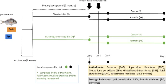
from: Cornish et al. (2017))………..21 Figure 7: Gilthead seabream Sparus aurata (adapted by Desouky and Jover (2016))………23 Figure 8: Global aquaculture production of Sparus aurata (tonnes) (Adapted from: FAO FishStat cited in FAO, 2015)……….……….24 Figure 9: Reaction between the formalin and the adenine base (adapted from Leal et al, 2016)……….26 Figure 10: Design of the experiment with gilthead seabream (Sparus aurata). Prior to formalin exposure, the fish were acclimatized during 3 weeks and after that the fish were divided in two groups and fed with a standard diet (S) and a macroalgae-enriched diet (A) during 2 months (dietary background settlement). The fish were exposed in two separated times, in the end of 2 month and 2 days later. Thereafter, the fish eyes and brain of the four treatments (S, SF, A, AF) are collect 4 and 18 days after the first formalin exposure. Eyes and brain samples were collected for antioxidants analyses, damage indicators and for the determination of acetylcholinesterase (AChE) activity………...31 Figure 11: Schematization of the sample preparation procedure for further biochemical quantification ……….………..…....32 Figure 12: Activities of catalase (CAT), superoxide dismutase (SOD), glutathione peroxidase (GPx), glutathione S-transferase (GST) and glutathione reductase (GR), together with the levels of total glutathione (GSHt), in the eyes of gilthead seabream (Sparus aurata) following 2 months of macroalgae dietary supplementation, as well as 4 and 18 days after a subsequent formalin treatment. Algae-supplemented groups (A) correspond to green bars, while groups fed with standard feeds (S) correspond to gray bars. Experimental groups submitted to formalin treatment are identified by abbreviations with an “F” after the letter identifying the dietary profile (i.e. SF and AF). The grey color represents the standard diet and the green color represents the macroalgae-enriched diet. Data were represented as mean ± standard error. Statistically significant differences between fish exposed to formalin and the respective unexposed group (for each feeding groups) are indicated by * (p < 0.05), *** (p< 0.001). Statistically significant differences between different dietary backgrounds (within formalin exposed fish and non-exposed fish) are identified by “d” (p < 0.05)……….38
3 Figure 13: Levels of lipid peroxidation (LPO) and protein oxidation (PO), in the eyes of gilthead seabream (Sparus aurata) following 2 months of macroalgae dietary supplementation, as well as 4 and 18 days after a subsequent formalin treatment. Algae-supplemented groups (A) correspond to green bars, while groups fed with standard feeds (S) correspond to gray bars. Experimental groups submitted to formalin treatment are identified by abbreviations with an “F” after the letter identifying the dietary profile (i.e. SF and AF). The grey color represents the standard diet and the green color represents the macroalgae-enriched diet. Data were represented as mean ± standard error. Statistically significant differences between fish exposed to formalin and the respective unexposed group (for each feeding groups) are indicated by * (p < 0.05). Statistically significant differences between different dietary backgrounds (within formalin exposed fish and non-exposed fish) are identified by “d” (p < 0.05)……….40 Figure 14: Activities of acetylcholinesterase (AChE) in the eyes of gilthead seabream (Sparus aurata) following 2 months of macroalgae dietary supplementation, as well as 4 and 18 days after a subsequent formalin treatment. Algae-supplemented groups (A) correspond to green bars, while groups fed with standard feeds (S) correspond to gray bars. Experimental groups submitted to formalin treatment are identified by abbreviations with an “F” after the letter identifying the dietary profile (i.e. SF and AF). The grey color represents the standard diet and the green color represents the macroalgae-enriched diet. Data were represented as mean ± standard error. Statistically significant differences between fish exposed to formalin and the respective unexposed group (for each feeding groups) are indicated by * (p < 0.05), ** (p < 0.01). Statistically significant differences between different dietary backgrounds (within formalin exposed fish and non-exposed fish) are identified by “d” (p < 0.05)……….41 Figure 15: Activities of catalase (CAT), superoxide dismutase (SOD), glutathione peroxidase (GPx), glutathione -s-transferase (GST), together with the levels of total glutathione (GSHt), in the brain of gilthead seabream (Sparus aurata) following 2 months of macroalgae dietary supplementation, as well as 4 and 18 days after a subsequent formalin treatment. Algae-supplemented groups (A) correspond to green bars, while groups fed with standard feeds (S) correspond to gray bars. Experimental groups submitted to formalin treatment are identified by abbreviations with an “F” after the letter identifying the dietary profile (i.e. SF and AF). The grey color represents the standard diet and the green color represents the macroalgae-enriched diet.Statistically significant differences between fish exposed to formalin and the respective unexposed group (for each feeding groups) are indicated by * (p < 0.05), ** (p < 0.01), *** (p< 0.001). Statistically significant differences between different dietary backgrounds (within formalin exposed fish and non-exposed fish) are identified by “d” (p < 0.05)………....43 Figure 16: Activities of lipid peroxidation (LPO) in the brain of gilthead seabream (Sparus aurata) following 2 months of macroalgae dietary supplementation, as well as 4 and 18 days after a subsequent formalin treatment. Algae-supplemented groups (A) correspond to green bars, while groups fed with standard feeds (S) correspond to gray bars. Experimental groups submitted to formalin treatment are identified by abbreviations with an “F” after the letter identifying the dietary profile (i.e. SF and AF). The grey color represents the standard diet and the green color represents the macroalgae-enriched diet. Data were represented as mean ± standard error. Statistically significant differences between fish exposed to formalin and the respective unexposed group (for each feeding groups) are indicated by * (p < 0.05). Statistically significant differences between different dietary backgrounds (within formalin exposed fish and non-exposed fish) are identified by “d” (p < 0.05)………..………..44
4 Figure 17: Activities of acetylcholinesterase (AChE) in the brain of gilthead seabream (Sparus aurata) following 2 months of macroalgae dietary supplementation, as well as 4 and 18 days after a subsequent formalin treatment. Algae-supplemented groups (A) correspond to green bars, while groups fed with standard feeds (S) correspond to gray bars. Experimental groups submitted to formalin treatment are identified by abbreviations with an “F” after the letter identifying the dietary profile (i.e. SF and AF). The grey color represents the standard diet and the green color represents the macroalgae-enriched diet. Data were represented as mean ± standard error. Statistically significant differences between fish exposed to formalin and the respective unexposed group (for each feeding groups) are indicated by * (p < 0.05), ** (p < 0.01). Statistically significant differences between different dietary backgrounds (within formalin exposed fish and non-exposed fish) are identified by “d” (p < 0.05)……….………...45
5
List of tables
Table 1: Compounds of marine macroalgae with neuroprotective related properties………...8 Table 2: Mean values (± standard error) of weight, length and estimated condition factor (K) in Sparus
aurata at the different sampling moments (i.e. dietary background corresponds to a sampling after 2
months of macroalgae dietary supplementation; 4 and 18 days corresponds to the days after the first formalin bath). S represents the standard feed; A indicates a macroalgae-enriched feed; experimental groups submitted to formalin treatment are identified by abbreviations with the “F” letter. The grey color indicates the treatments with the standard diet and the green color indicates the treatments with macroalgae-enriched feed………35 Table 3: Synopsis of the eyes brain oxidative stress profiles upon fish exposure to formalin. Significant alterations are marked by up and down arrows meaning increased or decreased levels of the parameter, respectively. Grey arrows represent the standard diet and the green arrows represent the MM-enriched diet……….…….57 Table 4: Mann-Whitney U test for the weight, length and condition factor between the different treatments in dietary background, 4 days and 18 days. The grey color represents the standard diet and the green color represents the macroalgae-enriched diet……….77 Table 5: Mann-Whitney U test for the dietary background and Kruskal-Wallis one way analyses of variance for the (CAT), superoxide dismutase (SOD), glutathione peroxidase (GPx), glutathione S-transferase (GST), glutathione reductase (GR), total glutathione (GSHt), lipid peroxidation (LPO), protein oxidation (PO) and acetylcholinesterase (AChE) at the end of the 4 and 18 days after the formalin bath between the different treatments in the eyes. Significative results are marked in bolt. The grey color represents the standard diet and the green color represents the macroalgae-enriched diet………77 Table 6: Mann-Whitney U test for the dietary background and Kruskal-Wallis one way analyses of variance for the (CAT), superoxide dismutase (SOD), glutathione peroxidase (GPx), glutathione S-transferase (GST), total glutathione (GSHt), lipid peroxidation (LPO) and acetylcholinesterase (AChE) at the end of the 4 and 18 days after the formalin bath between the different treatments in the eyes. Significative results are marked in bolt. The grey color represents the standard diet and the green color represents the macroalgae-enriched diet……….78 Table 7: Bioactive compounds with antioxidant capacity present in the three marine macroalgae classes………..79
6
1. INTRODUCTION
1.1. Marine macroalgae as a source of potential health-promoting compounds
Marine macroalgae (MM) can be subdivided in 3 different groups based on their pigmentation,
i.e, brown (Phaeophyta), red (Rhodophyta), or green
(Chlorophyta) (Figure 1). The pigment responsible for the brown color of the Phaeophyta species is fucoxanthin, the red color of the Rhodophyta species comes from phycobilins, whiles there are several pigments responsible for the green color (e.g., chlorophyll a and b, carotenes and xanthophylls) in the Chlorophyta species (Øverland et al., 2018). MM have been largely investigated both for novel ingredients and as a health-promoting food (Patarra et al., 2011; Mohamed et al., 2012). Interestingly, archaeological studies underpinned that in southern Chile, MM have been consumed since the 14,000 years, while in the 50s of the 20th century the MM production in
aquaculture had a breakthrough in Asia (Kim et al., 2017). In Europe, MM aquaculture is under development since 90’s of the last century with a current production of 54,000 tons per year, almost exclusively directed to human consumption, whereas just a small portion is used in fish diet (FAO, 2017). Worldwide, the MM aquaculture production represents 20 % of the total marine aquaculture production by weight (Bjerregaard et al., 2016).
Marine macroalgae can be considered a functional food, meaning that they have ingredients that confer an additional function to the food (usually related to health-promotion or disease prevention) (Kaur and Das, 2011; Wells et al., 2016). Indeed, MM have a number of characteristics that make them valuable as a functional food. They have low cytotoxicity, anti-inflammatory and antioxidant capacities, and are able to hinder cellular death, while having a low production cost (e.g. Pangestuti and Kim, 2011). Besides supporting the exploitation of MM by the food industry (about 80 % of the world production), those properties are esteemed to the pharmaceutical and cosmetic production, where currently MM have also been widely used (Pangestuti and Kim, 2011).
Gracilaria gracilis Fucus vesiculosus
Ulva rigida
Figure 1: Marine macroalgae
species, potentially used in a macroalgae enriched-diet or as a source of functional compounds. (http://www.algaebase.org)
7 In general, MM are rich in soluble dietary fibres, proteins, antioxidants, vitamins and polyunsaturated fatty acids (PUFAs), while having a low caloric value (Rupérez and Saura-Calixto, 2001). Marine macroalgae have a wide range of antioxidant compounds, but there is still a poor knowledge on the hypothetical extension of their benefits to organisms upon consumption, namely in humans (Wells et al., 2016). Hypothetically, MM antioxidants could act by limiting reactive oxygen species (ROS) in the digestive tract, thus decreasing oxidative stress on the gut microbiome and epithelial cells. Additionally, MM antioxidants could be transported into the blood for distribution throughout the body. In humans, the evidence of a direct transport is very limited, since there are no systemic studies on the digestive uptake of these compounds. In fish, it was showed that MM bioactive compounds affect the gut morphology and thickness of the goblet cells inducing changes in the digestion and absorption of nutrients in Nile tilapia (Silva et al., 2015) and rainbow trout (Heidarieh et al., 2012). However, there are no evidences in the way of how and if the enzymatic antioxidants are absorbed. Lipids are essential for all living organisms as components of membranes, energy storage compounds, and as cell signaling molecules (Eyster, 2007; Muro et al., 2014). MM could be a source of lipids to other organisms, namely of the long-chain polyunsaturated fatty acids and carotenoids. The most important PUFAs are the essential fatty acids eicosapentaenoic acid (EPA) and docosahexaenoic acid (DHA), as well as, their precursors α-linolenic acid (ALA) and docosapentaenoic acid (DHA) (Cottin et al., 2011). However, humans and other animals cannot convert ALA to EPA and DHA at required level. Thus, they need a food source rich in these essential fatty acids (Wells et al., 2016). Furthermore, there are evidences that enhanced DHA intake may improve the child cognitive performance, as well as, the visual acuity (Jensen et al. 2005, 2010; Imhoff-Kunsch et al. 2011). Also, DHA present in the MM can also produce cardiovascular protection in humans, thought the alteration of the plasma lipoproteins (2 g algal DHA day−1 over 4.5 month) (Neff et al., 2011). The carotenoids present in the MM are capable of free radical scavenging activity in vitro and in vivo (Wells et al., 2016).
There are a wide number of beneficial effects described for MM, namely a reduction on the human blood pressure (Wada et al., 2011) and an improvement of the immune system (Zhang et al., 2017). Specifically, the fucoidans of a brown MM Laminaria saccharina, Fucus vesiculosus and Fucus
spiralis demonstrated the capacity to induce apoptosis in cancer cell lines and to promote
macrophage-induced tumor cell death (Cumashi et al., 2007). A sulphate polysaccharide of a red MM Gigartina
skottsbergii revealed antiviral properties (Ahmadi et al., 2015), while phenols of a green MM Ulva lactuca have the capacity of scavenging the ROS and to confer protection against synaptic loss
8 (Mohamed et al., 2012). Indeed, the neuroprotective properties of MM have been also demonstrated, mostly related with their antioxidant properties, anti-neuroinflammation, together with a modulation of the cholinesterase activity (Pangestuti and Kim, 2011). Table 1 describes a number of compounds identified in MM with the previous properties in mammal models (e.g. rodents; mammalian cell lines). For example, fucoxanthin of Laminaria japonica afforded protection to rabbits’ retina against the oxidative damage inflicted by the visible light (Liu et al., 2016), while positive interferences with the cholinergic, dopaminergic and serotonergic systems were also described in humans and rodents (Rico et al., 2011). Moreover, a study with rats demonstrated that fucoidan administration can act as an anti-adhesion of leukocyte-endothelial attenuating the hypoxia-ischemia that will lead to brain damage (Uhm et al., 2003).
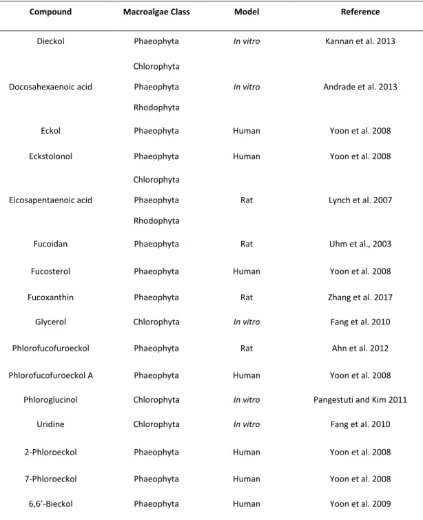
Overall, various MM species have valuable bioactive compounds such as vitamins, minerals, proteins, polysaccharides, steroids, dietary fibers, carotenoids, saturated and polyunsaturated fatty acids (Peixoto et al., 2016) that may have several phytochemicals with potential advantages for neurodegenerative conditions (Table 1).
9
Table 1: Compounds of marine macroalgae with neuroprotective related properties
1.2. Fish sensory organs and brain: considerations on biochemical condition and physiology
Most fish have developed highly sensory organs/structures, comprising different types of receptors/sensors, namely: (i) chemoreceptors (responsible, for example, for gustation and olfaction);
Compound Macroalgae Class Model Reference
Dieckol Phaeophyta In vitro Kannan et al. 2013
Docosahexaenoic acid
Chlorophyta Phaeophyta Rhodophyta
In vitro Andrade et al. 2013
Eckol Phaeophyta Human Yoon et al. 2008
Eckstolonol Phaeophyta Human Yoon et al. 2008
Eicosapentaenoic acid
Chlorophyta Phaeophyta Rhodophyta
Rat Lynch et al. 2007
Fucoidan Phaeophyta Rat Uhm et al., 2003
Fucosterol Phaeophyta Human Yoon et al. 2008
Fucoxanthin Phaeophyta Rat Zhang et al. 2017
Glycerol Chlorophyta In vitro Fang et al. 2010
Phlorofucofuroeckol Phaeophyta Rat Ahn et al. 2012
Phlorofucofuroeckol A Phaeophyta Human Yoon et al. 2008
Phloroglucinol Chlorophyta In vitro Pangestuti and Kim 2011
Uridine Chlorophyta In vitro Fang et al. 2010
2-Phloroeckol Phaeophyta Human Yoon et al. 2008
7-Phloroeckol Phaeophyta Human Yoon et al. 2008
10 (ii) photoreceptors (visual information); (iii) nociceptors (detection of noxious tissue-damaging stimuli); (iv) mechanoreceptors (mechanosensory lateral line system); (iv) electroreceptors (electrosensory lateral line system). The mechanosensory lateral line together with the auditory sense, compose the mechanical sense system that is involved in the control of fish body position.
The fish eyes are an important and highly specialize sensory organ that converts light/images into neural signals. This conversion is possible due to the existence of photoreceptors that are located in the retina (Ali et al., 1978). The eyes are located symmetrically or in the same side of the fish head and present a direct contact with the surrounding environment (Randall et al., 1997). This organ is mainly divided into: (i) cornea (outermost transparent layer of the eye); (ii) iris (controls the amount of light entering the eyes); (iii) lens (focuses light on the retina); (iv) sclera (forms the outer layer of the eyes and protects the inner structures of the eyes); (v) choroid (highly vascularized region between the sclera and the retina); (vi) retina (transparent laminar structure located at the back of the eyes) (Figure 2). The retina structure presents a colorful pigment that is called carotenoid, with the capacity to provide protection against several stressors, including UV radiation or ROS (de Carvalho and Caramujo, 2017). Carotenoids are only synthesized by macroalgae, plants, fungi and bacteria, while other organisms only obtain the necessary pool of these compounds through the diet (de Carvalho and Caramujo, 2017) (Table 1). The dietary supplementation with polyunsaturated fatty acids showed relevant benefits for the health of the eyes, in order to prevent diseases, like glaucoma and macular degeneration (Saccà et al., 2018). Furthermore, eyes are a tissue that can accumulate lipids, but the percentage of accumulated lipids, such as DHA, may vary between different fish species (Stoknes et al., 2004). In the same line, Hong et al. (2014) showed that freshwater fish can accumulate more lipids than the muscle tissue. Vitamins are a complex organic compounds present in the fish eyes structure and are essential to the normal metabolism and the lack of these compounds can lead to diseases (McDowell and Cunha, 1989). The vitamin C or ascorbic acid presents an effective antioxidant capacity (Bendich et al., 1986), which has been demonstrated in several experiments in vitro studies with several species, including fish. For example, vitamin C can reduce the formation of ROS, resulting in the reduction of the lipid peroxidation in in vitro cells (Padayatty et al., 2003).
Fish brain is relatively small in comparison with other vertebrates (generally one-fifteenth the brain mass of a similarly sized mammal) (Bone and Moore, 2009). The fish brain has main divisions that extend rostrocaudally, as following (examples of minor divisions/structures are indicated in brackets together with their function) (Evans and Claiborne, 1997) (Figure 2): (i) telencephalon (e.g. bulbus
11 olfactorius; mostly involved in olfaction) and diencephalon (e.g. epithalamus, thalamus and hypothalamus; mainly involved in the correlation of afferent and efferent impulses and modulation of the endocrine system); (ii) mesencephalon or midbrain (e.g. optic tectum and tegmentum; mainly involved in the vision and learning); (iii) metencephalon (cerebellum; mostly implicated in the coordination of muscular activities during swimming) and myelencephalon (medulla oblongata; mainly involved in sensory functions such as gustation and audition). The medulla oblongata and the tegmentum are collectively referred as brainstem (Evans and Claiborne, 1997). The spinal cord extends along the fish body. The cerebellum (motor learning and coordination, and, probably, cognition), optic tectum (orientation tasks, such as object identification and location) and telencephalon (olfaction) are examples of integrative centers in fish brain (Evans and Claiborne, 1997).
Figure 2: Schematic images of fish eyes (https://bit.ly/2Am0BFh) and brain (with the identification of the main areas
distributed rostrocaudally) (https://bit.ly/2Ri985Y).
Fish brain is composed by essential fatty acids that are important for the function maintenance and may represent two-thirds of the brain weight (Singh, 2005). Docosahexaenoic acid (DHA) is an essential fatty acid distributed in the cerebral cortex (Singh, 2005). This fatty acid is important for the growth and functional development of the brain and the DHA deficiency can be associated with deficits in learning (Horrocks and Yeo, 1999). Eicosapentaenoic acid (EPA) and docosapentaenoic acid (DPA) are other essential fatty acids that are important for the brain health, although less abundant than DHA (Dyall, 2015). In general, these three fatty acids (DHA, EPA and DPA) present important functions
12 against the cognitive decline or depressive symptoms, having a vital role in the neuroprotective capacity (Dyall, 2015).
Neurotransmission is the basis of neuronal communication, comprising a set of biochemical processes that are vulnerable to environmental toxicants exposure. The major neurotransmitter systems identified in fish were thoroughly reviewed by (Horzmann and Freeman, 2016) and are associated to the following classical transmitter substances, namely: (i) glutamate (the primary excitatory neurotransmitter and the most common in the bony fish brain); (ii) gamma aminobutyric acid (GABA - the major inhibitory neurotransmitter in the CNS); (iii) catecholamine neurotransmitters [dopamine (DA), norepinephrine (NE) and epinephrine - modulatory neurotransmitters]; (iv) serotonin (5-HT – a modulatory neurotransmitter); (v) acetylcholine (ACh - the major neurotransmitter in the parasympathetic nervous system); (vi) histamine (a non-synaptic neuromodulator); (vii) glycine (an inhibitory neurotransmitter).
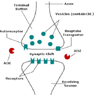
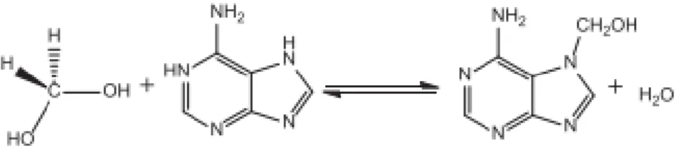
In detail, acetylcholine (ACh) is a fast-acting neurotransmitter at the neuromuscular junction and in the autonomic ganglia (Figure 3). Furthermore, there is an anatomical mismatch between the sites of ACh release and the location of cholinergic receptors (Picciotto et al., 2012). This neurotransmitter has the capacity to modulate the neurological function in the brain (Picciotto et al., 2012). Acetylcholinesterase is an important part of the cholinergic nervous system in fish (Whitehead et al., 2005). Furthermore, there is a necessity for maintenance of acetylcholine levels and, acetylcholinesterase (AChE) (Figure 3) is the enzyme responsible for that process, through the break of acetylcholine into acetate and choline (Soreq, 2001; Wilkinson et al., 2004). This enzyme is a target for several toxins, contaminants or pharmaceutical that may inhibit or reactivate its function, although, the main effect is the inhibition of the AChE (Araújo et al., 2016).
13
Figure 3: Basic functioning of cholinergic neurotransmission (adapted from Randall et al. (1997)).
The knowledge of the differences and similarities in the functional organization of nervous system between fish and other vertebrate groups (mammals, including) remains elusive (Evans and Claiborne, 1997). Several research challenges remain unsolved, namely regarding the homology of particular nuclei, neuronal connections, neurotransmitter distribution, as well as sensory pathways. Despite that, remarkable progresses have been made due to the investigation on the zebrafish neurobiology (Parng et al. 2002; Gerlai 2011; Rico et al. 2011; Kalueff et al. 2014; Alshabani et al. 2016). This model species shares the main neurotransmitter pathways with mammals and has similar neuroanatomy in many areas (e.g. spinal cord, hindbrain and retina), while some of the classical regions of the mammalian brain are not present (e.g. hippocampus, amygdala, and substantia nigra) with that organization (Horzmann and Freeman, 2016).
14 1.3. Oxidative stress and neurodegeneration in fish
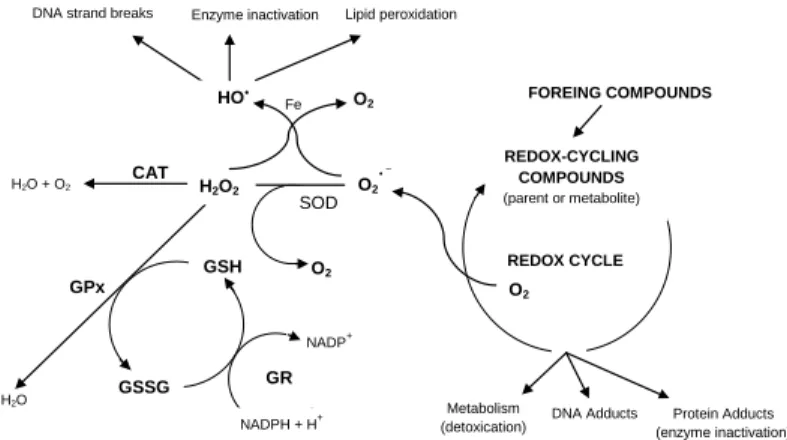
Oxygen is vital for all living cells, while it is potentially dangerous in excess. Therefore, oxygen is kept under a tight check by a complex system that regulates and monitors the usage and uptake of this element. Reactive oxygen species are produced in cells, primarily as a result of the aerobic metabolism. Numerous studies have been showing the advantageous biological effects of some ROS, such as superoxide anion radical (O2-), hydrogen peroxide (H2O2) and peroxyl radical (ROO). Important
physiological functions involving ROS include the following: 1) regulation of vascular tone; 2) sensing of oxygen tension and regulation of functions that are controlled by oxygen concentration; 3) enhancement of signal transduction from various membrane receptors; 4) oxidative stress responses that ensure the maintenance of redox homeostasis (Dröge 2003). While O2- is formed through
one-electron reduction of O2, H2O2 can be produced by the dismutation of O2- (catalysed by superoxide
dismutases) via the hydroperoxyl radical (HO2-) (Figure 4). OH is probably the most reactive and toxic
form, which is produced by the metal ion catalysed decomposition (e.g. iron or copper) of H2O2. In
mitochondria, oxygen takes part in glucose breakdown through oxidative phosphorylation, generating energy (in the form of ATP). Over 90% of cellular oxygen is consumed in mitochondria of unstressed cells, and therefore it is considered a major site of aerobic cellular ROS production (Han et al., 2001). Moreover, ROS generation occurs also by microsomal systems of the endoplasmatic reticulum (Winston et al. 1996).
Figure 4: Antioxidant defenses dynamic
in relation to ROS production under a scenario of exposure to exposure to a foreign compound to the cell with potential toxic action. SOD: superoxide dismutase; CAT: catalase; GPx: glutathione peroxidase; GR: glutathione reductase; GSH: reduced glutathione; GSSG: oxidised glutathione (adapted from Stegeman et al., 1992).
FOREING COMPOUNDS REDOX-CYCLING COMPOUNDS (parent or metabolite) O2 REDOX CYCLE Metabolism
(detoxication) DNA Adducts (enzyme inactivation) Protein Adducts
GPx H2O SOD O2 O2 GSH GSSG
DNA strand breaks Enzyme inactivation Lipid peroxidation
H2O2 HO O2 H2O + O2 CAT Fe GR NADPH + H+ NADP+
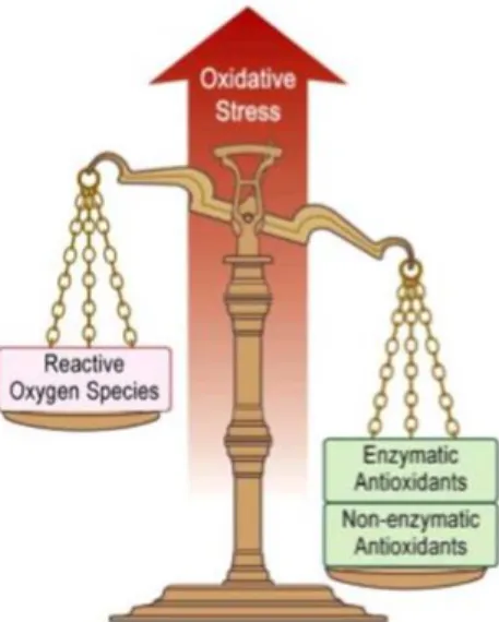
15 A regulated production of free radicals and the maintenance of ‘‘redox homeostasis’’ are essential for the physiological health of organisms (Ames et al., 1993). Despite that, a small proportion (2–3%) of free radicals may escape from the protective shield of antioxidant mechanisms, causing oxidative damage to biomolecules (DNA, proteins and lipids) (Halliwell and Gutteridge, 1999). The imbalance between generation and neutralization of ROS by antioxidant mechanisms is called oxidative stress (Davies, 1995) (Figure 5). During evolution, biological systems have been developing adequate enzymatic and non-enzymatic antioxidant mechanisms to protect their cellular components from oxidative damage. These include antioxidant enzymes, such as: i) superoxide dismutase (SOD); ii) catalase (CAT); iii) glutathione peroxidase (GPx); iv) glutathione reductase (GR); v) glutathione-S-transferase (GST) (Figure 4). Other molecules with antioxidant action such as glutathione, uric acid and ascorbate play also an important role in counteracting ROS (Martínez-Álvarez et al., 2005).
Figure 5: Oxidative stress occurs when the balance highly reactive radicals (oxidants) and antioxidants tips towards the
oxidants (Adapted from Lee et al., 2010).
In detail, CAT is a heme-containing enzyme that facilitates the removal of H2O2, which is
decomposed to O2 and water (Figure 4) (Schrader and Fahimi, 2006). CAT employs one molecule of
H2O2 as donor in the reduction of another H2O2, while peroxidases use other reductants. In animal cells,
the principal peroxidase is a selenium-dependent tetrameric cytosolic enzyme (GPx) that employs reduced glutathione (GSH) as a cofactor. GPx catalyses the metabolism of H2O2 to water with the
16
Figure 4) (Halliwell and Gutteridge, 1999). GSTs may play a dual protective role associated to their activity on conjugation of electrophilic compounds (or phase I metabolites) with GSH (Oost et al., 2003) and can also employ GSH in the reduction of a broad range of organic hydroperoxides, but it cannot reduce H2O2 (Wang and Ballatori, 1998). GR catalyses the transformation of GSSG to GSH with
the concomitant oxidation of NADPH to NADP+ (
Figure 4). Therefore, GR maintains the GSH/GSSG homeostasis under oxidative stress conditions (Winston and Giulio 1991). Glutathione represents the bulk of the non-protein thiols of the cells. Specifically, GSH (a tripeptide of glutamine acid, cysteine and glycine) may have a dual role in detoxification, namely: (i) as a key conjugate of electrophilic intermediates (mainly via GST activity in phase II metabolism); (ii) as an important antioxidant (Stegeman et al., 1992). Exposure of aquatic organisms to environmental toxic compounds may lead either to an increase or decrease of those enzyme activities, as well as of GSH. Specifically, in fish eyes and brain it have been described significative alterations in the antioxidant system of Liza aurata eyes when exposed to mercury (Pereira et al. 2016). In the same species, it has already been shown variations in the brain antioxidants when the fish was exposed to mercury (Cardoso et al., 2017). Furthermore, it was recorded an alteration in GSH content in Liza aurata subject to an accumulation of mercury (Mieiro et al., 2011). Furthermore, a glyphosate-base herbicide roundup transorb (RDT) can alter the antioxidant defenses in the neotropical fish Prochilodus lineatus (Modesto and Martinez, 2010). Besides that, the antioxidant system of juvenile common carps can be affected by the hexachlorobenzene (HCB) (Song et al., 2006). Overall, these findings pointed out that these antioxidants are able to underpin a challenging condition to fish.
17 The biochemical and physiological effects provoked by xenobiotics in the cells have been associated with increased fluxes of oxyradicals, which may reflect the emergence of lipid and protein damage (Oost et al., 2003). The process of lipid peroxidation comprises a set of chain reactions which can influence the PUFA, given that due to the double bounds, they are very sensitive to reactions by ROS. Lipid peroxidation products may form DNA adducts giving rise to mutations and altered patterns of gene expression (Marnett, 1999). Peroxidized membranes become rigid, with the consequence of loosing permeability and integrity. Furthermore, the proteins are one of the major targets ROS (Davies, 2016). The ROS leading to protein oxidation include radical species such as hydrogen peroxide, hypochlorous acid (HOCl), ozone (O3) and peroxynitrite (ONOO-) (Ahmad, 2017). Carbonyl groups are
produced on protein side chains when the proteins are oxidized (Ahmad, 2017). However, can be also introduced into proteins by secondary reaction on the nucleophilic side chains of cysteine, histidine, and lysine side chains, with aldehydes produced during lipid peroxidation (Dalle-Donne et al., 2003).
There are many natural sources of oxidative stress, such as UV radiation, heat shock and inflammation. On another hand, there are endogenous sources of ROSin cells, such as oxidizing enzymes (e.g. cytochrome P450 that can produce O2). Additionally, ROS production may increase by
cellular exposure to a wide range of toxicants, including organic contaminants and metals (Halliwell and Gutteridge, 1999). At this light, oxidative stress has been extensively considered in any research areas, ranging from aquatic toxicology to biomedical sciences.
Neuronal and glial cells are particularly vulnerable to the attack of ROS, which can eventually lead to neuronal damage and neurodegeneration in several species (review in Uttara et al. 2009). The reason for neuronal cell hypersensitivity towards oxidative stress is related both with anatomic and metabolic factors. For example, glial cells in the brain require more oxygen and glucose consumption to generate the ATP necessary for the normal brain functioning, associated with its demanding activity (in general terms, the brain keep all other organs active and under control). This makes the glial cells more susceptible towards oxygen overload, and thus free radical generation (review in Uttara et al., 2009). Just a small portion of oxygen is converted in ROS, as previously described. However, in an aged brain this percentage goes up related with the reduced surveillance of antioxidants and low regenerative capacity (review in Uttara et al., 2009). Moreover, the brain contains a high level of fatty acids, which are highly susceptible to peroxidation, while not being particularly enriched in antioxidant defenses. Actually, the brain has lower antioxidant activity in comparison with other tissues (e.g. it has just 10% of liver antioxidant activity) (review in Uttara et al., 2009).
18 1.4. Advantages provided by marine macroalgae-enriched feeds to farm fish: emphasis on antioxidant protection and neurotransmission
The use of different MM as a supplementary feed resource in animal production is not recent (Evans and Critchley 2014; Angell et al. 2016; Garcia-Vaquero and Hayes 2016; Makkar et al. 2016). So far, there are only a couple of studies that had investigated the role of MM dietary supplementation on antioxidant responses in fish. Peixoto et al. (2016) had assessed the benefits of MM enriched feeds (a mixture of Gracilaria, Fucus and Ulva in a total percentage of 7.5 %) on the liver antioxidant responses of Dicentrarchus labrax, as well as the individual benefits of a Gracilaria-supplemented diet. The MM mix diet increased GR activity when compared with fish under a control diet, while the Gracilaria-enriched feeds had increased lipid peroxidation together with GST activity. However, Fazio et al. (2016) showed that lipid peroxidation can be influenced by the fish feeding habits. According to these authors, herbivorous fish tend to have a lower lipid peroxidation than carnivores or omnivores fish. This information can possibly indicate that a feed supplemented with MM may reduce the levels of lipid damage. Even because, Magnoni et al. (2017) results, indicating that fish fed with 5 % of Gracilaria or Ulva had lower levels of lipid peroxidation when exposed to an acute hypoxia. The alterations in the antioxidant system were observed without a compromise of the fish growth in both tested alternative diets.
The increase of lipid peroxidation in liver of fish feed with Gracilaria is intriguing in the way that its suggests that supplementation with this MM increases the degradation of the lipid layer, as described in Peixoto et al. (2016). However, this observation was in accordance with a previous study that revealed a dose-dependent inhibition of the lipid accumulation in cells treated with Gracilaria
verrucosa extracts (Woo et al., 2013). Overall, Peixoto et al. (2016) had attributed the differences found
in antioxidant enzyme activities in seabass fed with a mix diet as the result of a synergistic effect between Gracilaria, Ulva and Fucus, even if this hypothesis remained unclear due to the lack of research on the effect of Ulva and Fucus on the antioxidant system in fish. Carotenoids that can be found in the MM, showed the ability to change the antioxidant system, through the decrease of SOD activity with increasing of the carotenoid concentration in the diet of Hyphessobrycon callistu (Wang et al. 2006).
As detailed in the previous section, the chief enzymes that restrict oxidative damage are the SOD, CAT and peroxidases that convert hydrogen peroxide to water. In eukaryotic algae, the superoxide
19 dismutase has Mn or Fe as cofactors, or some combination of Fe, Mn and Cu+Zn. Moreover, catalase has a Fe-containing heme cofactor, while peroxidases use a reductant to convert hydrogen peroxide to water. Some of these enzyme cofactors, such as Cu and Zn, and particularly Fe, are used in numerous metabolic pathways (Wells et al., 2016). Since the ingested antioxidant enzymes are digested in the intestine (Figure 6), it is believed that the only effect that the MM antioxidant enzymes can have is through the uptake of the associated metal cofactors across the intestinal epithelium (Wells et al., 2016). Nevertheless, the possible effects on the intestinal microbiome of any undigested enzyme, or of the released metal cofactors, have not been investigated yet. Additionally, MM can be a source of selenium (Schiavon et al., 2017) that is an essential element for Se-requiring glutathione peroxidase. However, the knowledge of the factors regulating Se content of algal foods and its availability to the organisms that consume MM remains elusive. Moreover, algae are composed by a wide variety of molecules capable of scavenging ROS, as observed in vitro and in vivo cells. These molecules comprise mainly: i) the water-soluble ascorbate (vitamin C); ii) the lipid-soluble α-tocopherol (vitamin E); iii) carotenoids such as astaxanthin; iv) phenolic compounds; v) sulfated polysaccharides (Halliwell and Gutteridge, 1999). Fish can absorb non-enzymatic antioxidants, such as, sterols or peptides present in the MM that in turn may influence the production of enzymatic antioxidants. For example, sterols, such as fucosterol form provided by several brown MM to fish showed antioxidant activity, resulting in increased activities of CAT, GPx or SOD (Kristinsson, 2014). Instead of serving to facilitate the control of ROS, some MM components can inhibit their production, but most studies do not properly distinguish between the decreased production and increased removal of ROS (Wells et al., 2016). There are substantial knowledge gaps on the efficacy of antioxidant properties of MM at several levels, ranging from the characterization among species, through the effects on gut microbiota and transport across the gut lumen to their impacts on fish physiology.
One of the most interesting neurological protective effects of the MM is the influence in the cholinergic activity, as Suganthy et al. (2010) reported the neuroprotective effect of eight different MM (Enteromorpha intestinalis, Dictyota dichotoma, Ulva reticulata, Gracilaria edulis, among others). The compounds in the eight MM were capable to inhibit the cholinesterase activity. Hodges (2006) demonstrated that inhibition of AChE plays a key role enhancing the cholinergic neurotransmission in the brain and reduce the aggregation of β-amyloid. DHA is critical for visual acuity, while it is implicated in the activities that underlie cognitive development, such as modulating synaptic efficiency, transmission speed, and myelination processes, as described in section 1.2. Generally, the dietary
20 sources of DHA to farmed fish are in the form of fish meal and fish oil that are dominant in the compounds feed for fish (Naylor et al., 2000). Marine macroalgae can be an alternative source of DHA to fish, as demonstrated in Kumari et al. (2010). The dietary intake of DHA showed benefits in the neurovascular and neurological health in zebrafish (Sierra et al., 2012). Furthermore, studies with EPA and DHA applied extracellularly raise the stimulatory thresholds of CA1 neurons in hippocampal slices (Xiao and Li, 1999) and the arachidonic acid (ARA) inhibits sodium currents and synaptic transmission (Fraser et al., 1993). Furthermore, DHA treatments in zebrafish can modulate the brain to be more resistance to neurotoxic insults, such as contaminants of pharmaceuticals (Sierra et al., 2012).
The benefits of MM enriched aquafeeds on fish brain and sensory organs were not addressed, yet. Despite that, neuroprotection afforded by MM to fish can be speculated at the light of mammals’ findings (details in section 1.1 and Table 1), which is also supported by the homology between fish and mammals regarding the main neurotransmitter pathways and neuroanatomy (section 1.2). Since MM are at the base of the aquatic trophic chains, representing an important natural food source to wild fish, particularly for some species that seem to be well adapted to its consumption (Norambuena et al., 2015). Sparus aurata is probably a good example, since in the wild it was found that its diet was composed by small crustaceans and molluscs, however, will also consume algae (Alarcón et al., 2001). Only a few studies had been focused on the beneficial effects of MM on fish sensory organs and brain. Marine macroalgae had lutein and zeaxanthin, two carotenoids, with beneficial effects (Ma and Lin, 2010). Abdel-Aal et al. (2013) described the existence of this two compounds in the retina cells. Lutein and zeaxanthine are transported into retina in the same ratio that to the plasma, and then transferred to macula where lutein is preferentially converted into meso-zeaxanthine, a non-dietary carotenoid that is not found in the serum, but only in the retina. These evidences suggest the importance of lutein, zeaxanthin and meso-zeaxanthin in the good eyes health. Furthermore, carotenoids present in green and brown MM have a beneficial effect in the fish eyes (Abdel-Aal et al., 2013).
Farmed fish can have smaller brain in relation with their wild conspecifics, as far as was reported for Oncorhynchus mykiss (Marchetti and Nevitt, 2003; Kihslinger, 2005). Due to the existence of bioactive compounds in the MM that are vital for the good brain functioning and development, such as DHA and EPA (already described above), it is expected that feed the farm fish with MM enriched-meals to farmed fish could counterbalance this problem.
21
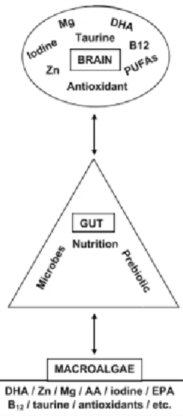
Figure 6: Schematic illustration of the microbiota–gut–brain axis and how the essential brain nutritional elements and
antioxidants are related to the contents of marine macroalgae. AA: Amino acid; B12: Vitamin B12; DHA: Docosahexaenoic acid;
EPA: Eicosapentaenoic acid; Mg: Magnesium; Zn: Zink (Adapted from: Cornish et al. (2017)).
The advantages of using MM can also include high growth rate, potential cultivation in saltwater, together with an independence of arable land and industrial fertilization (Øverland et al. 2018). In fact, MM are starting to be used as a novel food ingredient in pisciculture (Batista, 2008) with the main aim of enhancing fish health and aquaculture productivity. Fish aquaculture has been increasing its production, which is followed by the need of improving organism’s welfare. The use of MM, under this context, has an added value particularly considering several farming protocols that can severely affect fish physiology, as already documented for the immune system (Queiroz et al., 2014; Peixoto et al., 2016). In vitro, all three groups of macroalgae (i.e., red, green and brown) have shown antimicrobial properties and inhibitory effects against fish pathogen (Øverland et al. 2018). Conversely, still there is limited information on the effect of dietary macroalgae supplementation on health of farmed fish in vivo (Øverland et al. 2018). Even though, there is an increasing interest on the use of MM as a bioactive component in functional feeds for fish. In this direction, it was reported an improvement
22 in growth performance and a lower lipid content in the carcass of Nile tilapia feed with 5 % of Ulva (Ergün et al., 2009). Additionally, previous studies suggested that feed supplemented with MM could mitigate fish stress responses and improve vitality (Mohamed et al., 2012), while increasing illness (Araújo et al. 2016) resistance together with the flesh quality (Valente et al., 2016), representing unquestionable advantages for the aquaculture industry (Luna-Acosta et al., 2011).
1.5. Cultivation and nutrition of gilthead seabream (Sparus aurata)
The gilthead seabream, Sparus aurata (Figure 7), is a teleost species belonging to the Sparidae family. This species can be found in the Atlantic Ocean, from the British islands, Gibraltar Strait to Cape Verde, around the Canary Islands, and in all the Mediterranean Sea. It is an eminently coastal species, living on rocky or sandy bottoms. Gilthead seabream is a sedentary fish that migrates alone or in small aggregations, moving in early spring towards protected coastal waters, in order to find abundant food and mild temperatures. The feeding habits are based on shellfish (bivalves and gastropods) and crustaceans, although can also feed on small fish and macroalgae (Madeira et al., 2016).
Figure 7: Gilthead seabream Sparus aurata (adapted by Desouky and Jover (2016))
The Sparus aurata is a very suitable species for aquaculture in the Mediterranean region, as well as in Portugal, due to their good market price, high survival rate and feeding habits (which are relatively low in the food chain), as well as due to the fact that it is possible to control their whole life in captivity (FAO, 2004). In the early years of farm fish, gilthead seabream was traditionally cultured extensively in coastal lagoons and saltwater ponds (FAO, 2004). In this type of systems, it is generally reared with mullets, seabass or ells and feed naturally. However, this species can also be reared in the semi-intensive or intensive systems. In semi-intensive conditions, the natural diet is supplemented with
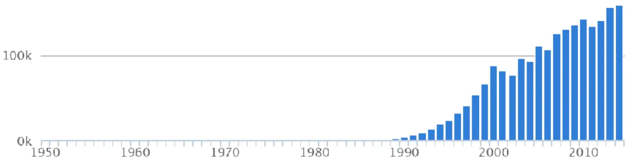
23 a commercial feed. In fact, the commercial food makes it possible the creation of polycultures. Furthermore, Sparus aurata, are widely used in Portugal as an accessorily herbivorous in polyculture system with european seabass (Dicentrarchus labrax), in order to prevent the excessive growth of macroalgae in the pounds, a frequent problem in seabass monoculture pounds (Afonso, 2016). In the intensive systems, gilthead seabream is fed exclusively with commercial pellets (European commission, 2012). Sparus aurata has been highly farmed in Europe, being its production growing globally (FAO, 2015) (Figure 8).
1.6. Potential hazards to fish in aquaculture: the formalin case
It has been estimated that fisheries and aquaculture supplied the world with around 110 million metric tons of food fish per year (FAO, 2010). Aquaculture production supports 47% of this supply, with intensive aquaculture gaining a chief importance to meet the population needs. Intensive aquaculture is a system characterized by a high density of production of aquatic species (level of production up to 200 tonnes ha-1 year-1), with a high level of control, technology and high production efficiency and
nutritionally complete feeding, using man-made culture systems (FAO, 1988). Maintaining the high level of production is highly demanding, which had led to the use of several chemicals along the process. In fact, there is a variety of chemicals that are frequently used in the aquaculture production of fish, including: (i) disinfectants (e.g., hydrogen peroxide, malachite green, formalin); (ii) antibiotics