RESUMO.- [Nefrite intersticial em suínos abatidos no Estado de Mato Grosso.] O propósito desse estudo foi ava-liar as lesões histológicas observadas em rins condenados por nefrite pelo Serviço de Inspeção Federal, em dois fri-goríicos de Mato Grosso, Brasil. Foram coletados 400 rins condenados por nefrite e submetidos aos exames de histo-logia, imuno-histoquímica (IHC) para Circovirus suíno Tipo 2 (PCV2), imunoluorescência direta (IF) para Leptospira sp. e reação em cadeia pela polimerase (PCR) para detecção
1 Received on August 30, 2011.
Accepted for publication on November 28, 2011.
2 Laboratório de Microbiologia Veterinária e Biologia Molecular,
Facul-dade de Agronomia e Medicina Veterinária (Famev), UniversiFacul-dade Federal de Mato Grosso (UFMT), Avenida Fernando Corrêa da Costa 2367, Bairro Boa Esperança, Cuiabá, MT 78060-900, Brazil. *Corresponding author: [email protected]
3 Embrapa Suínos e Aves, BR153 Km 110, Vila Tamanduá, Cx. Postal 21,
Concórdia, SC 89700-000, Brazil.
4 Laboratório de Patologia Veterinária, Famev-UFMT, Cuiabá, MT.
Interstitial nephritis of slaughtered pigs in the State of Mato
Grosso, Brazil
1João X. Oliveira Filho2, Daphine A.J. de Paula2, Nelson Morés3, Caroline A. Pescador4,
Janice R. Ciacci-Zanella3, Arlei Coldebella3, Valéria Dutra2 and Luciano Nakazato2*
ABSTRACT.- Oliveira Filho J.X., De Paula D.A.J., Morés N., Pescador C.A., Ciacci-Zanella J.R., Coldebella A., Dutra V. & Nakazato L. 2012. Interstitial nephritis of slaughtered pigs in-the State of Mato Grosso, Brazil. Pesquisa Veterinária Brasileira 32(4):303-318. Laborató-rio de Microbiologia Veterinária e Biologia Molecular, Faculdade de Agronomia e Medici-na Veterinária, Universidade Federal de Mato Grosso, Av. FerMedici-nando Corrêa da Costa 2367, Bairro Boa Esperança, Cuiabá, MT 78060-900, Brazil. E-mail: [email protected]
This study evaluated histological lesions in kidney samples from pigs with nephritis in two slaughterhouses in the State of Mato Grosso, Brazil. Four hundred samples were sub-jected to histology, anti-porcine circovirus type 2 (PCV2) immunohistochemistry (IHC), an-ti-Leptospira sp. immunoluorescence (IF), and polymerase chain reaction (PCR) for PCV2, porcine parvovirus (PPV), and Torque teno virus type 1 and 2 (TTV1, TTV2) detection. His-tological lesions were found in 81% of the samples, and mononuclear interstitial nephritis was the most frequent lesion (77.50%). A follicular pattern was observed in 40.97% of the interstitial nephritis lesions. PCV2, PPV, TTV1, and TTV2 were identiied in the kidneys by PCR in 27.25%, 28.50%, 94%, and 87.5% of the samples, respectively. Leptospira sp. was not detected through IF. Infection by PCV2 (PCR) and the presence of histological lesions (P=0.008) and giant cells (P=0.0016) were signiicantly associated. An association was observed between the TTV2-TTV1 co-infection (P<0.0001) and the risk for pathogenesis. These indings indicated that PCV2, PPV, TTV1, and TTV2 were widely distributed among pigs in the local farms and that the presence of these agents should be considered in the differential diagnosis of kidneys with interstitial nephritis in pigs.
INDEX TERMS:Pigs, interstitial nephritis, porcine circovirus type 2, torque teno virus.
de PCV2, Parvovirus suíno (PPV) e Torque teno vírus Tipo 1 e 2 (TTV1 e TTV2). Foram observadas lesões histológi-cas em 81% das amostras, sendo nefrite intersticial mono-nuclear a mais freqüente (77,50%). Das lesões de nefrite intersticial encontradas, 40,97% apresentaram padrão folicular. Através da PCR foi observada ampla distribuição dos agentes (PCV2, PPV, TTV1 e TTV2) nas propriedades e municípios, com ocorrência de 27,25%, 28,50%, 94% e 87,50%, respectivamente. Leptospira sp. não foi detectada através da IF. Houve associação signiicativa da infecção do PCV2 com presença de lesão histológica (P=0,008) e de cé-lulas gigantes (P=0,0016). Também houve associação en-tre a co-infecção TTV2 e TTV1 (P<0,0001). Esses achados indicam que os vírus PCV2, PPV, TTV1 e TTV2 devem ser considerados no diagnóstico diferencial de rins com nefrite intersticial em suínos.
INTRODUCTION
Mononuclear interstitial nephritis (MIN) is character-ized by the presence of “white spotted areas” on the renal surface and represents the main cause for condemning kidneys in clinically healthy slaughtered pigs (Drolet et al. 2002). However, numerous etiologic agents may be in-volved (Drolet et al. 2002, Martinez et al. 2006).
Viral agents, such as the porcine reproductive and res-piratory syndrome virus (PRRSV), porcine circovirus type 2 (PCV-2), and porcine parvovirus (PPV) might be involved in the etiology of MIN (Cooper et al. 1997, Drolet et al. 2002). Bacterial agents might also be related, for example, Leptos-pira interrogans, which represents an important potential risk to the health of workers in the production line (Baker et al.1989).
Considering the risks towards public health and econo-mic losses in the pig industry resulting from decreased ani-mal performance and organ condemnation, the association between histological lesions and etiologic agents in kid-neys that were condemned by the Brazilian Federal Inspec-tion Service (SIF), based on the presence of nephritis were evaluated in two slaughterhouses in Mato Grosso, Brazil.
MATERIALS AND METHODS Collection of samples.
Four hundred kidneys, condemned by the SIF due to the pre-sence of nephritis, were sampled in two slaughterhouses (FA and
FB) located in North and Southeast meso-regions of Mato Grosso--Brazil, respectively. The sample population was deined accor-ding to the method described by Thrusield (2004) with a coni-dence level of 95%, expected prevalence of 50%, and a maximum margin of error of 5%.
The average daily slaughter, from June 2003 to December 2008 in FA, and from January 2006 to December 2008 in FB, was 927 and 2,341 pigs, respectively. Random sampling was simple and proportional to the number of animals slaughtered in each slaughterhouse, totalling 152 kidneys from FA (19 kidneys/col-lection day) and 248 kidneys in FB (31 kidneys/colkidneys/col-lection day). All samples were obtained from 33 different farms involved in in-tensive production; the pigs were slaughtered at approximately the average weight of 125 kg and 180 days of age.
The kidneys were classiied in grades from 0-3 according to the presence of white spots on the renal surface: grade 0 (zero), normal kidney; grade 1, mild lesions with the presence of up to ive grayish-white spots; grade 2, moderate lesions with the pre-sence of six to 10 grayish-white spots; and grade 3, serious lesions presenting more than 10 grayish-white spots, and/or enlarged kidneys, and/or diffused ibrosis in the capsule with irregular re-nal surface (Fig.1).
Histological and immunohistochemical (IHC) analyses.
Histological examination covered the areas in which the white spots were seen, from cortical to medullary portions. Samples with microscopic alterations (n=324) were subjected to immuno-histochemistry (IHC) for PCV2, as described by Ciacci-Zanella et al. (2006). The anti-PCV2 ORF2 region polyclonal antibody was used as the primary antibody at the dilution of 1:1500. This
body was produced in rabbits (prepared at the Iowa State Univer-sity and kindly provided by Professor David Driemeier, UFRGS). Lymph node samples from speciic pathogen free animals (SPF) served as negative controls. Lymph node samples from PMWS af-fected pigs (conirmed by nested-PCR) were positive controls.
Immunoϐluorescence (IF) for Leptospira sp.
Imprints, taken during the collection of samples were subse-quently ixed in acetone p.a. for IF according to Ellis et al. (1982). The LEP-FAC (NVSL) polyclonal antibody, prepared in rabbits and conjugated with luorescein isothiocyanate, was used at the dilu-tion of 1:2500. Smears of pure cultures of L. interrogans serovar Pomona ixed in heat and acetone p.a. were positive controls.
Polymerase Chain Reaction (PCR).
Genomic DNA was extracted from 100 mg of kidney tissue af-ter Proteinase K digestion, as described by Sambrook & Russell (2001). Detection of viral DNA through PCR followed the methods described by Fenaux et al. (2000) (PCV2) and Kim & Chae (2004) (PPV), and through Nested-PCR described by Kekarainen et al. (2006) (TTV2 and TTV1). Samples with proven identity through genome sequencing were the positive controls and ultra pure wa-ter samples were the negative controls. Ampliied PCR products were separated by agarose gel electrophoresis (1.0%), stained with ethidium bromide, and analyzed in an UV transilluminator.
Statistical analysis.
Data related to the presence of agents and histological lesions were statistically analyzed. Initially, a descriptive analysis of the data by frequency tables was executed. The association between variables was calculated using the Yate’s corrected Chi-square test (χ2) and the Fisher's exact test. Associations with 5% probability
level were considered signiicant. The odds ratios (OR) were cal-culated with a conidence interval (IC) of 95% for the signiicant associations. These analyses were performed in the GraphPad InStat software for Windows.
RESULTS Pathological evaluation
Thesamples classiied as grade 3 presented the higher frequency of nephritis (190/400 - 47.50%) in the macros-copic evaluations, followed by grade 1 (128/400 - 32%) and grade 2 samples (82/400 - 20.50%). In the histological evaluations, 19% (76/400) of the kidneys showed no ne-phritis changes and were therefore normal. There was no association between the macroscopic degree of lesions and the presence of microscopic lesions (P=0.1175).
MIN showed the highest frequency (310/400 - 77.50%) among the samples with histological lesions (324/400 - 81%) (Table 1). Among these, 127/310 (40.97%) psented a follicular pattern, and the inlammatory re-sponse occurred in the form of lymphoid follicles (Fig.2). Other inlammatory changes such as giant cells (13/400 - 3.25%), glomerulitis (7/400 - 1.75%), and membrano-proliferative glomerulonephritis (5/400 - 1.25%) were also observed.
Histological lesions were mostly observed in the corti-cal region (312/400 - 78%), followed by those presented in the medullary region (9/400 -.25%) and renal pelvis (3/400 - 0.75%). Kidney lesions were predominantly of multifocal distribution (247/400 - 61.75%).
Aetiological ϐindings
Viral antigens and nucleic acids from PCV2, PPV, TTV1, and TTV2 were detected in 109/400 (27.25%), 114/400 (28.50%), 376/400 (94%), and 350/400 (87.5%) in the analyzed kidneys, respectively. Positive samples for Leptos-pira sp. were not detected. PCV2 and PPV positive samples were identiied in 23/33 (66.67%) and 27/33 (81.82%) in the samples from the farms and in 11/13 (84.62%) and 12/13 (92.34%) in the samples from the municipalities, respectively. TTV1 and TTV2 positive samples were detec-ted in 33/33 (100%) in the samples from the farms and municipalities (Table 2).
Table 1. Histological lesions in porcine kidneys condemned by the Federal Inspection Service, as presenting nephritis
(n=400), in the state of Mato Grosso, Brazil, from March to April 2009
Histological alteration Presence
(n) (%)
Without alteration 76 19.00%
With alteration 324 81.00
MINa 183 45.75
MINa with follicular pattern 127 31.75
Interstitial ibrosis 27 6.75
Presence of protein inside renal tubules 22 5.50
Presence of giant cells 13 3.25
Tubular degeneration 9 2.25
Glomerulitis 7 1.75
Membranoproliferative glomerulonephritis 5 1.25
Eosinophilic iniltrate 3 0.75
Infarct 1 0.25
a MIN = Mononuclear interstitial nephritis.
Table 2. Geographic distribution of Porcine Circovirus Type 2 (PCV2), swine Parvovirus (PPV), and Torque teno virus Type 1 and 2 (TTV1 and TTV2) in pig kidneys condemned by the Federal Inspection Service, as presenting nephritis in
Mato Grosso, Brazil
Mesoregion Municipalities Properties Number of positive farms PCV2 PPV TTV1 TTV2
North Diamantino 01 1 1 1 1
Lucas do Rio Verde 04 3 4 4 4
Nova Mutum 04 1 3 4 4
Novo Horizonte 01 0 1 1 1
Santa Rita do Trivelato 01 1 1 1 1
Sorriso 05 4 3 5 5
Tapurah 09 6 7 9 9
Vera 01 1 1 1 1
Southeast Campo Verde 02 2 2 2 2
Itiquira 01 1 1 1 1
Pedra Preta 01 1 1 1 1
Poxoréu 02 2 2 2 2
Rondonópolis 01 0 0 1 1
Total N 33 23 27 33 33
% 100 66.67 81.82 100 100
with histological lesions to be positive for PCV2 by IHC was 6.5 times higher when these samples were also positive for PCV2 by PCR (Table 3).
A signiicant association was also observed between the presence of PCV2, detected by PCR in samples with mi-croscopic lesions (P=0.008), and the presence of giant cells (P=0.0016). The probabilities of kidney samples to present histological lesions and giant cells were 2.57 and 6.45 times higher when they were positive for PCV2 by PCR, respectively.
Signiicant associations were observed between the pres-ence of TTV1 and the prespres-ence of glomerulitis (P=0.027) and tubular degeneration (P=0.005); similarly, between the presence of TTV2 and the presence of protein in the renal tubules (P=0.012) and tubular degeneration (P=0.0002). Co-infection with TTV1 and TTV2 was observed in 84.25% (337/400) of the samples showing signiicant association (P<0.0001). In the studied samples, the probability of TTV2 infection was 7.31 times greater when the animal was also infected by TTV1 (χ 2 =22.7; CI 95%: 3.0-17.4).
DISCUSSION
The present study contributes to the pathological and aetio-logical characterization of nephritis lesions identiied and condemned by SIF in healthy pigs from slaughterhouses. The number of healthy kidneys condemned as presenting nephritis, but that were considered normal in the histolo-gical evaluation was signiicant (19%). The absence of as-sociation between the macroscopic degree of lesions with the presence of microscopic lesions, and the frequency of kidneys classiied as presenting nephritis without histolo-gical lesions explain the dificulty in distinguishing nephri-tis from vascular disorders, which may cause alterations in the colour of the organ’s surface and thus, confuse the evaluator in the slaughter line.
Different results were obtained by Martínez et al. (2006) in a study with kidneys from pigs from a slaughter in eastern Spain. They reported that 6.9% of the kidneys considered normal in the histological analysis were classi-ied as presenting nephritis in the macroscopic assessment resulting in signiicant association between nephritic ma-croscopic lesions and the presence of mima-croscopic lesions (p < 0.005). These results indicate the need to enhance the technical criteria for assessing such macroscopic evalua-tions, as well as the training and monitoring of professio-nals responsible for the inspection. Furthermore, the re-sults supported the importance in conducting pathological studies in order to characterize lesions observed in slau-ghtered animals.
Mononuclear interstitial nephritis was the main mi-croscopic lesion detected in this study (77.50%), 40.97% of which presented a follicular lesion pattern. Similar results
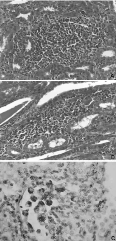
Fig.2. (A) Mononuclear nephritis with moderate focal follicular pattern in pig. HE, obj.40x. (B) Moderate focal mononuclear interstitial nephritis in pig. HE, obj.40x. (C) Positive PCV-2 immunolabeling within the renal tubule. Avidin-streptavidin-peroxidase, obj.40x.
Table 3. Association between the presence of PCV2 detected by PCR and the presence of PCV2 detected by
immunohis-tochemistry and histological changes in porcine kidneys condemned by the Federal Inspection Service as presenting
nephritis in two slaughterhouses in Mato Grosso, Brazil
Variable PCV2 – PCR P χ 2 ORb CIc 95%
Samples Positive (%)
PCV2-immunohistochemistry
Negative 283 70 (24.73%) < 0.0001 30.17 6.5 3.2 – 13.3 Positive 41 28 (68.29%)
Histological lesion
Negative 76 11 (14.5%) 0.008 6.95 2.56 1.29– 5.0 Positive 324 98 (30.35%)
Presence of giant cells
Negative 387 100 (25.84%) 0.0016 10.38a 6.45 1.94 – 21.43 Positive 13 9 (69.23%)
were previously observed and the reported prevalence ranges from 61.87 to 100% (Neves 1985, Drolet et al. 2002, Martínez et al. 2006). PCV2 infection has been associated with MIN (Segalés et al. 2004) and lymphohistiocytic inil-trate has been linked to the presence of giant cells (Sega-lés & Domingo 2002). Similarly, in the present study, PCV2 infection was associated with the presence of giant cells (P=0.0016; OR=2.56).
Leptospira sp. was not detected through bacterial iso-lation in the analyzed samples; Drolet et al. (2002) report similar results. This may be a result of infection control programs in the farms through mass vaccinations, better condition of hygiene in the premises, and the use of antibio-tics (Ribotta et al. 1999). Martínez et al. (2006) observed only two positive samples for Leptospira sp. by IF in a simi-lar study. However, in some studies executed in the 1980s, researchers showed an association between the presence of this bacterium and the occurrence of MIN using serology, immunoluorescence techniques, and culture of pig sam-ples (Grégoire et al. 1987, Baker et al. 1989). The results presented here suggested that a signiicant improvement in the control of this important zoonosis in swine culture has taken place in the recent decades.
This study indicated that the analyzed agents were widely distributed in Mato Grosso (Brazil). It is important to emphasize that the number of samples infected with PCV2 may be underestimated because the agent may no longer be present in chronic lesions (Maxie & Newman 2007). Furthermore, kidneys have not been the targeted organs for PCV2 infection (Segalés & Domingo 2002), ex-cept in porcine dermatitis and nephropathy syndrome cas-es (Choi et al. 2002).
Even with the lower sensitivity of the IHC technique to detect PCV2 in comparison with PCR, IHC is useful to loca-lize the agent in the tissue and demonstrate the pathogenic role of PCV2 in the occurrence of porcine nephritis. PCV2 was observed in the interstitium, renal tubules, glomeruli, in the giant cells, and in the cytoplasm of histiocytes. These results support the assumption that PCV2 can replicate in the functional structures of the kidneys (tubules and ne-phrons) as it has also been reported by Sarli et al. (2008). Hence, PCV2 might be involved in the occurrence of MIN.
The presence of PPV in the samples was relevant; how-ever, no signiicant association was observed between other agents and the presence of histological lesions. This result is contrary to the results reported by Drolet et al. (2002) who demonstrated signiicant association between interstitial nephritis lesions and the presence of PCV2 (P=0.0074) and PPV (P<0.0001) with an OR=3.4 and 7.5, respectively. These authors also observed that the OR in-creased to 22.7 (P<0.0001) when there was co-infection with both viruses. Other studies reported association be-tween PCV2 and PPV and the development of diseases such as post weaning multisystemic wasting syndrome in pigs (PMWS), renal lesions, and reproductive failures (Allan et al. 1999, Kim & Chae 2004, Fernandes et al. 2006, Pescador et al. 2007).
The high occurrence of TTV infection (20-73%) obser-ved in these pigs agrees with results previously reported
(Leary et al. 1999, Bigarré et al. 2005). Such a high pre-valence can be explained by the containment conditions that facilitates transmission (Deng et al. 2000, Gerner et al. 2000, Martínez-Guinó et al. 2009) and the ubiquitous cha-racteristic of TTV.
Signiicant associations were observed between infec-tion by TTV1 and the presence of glomerulitis and tubu-lar degeneration, and between infection by TTV2 and the presence of protein in renal tubules and tubular degene-ration. These results are not biologically plausible because infections by TTV1 and 2 were considered as a protective factor in the occurrence of these lesions in the odds ratio analyses. In fact, in this study, the high occurrence of TTV was associated with a low occurrence of these lesions.
Associations between TTV and the other analyzed agents were not observed. It was observed that the infec-tion by a genogroup of TTV represented a risk factor for the occurrence of co-infection with another genogroup (OR=7.31) and it might be associated with the pathogeni-city of one of the agents. However, Kekarainen et al. (2006) compared the TTV infection in pigs with and without PMWS, and observed association (P<0.05) between TTV infection and the presence of PMWS. These authors also demonstrated associations between TTV2 and the presen-ce of PMWS and between TTV1 and pigs with no syndrome. The TTV viral load in pigs, which was not analyzed in the present study, can be another factor related to the occur-rence of lesions because of the ability of TTV to modulate the immune system through the inhibition of INF-α; a gre-ater effect was observed with 1000 ng of TTV DNA when compared with 500ng (Martínez-Guinó et al. 2010).
The present study demonstrated that a signiicant num-ber of kidneys, considered as presenting nephritis, did not present histological lesions (19%). In addition, this study demonstrated that several histological changes observed in the kidneys with MIN were associated with the infec-tion of viral agents such as the PCV2, PPV, TTV1, and TTV2. Although Leptospira sp. was not detected in the analyzed samples, constant monitoring is important because of its zoonotic potential and the health problems that can result from this infection.
Acknowledgements.- To the slaughterhouses’ owners/managers and to SIF-MT, for enabling the execution of this study, and to CAPES for the scholarship granted. The authors are also thankful to the technical assis-tance in the immunohistochemistry analyses provided by Kelen R.A. Baldi, Animal Health Diagnostic Center (CEDISA), Brazil, and to Neide L. Simon and Giseli Ritterbusch for the technical assistance in the Torque Teno Vi-rus PCR detection, Embrapa Swine and Poultry Research Center (CNPSA), Concórdia, SC.
REFERENCES
Allan G., Kennedy S., Mcneilly F., Foster J.C., Ellis J., Krakowka S., Meehan B.M. & Adair B.M. 1999. Experimental reproduction of severe wasting disease by co-infection of pigs with porcine circovirus and porcine par-vovirus. J. Comp. Pathol. 121:1-11.
Baker T., McEwen S., Prescott J. & Meek A. 1989. The prevalence of leptos-pirosis and its association with multifocal interstitial nephritis in swine at slaughter. Can. J. Vet. Res. 53:290-294.
Choi C., Kim J., Kang I. & Chae C. 2002. Concurrent outbreak of PMWS and PDNS in a herd of pigs in Korea. Vet. Rec. 151:484-485.
Ciacci-Zanella J.R., Morés N., Simon N.L., Oliveira S.R. & Gava D. 2006. Iden-tiicação do circovírus suíno tipo 2 por reação em cadeia da polimerase e por imunoistoquímica em tecidos suínos arquivados desde 1988 no Brasil. Ciência Rural 36(5):1480-1485.
Cooper V., Hesse R. & Doster A. 1997. Renal lesions associated with expe-rimental porcine reproductive and respiratory syndrome virus (PRRSV) infection. J. Vet. Diagn. Invest. 9:198-201.
Deng X., Terunuma H., Handema R., Sakamoto M., Kitamura T., Ito M. & Akahane Y. 2000. Higher prevalence and viral load of TT virus in saliva than in the corresponding serum: Another possible transmission route and replication site of TT virus. J. Med. Virol.62:531-537.
Drolet R., D’Allaire S., Larochelle R., Magar R., Ribotta M. & Higgins R. 2002. Infectious agents identiied in pigs with multifocal interstitial nephritis at slaughter. Vet. Rec. 150:139-143.
Ellis W., O’Brien J., Neill S., Ferguson H. & Hanna J. 1982. Bovine leptospi-rosis: microbiological and serological indings in aborted fetuses. Vet. Rec.110:147-150.
Fenaux M., Halbur P., Gill M., Toth T. & Meng X. 2000. Genetic characteri-zation of type 2 porcine circovirus (PCV-2) from pigs with postweaning multisystemic wasting syndrome in different geographic regions of Nor-th America and development of a differential PCR-restriction fragment length polymorphism ssay to detect and differentiate between infec-tions with PCV-1 and PCV-2. J. Clin. Microbiol.38:2494-2503.
Fernandes L.T., Ciacci-Zanella J.R., Sobestiansky J., Schiochet M.F. & Trom-betta C. 2006. Coinfecção experimental de circovírus suíno tipo 2 iso-lado no Brasil e parvovírus suíno em suínos SPF. Arq. Bras. Med. Vet. Zootec. 58:1-8.
Gerner P., Oettinger R., Gerner W., Falbrede J. & Wirth S. 2000. Mother-to--infant transmission of TT virus: prevalence, extent and mechanism of vertical transmission. Pediatr. Infect. Dis. J. 19:1074-1077.
Grégoire N., Higgins R. & Robinson Y. 1987. Isolation of leptospires from nephritic kidneys of beef cattle at slaughter. Am. J. Vet. Res. 48:370-371.
Kekarainen T., Sibila M. & Segalés J. 2006. Prevalence of swine Torque teno virus in post-weaning multisystemic wasting syndrome (PMWS)-affec-ted and non-PMWS-affec(PMWS)-affec-ted pigs in Spain. J. Gen. Virol. 87:833-837. Kim J. & Chae C. 2004. A comparison of virus isolation, polymerase chain
reaction, immunohistochemistry, and in situ hybridization for the
de-tection of porcine circovirus 2 and porcine parvovirus in experimentally and naturally coinfected pigs. J. Vet. Diagn. Invest. 16:45-50.
Leary T., Erker J., Chalmers M., Desai S., Mushahwar I. 1999. Improved detection systems for TT virus reveal high prevalence in humans, non--human primates and farm animals. J. Gen. Virol. 80:2115-2120. Martinez J., Segalés J., Aduriz G., Atxaerandio R., Jaro P., Ortega J., Peris B.
& Corpa J.M. 2006. Pathological and aetiological studies of multifocal interstitial nephritis in wasted pigs at slaughter. Res. Vet. Sci. 81:92-98. Martínez-Guinó L., Kekarainen T. & Segalés J. 2009. Evidence of
Tor-que teno virus (TTV) vertical transmission in swine. Theriogenology 71:1390-1395.
Martínez-Guinó L., Mccullough K.C., Summerield A., Segalés J. & Kekara-ine T. 2010. Impact of Torque teno virus (TTV) on porcKekara-ine dendritic cell function and activity. Proc. 21st International Pig Veterinary Society
Con-gress, Vancouver, Canada, p.879.
Maxie M.G. & Newman S.J. 2007. Urinary System, p.425-522. In: Juub, K.V.F., Kennedy, P.C. & Palmer, N. (Eds), Patology of Domestic Animals. Vol 2. 5th ed. Saunders Elsevier, Philadelphia.
Neves D.S. 1985. Patologia Renal de Suínos Abatidos. Minas Gerais, Brasil. Dissertation of Master’s Degree, Universidade Federal de Minas Gerais, Belo Horizonte. 69p.
Pescador C.A., Bandarra P.M., Castro L.A., Antoniassi N.A.B., Ravazzolo A.P., Sonne L., Cruz C.E.F & Driemeier D. 2007. Co-infection by porcine cir-covirus type 2 and porcine parvovirus in aborted fetuses and stillborn piglets in southern Brazil. Pesq. Vet. Bras. 27:425-429.
Ribotta M., Higgins R. & Perron D. 1999. Swine leptospirosis: low risk of exposure for humans?. Can. Vet. J. 40:809-810.
Sarli G., Mandrioli L., Panarese S., Brunetti B., Segalés J., Domínguez J. & Marcato P.S. 2008. Characterization of interstitial nephritis in pigs with naturally occurring postweaning multisystemic wasting syndrome. Vet. Pathol. 45:12-18.
Sambrook J. & Russell D.W. 2001. Molecular cloning: a laboratory manual. 2nd ed. Ed Press CsHL, New York, p.2.56-2.60.
Segalés J. & Domingo M. 2002. Postweaning multisystemic wasting syn-drome (PMWS) in pigs: a review. Vet. Quart. 24:109-124.
Segalés J., Rosell C. & Domingo M. 2004. Pathological indings associated with naturally acquired porcine circovirus type 2 associated disease. Vet. Microbiol. 98:137-149.