Influence of prolonged flaxseed (
Linum usitatissimum
)
consumption over epididymis and testicle histoarchitecture
of Wistar rats
1Lanna B.N.S. Corrêa2, Ludmila F.M. de F. Cardozo3, Ilma C. de A. Ribeiro2, Gilson T. Boaventura3 and Maurício A. Chagas2*
ABSTRACT.- Corrêa L.B.N.S., Cardozo L.F.M.F., Ribeiro I.C.A, Boaventura G.T. & Chagas M.A. 2017. Influence of prolonged flaxseed (Linum usitatissimum)consumption over ep
-ididymis and testicle histoarchitecture of Wistar rats. Pesquisa Veterinária Brasileira 37(6):650-656. Laboratório de Biomorfologia Celular e Extracelular, Instituto Biomédico, Universidade Federal Fluminense, Rua Prof. Hernani Melo 101, São Domingos, Niterói, RJ 24210-130, Brazil. E-mail: chagas.m@gmail.com
Flaxseed is considered a functional food with several health benefits. However, because of its high phytoestrogen content, flaxseed influences hormone metabolism and affects the
gonadal biomorphology. In this study, computerized histomorphometry was used to evalua-te seminiferous and epididymal tubules, considering the different regions of the epididymis
(head, body and tail) of rats subjected to a prolonged diet of flaxseed. Young adult male Wis -tar rats (n=20) were divided into 2 groups during their lactation period: Control Group (CG),
fed casein-based meals and Flaxseed Group (FG), fed a 25% flaxseed meal. After 250 days of
continuous ingestion, the animals were euthanized and a blood sample was collected. The
testicles and epididymis were removed and fixed in buffered formalin solution. The samples were subjected to routine histological paraffin techniques and stained with hematoxilin and
eosin. Immunostaining was performed using an antivimentin antibody for Sertoli cell
identi-fication. For morphometry, images of the slides were scanned and analyzed using Image J to
determine the epithelial height, tubular and luminal diameter and tubular and luminal area. In the hormonal evaluation, FG had a higher serum concentration of estrogen (P=0.001), but no change was observed in the concentration of testosterone. The morphometric assay of
seminiferous tubules and epididymal regions revealed no significant differences between the analyzed groups. Similarly, Sertoli cell quantification showed no significant differences
in the FG (P=0.98). These results revealed that the continuous and prolonged intake of 25%
flaxseed meals from gestation to 250 days of age, even with a significant increase in serum
levels of estradiol, does not exert adverse effects on the testicular and epididymal structure or on the cells participating in the spermatogenesis of rats.
INDEX TERMS: Flaxseed, Linum usitatissimum, epididymis, testicle, Wistar rats, seminiferous tubules, Sertoli cells, phytoestrogens, histomorphometry, vimentin.
1 Received on June 24, 2016.
Accepted for publication on November 29, 2016.
2 Laboratório de Biomorfologia Celular e Extracelular, Departamento de
Morfologia, Universidade Federal Fluminense (UFF), Rua Hernani Mello 101, São Domingos, Niterói, RJ 24210-130, Brazil. E-mails: beatrizlan na09
RESUMO.- [Influência do consumo prolongado de se
-mente de linhaça (Linum usitatissimum)sobre a histo
-arquitetura dos testículo e epidídimo de ratos Wistar.] A semente de linhaça é considerada um alimento funcional
com vários efeitos benéficos à saúde. Entretanto, devido ao
@gmail.com, ilmavetuff@gmail.com. *Autor para correspondência: chagas.m@gmail.com
3 Laboratório de Nutrição Experimental (LabNE), Departamento de
Nutrição, Universidade Federal Fluminense (UFF), Niterói, RJ 24210-130, Brasil. E-mail: gilsontb@gmail.com, ludmila.cardozo@gmail.com
seu elevado teor de fitoestrógenos, esta semente pode in
-fluenciar no metabolismo hormonal e interferir na biomor -fologia gonadal. Neste estudo, utilizamos a
histomorfome-tria computadorizada para avaliar os túbulos seminíferos e
epi-dídimo (cabeça, corpo e cauda) de ratos submetidos a uma dieta prolongada de semente de linhaça. Foram utilizados
ratos Wistar machos adultos jovens (n=20) divididos em 2
grupos, durante o período de lactação: Grupo Controle (GC) a base de caseína e Grupo Linhaça (GL) alimentados com
25% de semente de linhaça. Ao final de 250 dias de inges
-tão contínua, os animais foram sacrificados e amostra de
sangue foi coletada. Os testículos e epidídimos foram
reti-rados e fixados em formol tamponado. As amostras foram
submetidas ao processamento histológico de rotina para
parafina e coradas em hematoxilina e eosina. Foi feita a
imunomarcação com anticorpo antivimentina para
identi-ficação das células de Sertoli. Para morfometria, as imagens
das lâminas foram digitalizadas e analisadas pelo software ImageJ para obtenção dos dados de altura epitelial, diâme-tro e área tubular e luminal. Na avaliação hormonal o GL teve maior concentração de estrógeno sérico (p=0,001), mas nenhuma mudança na concentração de testosterona
foi observada. Nos parâmetros morfométricos dos túbulos
seminíferos e das regiões epididimárias, não houve
dife-renças significativas entre os grupos analisados. Da mesma forma, a quantificação das células de Sertoli não apresenta
-ram diferenças significativas no GL (p=0,98). Estes resulta
-dos mostraram que o consumo contínuo e prolongado de
25% de semente de linhaça desde período gestacional até
250 dias de idade, mesmo com o aumento significativo nos
níveis séricos de estradiol, não exerceram efeitos adversos sobre a estrutura testicular e epididimária, assim como nas células participantes da espermatogênese em ratos. TERMOS DE INDEXAÇÃO: Semente, linhaça, Linum usitatissimum, testículo, epidídimo, ratos Wistar, túbulos seminíferos, células de Sertoli, fitoestrógenos, histomorfometria, vimentina.
INTRODUCTION
Flaxseed (Linum usitatissimum) is rich in lignans
(phyto-estrogen) and α-linolenic polyunsaturated fatty acids
(omega-3) that have crucial functions in the prevention of
cardiovascular diseases and the efficacy of chronic disea -se treatments (Kinniry et al. 2006, Kaithwas & Majumdar 2010a, 2010b). According to the United States
Agricultu-re Department (USDA 2015), flaxseeds can be included in
diets and consumed daily at 1% to 12% concentrations wi-thout any health risks.
The major phytoestrogen groups include lignans,
iso-flavones and coumestrol (Moutsatsou 2007). Flaxseed lig -nans are structurally similar to endogenous estrogen, as
are the isoflavones present in soybeans, which have been
described as an endocrine deregulator (Duncan et al. 2003,
Wisniewski et al. 2003), depending on the administra
-tion dosage and dura-tion. Exposure to isoflavones during
prenatal life can alter testosterone levels and fertility ra-tes (Benson et al. 2007, Eustache et al. 2009). McVey et al. (2004) reported reduced serum testosterone levels in rats that were fed soybean phytoestrogens for a long term. Tou
et al. (1999) reported that a 10% flaxseed diet administe -red during pregnancy and lactation increased estradiol and testosterone levels in adult male offspring.
Studies have reported that it is possible to verify
chan-ges in Sertoli cell populations by specifically staining their main intermediate filament, vimentin, which is present sin -ce their development stage (Aumuller & Peter 1986, Mali et al. 1987, Russell & Griswold 1993). Sertoli cells provide primary support to the seminiferous epithelium, creating
an adequate hormonal and nutritional environment for im -mature germ cell differentiation until spermatozoid (Allard et al. 1993). Vimentin expression is associated with com-plete spermatogenesis (De Miguel et al. 1997), allowing the
quantification and presumption of possible alterations in
germ cell populations (Clermont & Morgentaler 1955).
Re-cently, soybean isoflavones were reported to be capable of
exerting negative effects on vimentin, affecting Sertoli cell
functions and possibly spermatogenesis (Yin et al. 2014).
The administration of phytoestrogen-containing diets during pregnancy and lactation can induce metabolic pro-graming in the offspring because of its interference in the
hormone system (Cardoso & Bao 2007, Wisniewski et al.
2005, Troina et al. 2010, Zhang et al. 2013). A short-term
study has evaluated the effects of flaxseed phytoestrogens
on the reproductive tract morphology of rats from gestation until puberty (age, 13 months old) (Oliveira et al. 2011). Sprando et al. (2000a, 2000b) analyzed lactating and
de-veloping rats that were fed 20% and 40% flaxseed diets;
they did not report any alterations in the testicle structu-re or spermatogenesis following 70 days of consumption. No study has reported the effects of long-term exposure to
25% flaxseed diets on male reproductive systems, particu -larly testicular and epididymal morphometry. Current
stu-dies have focused on soybean isoflavones and their effects
on reproductive health. Because of their similarity to these
phytoestrogens, flaxseeds have been suggested to affect
reproductive development (Boberg et al. 2013). Therefo-re, the present study aims at evaluating the changes in the testicular and epididymal morphology of rats whose
mo-thers were administered flaxseed diets, from lactation to offspring adulthood (i.e., after 250 days of ingestion). We
evaluated the morphometric parameters of the testis and epididymis, dosage of sex hormones and Sertoli cell immu-nostaining in all study groups.
MATERIALS AND METHODS
Experimental protocol. The rats used in this study were kept at the vivarium of the Laboratory of Experimental Nutrition (La-bNE) of the Federal Fluminense University (UFF), Niterói, Brazil, under adequate conditions: temperature, 21°C to 23°C and 12-hour light/dark cycles with ad libitum water and food and a 25% flaxseed dietary supplement. The meals administered during lac -tation were prepared according to the diet for rodents recommen-ded by the American Institute of Nutrition (AIN 93M) (Reeves et al. 1993). The present study was approved by the Ethics Commit-tee on Animal Use of the UFF under the number 0583-14 and is a continuation of the study by Cardozo et al. (2012).
During lactation, the 90-day-old mothers, who were matched at a ratio of 3 female rats for every male rat, were administered a commercial meal (23% protein, Nuvilab®, Nuvital Ltda, Parana,
groups, according to their groups of origin: CG (n=10), fed a stan-dard diet (based, 10% protein) and FG (n=10), fed casein--based meals with 10% protein and an additional 25% flaxseed (AIN-93M).
After 250 days, the animals were euthanized with an intrape-ritoneal injection of 5% (0.15mL/100g c.p., i.p) Thiopentax®
(so-dium thiopental 1G, Cristalia Produtos Químicos Farmacêuticos Ltda, Brazil) for blood collection through cardiac puncture. The blood sample was collected in tubes with and withoutEDTA for hormone evaluation. The righttesticles and epididymis were re-moved for histological and immunohistochemical analyses. The lefttesticle was maintained in Karnovsky solution for further analysis through transmission electron microscopy.
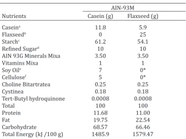
Experimental diet. Flaxseeds were ground in blenders into flour, weighed and immediately used in meal preparations. The FG group meals had a 25% flaxseed content to administer the recommended fiber intake. The ingredients of the experimental meals (Table 1) were weighed and homogenized using an
indus-trial blender (Hobart®, São Paulo, SP, Brazil) by using boiled water
for amid gelatinization. The obtained dough was transformed into pellets and dried in a ventilated oven (Fabbe-Primar® n171, São
Paulo, SP, Brazil) at 60°C for 24h and was refrigerated after iden -tification, until use. After weaning, the rats were administered a 10% protein diet in accordance with AIN-93M.
Biochemical methods. To determine hormone levels, speci-fic commercial kits were used for each hormone. The blood sam -ples were centrifuged (300rpm for 20 min at 4°C) to obtain the sera, which were individually stored at -20°C. Estradiol and tes -tosterone levels were determined using the chemiluminescence immunoassay method with the sera and ELISA kits (ABCAM Cat. Numb. ab108667/ab108666) with Immulite 2000/PPC/H2967, Siemens, Los Angeles, USA analyzer.
Histological processing. Testicles and epididymis were col-lected following euthanasia and immediately fixed in 10% buffe -red formalin solution. The material was cleaved and processed using standard paraffin embedding techniques, sliced (thickness, 5µm), and stained with hematoxylin and eosin (Bancroft & Cook 1994).
Immunohistochemical processing. The sections were sub-jected to an automated simultaneous dewaxing and rehydration process by using PT Link DAKO PT100 equipment and washed in phosphate buffered saline (PBS) for 5 minutes. The sections were subsequently treated at room temperature with 3% hydrogen pe -roxide solution in methanol to block any endogenous peroxidase. The slides were circled using a DAKO S2002 hydrophobic pen to avoid the diluted antibody solution from running. Immunos-taining was performed with an antivimentin monoclonal (mouse) antibody (Code IR630, clone v9, DAKO) associated with an EnVi-sion FLEX visualization system by using 4 µm-thick histological sections placed in silanized DAKO slides. Negative controls were incubated with PBS instead of the primary antibody (Fig.1A). Samples of a well-known tissue (palatine tonsil) with the antigen, were used as positive controls (Fig.1B). Subsequently, the pero -xidase label was visualized through reaction with DAB (diamino-benzidine tetrahydrochloride) solution (Sigma-Aldrich Co, St Lou-is, MO, USA) at room temperature. The slides were subsequently dehydrated in decreasing concentrations of alcohol and washed 4 times with xylol. The slides were assembled using an ALLKIMIA synthetic Canada Balsam for future microscopic analyses.
For histomorphometric analyses, the sections were analyzed using an Olympus BX-51 light microscope coupled to a DP72 di-gital camera, where the microscope fields were transferred to a LG Flatron x17527 screen and measured using Image J software, version 1.47t.
Table 1. Control and flaxseed diets composition of each 100g of diet
AIN-93M
Nutrients Casein (g) Flaxseed (g)
Caseina 11.8 5.9
Flaxseedb 0 25
Starchc 61.2 54.1
Refined Sugard 10 10
AIN 93G Minerals Mixa 3.50 3.50
Vitamins Mixa 1 1
Soy Oile 7 0*
Cellulosef 5 0*
Choline Bitartratea 0.25 0.25
Cystinea 0.18 0.18
Tert-Butyl hydroquinone 0.0008 0.0008
Total 100 100
Protein 11.68 11.00
Fat 19.75 22.54
Carbohydrate 68.57 66.46
Total Energy (kJ /100 g) 1485.9 1579.47
* The oil and flaxseed fiber diet are constituents to 25% of the added flax -seed. Ingredients used in diets preparation were provided by: a M. Cas-sab Comércio e Indústria Ltda (São Paulo/SP, Brazil), b Arma Zen
Pro-dutos Naturais Ltda (Rio de Janeiro/RJ, Brazil), c Maisena da Unilever
Bestfoods Brazil Ltda (Mogi Guaçu/SP, Brazil), d União (Rio de Janeiro/
RJ, Brazil), e Liza da Cargill Agricultura Ltda (Mairinque/SP, Brazil), f
Mi-crocel da Blanver Ltda (Cotia/SP, Brazil).
Computerized histomorphometric analysis of epididymal regions and seminiferous tubules. Right testicles and epididy-mis were separated by the initial segment and tail region of the epididymis. The testicles were transversally cleaved, whereas the epididymis were longitudinally cleaved to for visualizing epididy-mal regions: head, body and tail as reported by Serre & Robaire (1998). For the morphometric analysis, 50 round seminiferous tubules, stages VIII–X of the spermatogenic cycle (Leblond & Cler-mont 1952), were randomly obtained from 5 sections. In epididy-mal tubules, 30 tubules from the head and body regions and 20 from the tail region were evaluated.
Tubular area and epithelium height quantification of se-miniferous and epididymal tubules. The areas of the semini-ferous and epididymal tubules were measured using the Image J freehand tool by demarcating the apical portion of the cells on the lumen surface. By using the Image J straight tool, 4 lines were dra-wn and evenly distributed in the epithelium to measure its height. The epithelial height and seminiferous tubules and epididymis head areas were measured using a 40× objective lens. The areas of the remaining epididymis regions were measured using a 20× objective lens.
Seminiferous and epididymal tubule diameter quantifica-tion. Tubular and luminal diameters were measured using 2 strai-ght perpendicularly crossing lines originating from the borders of the tubules by using objective lenses that allowed visualizing the tubular structure.
Sertoli cell count. After immunostaining by using the antivi-mentin antibody, 25 tubules were randomly selected from each animal for Sertoli cell quantification. The tubules were visualized
at 40x magnification and the number of cells per tubule were counted using the plugin cell counter in Image J. The results were expressed as mean and standard deviation of the cells/tubules.
Statistical analysis. Statistical analyses were performed using GraphPad Instat 8.0 software. Data are shown as media ± standard deviation in a Microsoft Excel 2003 sheet. The normal distribution of the data was evaluated using the Kolmogorov– Smirnov test. After verifying data normality, the data were com-pared with independent samples by using the Student t test. For the results that did not show a normal distribution, the Wilcoxon nonparametric test was used. The significance level in all tests was established as P <0.05.
RESULTS Biochemical analysis
A significant increase was observed in 17β-estradiol
(17%) in the FG group. Testosterone levels were not
statis-tically significant (Table 2).
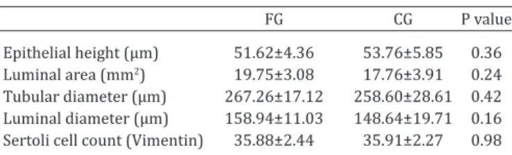
Histomorphometry of seminiferous tubules and Sertoli cell count
The height of the gametogenic epithelium, luminal area, tubular diameter and luminal diameter were not
statis-tically significant (Fig.2A and B). Sertoli cell counts using
vimentin staining (Fig.2C and D) did not show any
signi-ficant differences among the studied groups, as shown in
Table 3.
Histomorphometry of epididymal tubules. Epididy-mal histological analyses of the aniEpididy-mals administered 25%
flaxseed diets did not show any tissue architecture altera
-to their offspring during lactation, resulting in increased estradiol concentrations in these lactating offspring whi-ch remained high in their adulthood. Cardoso et al. (2010) observed an increase in estradiol after a prolonged intake
of 25% flaxseed, without changes in the penis morphology. Although 25% flaxseed was extrapolate recommended by
the USDA (2015), prolonged consumption at this concen-tration did not cause changes in reproductive morphology. Androgen suppression in neonates is associated with synthetic estrogen, which results in testicular morpholo-gical alterations. Soybean phytoestrogens, in high and low dosages, have been reported to reduce testosterone serum
concentrations (Weber et al. 2001, Caceres et al. 2014). Wisniewski et al. (2003) stated thatreduced testosterone biosynthesis can affect the reproductive physiology and behavior of phytoestrogen-fed rats. McVey et al. (2004)
evaluated rats fed with soybean isoflavones at different
concentrations and exposure times and reported that du-ring the prolonged period of 240 days, androgen levels were reduced in all the evaluated groups. In the present
study, continuous exposure to flaxseed lead to a nonsigni
-ficant decrease in serum testosterone levels, without mor -phologically altering the reproductive system of the tested
rats. These results suggest that flaxseed administered for
a prolonged period is more favorably assimilated by males than is soybean and does not lead to marked alterations in serum testosterone serum levels alterations.
No significant morphological changes were observed in
any of the regions of the epididymal tubules of either pre-
or post-natally flaxseed-administered rats. This behavior
was also observed with soybean-based diets. Piotrowska et
al. (2011) subjected rats to soybean isoflavones until sexu -al maturation and reported an increase in estradiol, reduc-tion in testosterone, and normal epididymal morphology.
In histomorphometry, seminiferous tubules should be analyzed because they are considered a reliable indicator of spermatic maturation (Schinckel et al. 1983, Tegegne et al. 1991). Moreover, tubular diameters have been used as a positive correlation parameter of testicular spermatogenic activity (Sinha Hikim et al. 1988). Sprando et al. (2000b) measured 100 seminiferous tubules per treatment group Table 2. 17-β estradiol (pg/mL) and testosterone (ng/dL)
serum concentration of 250-day-old rats
FG (n=10) CG (n=10) P Value
17 β-estradiol (pg/mL) 39.50±3.55 32.57±3.51 p=0.001*
Testosterone (ng/dL) 303.35±66.31 331.14±66.92 p=0.397
FG = Flaxseed Group, CG = Control Group. *Significance p<0.05.
Table 3. Histomorphometry of seminiferous tubules and Sertoli cell count of testes of 250-day-old rat
FG CG P value
Epithelial height (µm) 51.62±4.36 53.76±5.85 0.36 Luminal area (mm2) 19.75±3.08 17.76±3.91 0.24
Tubular diameter (µm) 267.26±17.12 258.60±28.61 0.42 Luminal diameter (µm) 158.94±11.03 148.64±19.71 0.16 Sertoli cell count (Vimentin) 35.88±2.44 35.91±2.27 0.98 Data show mean and standard deviation values of 10 rats per group. FG =
Flaxseed Group, CG = Control Group. * Significance p<0.05.
Table 4. Histomorphometry of tubules of epididymal regions of 250-day-old rats
FG Head CG Head P Value FG Body GC Body P Value FG Tail CG Tail P Value
Epithelial height (µm) 25.48±3.97 25.96±2.84 0.78 20.23±2.83 20.86±2.30 0.59 17.94±5.69 17.63±1.75 0.91 Tubular area (mm2) 6.88±1.35 7.21±1.77 0.67 37.68±9.72 38.91±10.80 0.79 162.00±32.94 158.63±33.57 0.87
Luminal area (µm) 91.43±8.79 92.72±9.49 0.78 211.49±28.03 216.93±32.60 0.69 413.15±66.94 340.75±80.88 0.16
Data show mean and standard deviation values of 10 rats per group. FG = Flaxseed Group, CG = Control Group. * Significance p<0.05.
tions compared with the control animals. Histological sec-tions of all regions showed some secsec-tions with round
tubu-les formed by pseudostratified columnar epithelium with
stereocilia and spermatozoa in its lumen.
The mean epithelial height and tubular and luminal are-as were smaller for the epididymal head and body regions of the experimental groups, unlike the tail region, which showed an increase in FG rats. However, the parameters evaluated in the different epididymal regions were not
sig-nificantly different (Table 4).
DISCUSSION
Flaxseed has been used in the prevention of cardiovascu-lar diseases (Hedelin et al. 2006) as well as in cancer risk reduction and cancer treatment because of the presence of
compounds that provide health benefits (Mason & Thomp -son 2014). Flaxseed lignans are structurally similar to
soy-bean isoflavones and endogenous estrogens and despite their benefits, can exert endocrine imbalances, affecting
the male reproductive system (Duncan et al. 2003, Boberg et al. 2013). Therefore, the concentrations of lignans and its derivatives have gained attention because of the possibili-ty of them affecting the gonadal morphology and hormone metabolism at certain administration doses and duration (Tou et al. 1999, 1998).
In our study, we used 25% flaxseed, attending the amount of dietary fiber recommended by the American Institute of Nutrition. After prolonged exposure to flaxse
-eds, serum estradiol levels significantly increased. Expo -sure to exogenous estrogen during gestation and lactation promotes local competition with endogenous estrogen, which can inhibittesticular androgen production (Sharpe & Skakkebaek 1993, Glover & Assinder 2006, Clark & Co-chrum 2007, Wohlfahrt-Veje et al. 2009), structurally alter developing reproductive tracts and alter the functional re-gulation of Sertoli cells (Lucas et al. 2011), thus resulting in infertility. Troina et al. (2010) analyzed endogenous serum estrogen levels in lactating rats (age, 21 days), adult rats (age, 150 days) and their mothers who had been
and did not report any significant differences in the short
--term results of rats fed with 20% and 40% flaxseed diets.
Oliveira et al. (2011) did not observe any statistical
diffe-rences in morphometric parameters among the flaxseed
--fed groups and concluded that the intake of a 12% flaxse
-ed flour diet does not exert negative effects on sperm and
morphometric analyses in rats. In this study, 500 tubules
were measured per group, a 25% flaxseed diet was admi -nistered and similar results were obtained. Continuous li-fetime exposure of rats does not result in long-term testicu-lar morphological alterations.
The spermatogenic capacity of adult animals depends
on the adequate development of Sertoli cells. Vimentin in
-termediate filaments expressed during the fetal life persist
in adulttesticles (Frojdman et al. 1989, Stosiek et al. 1990, Rogatsch et al. 1996, Bar-Shira Maymon et al. 2000) and support germ cells. Sprando et al. (2000b) evaluated
Serto-li cells in rats administered 20% flaxseed diets for 70 days and did not observe any significant differences. In our stu
-dy, even after a 250-day intake of the administered flaxseed
content, the number of Sertoli cells remained unchanged. A limitation of this study is that the sperm count was not evaluated. Motility, concentration, vigor and sperm patholo-gies can provide additional information about the
spermato-genic activity after the prolonged consumption of flaxseed.
CONCLUSION
Because of the absence of testicular and epididymal
mor-phometric alterations in the flaxseed-fed groups, we su -ggest that the continuous and prolonged consumption of this seed, even if initiated during gestation led to an incre-ase in serum estradiol levels, but did not interfere with the male gonad morphological development.
Acknowledgements.- This study was supported by FAPERJ, CNPq and
CAPES. The funders had no role in the study design, data collection and analysis, decision to publish, or manuscript drafting.
REFERENCES
Allard E.K., Johnson K.J. & Boekelheide K. 1993. Colchicine disrupts the cytoskeleton of rat testis seminiferous epithelium in a stage-dependent manner. Biol. Reprod. 48:143-153.
Aumuller G. & Peter S. 1986. Immunohistochemical and ultrastructural study of Sertoli cells in androgen insensitivity. Int. J. Androl. 9:99-108.
Bancroft J.D. & Cook H.C. 1994. Manual of Histological Techniques and
their Diagnostic Application. Churchill Livingstone, Edinburgh, p.35-67.
Bar-Shira Maymon B., Paz G., Elliott D.J., Hammel I., Kleiman S.E., Yogev L., Hauser R., Botchan A. & Yavetz H. 2000. Maturation phenotype of Sertoli
cells in testicular biopsies of azoospermic men. Hum. Reprod. 15:1537-1542.
Benson T.D., Akingbemi T.D., Kemppainen B.W., Hancock K.D., Sherrill J.D.,
Cook S.J., He X. & Supko J.G. 2007. Exposure to phytoestrogens in the perinatal period affects androgen secretion by testicular Leydig cells in the adult rat. Endocrinology 148:4475-4488.
Boberg J., Mandrup K.R., Jacobsen P.R., Isling L.K., Hadrup N., Berthelsen L., Elleby A., Kiersgaard M., Vinggaard A.M., Hass U. & Nellemann C. 2013. Endocrine disrupting effects in rats perinatally exposed to a dietary rel-evant mixture of phytoestrogens. Reprod. Toxicol. 40:41-51.
Caceres S., Silvan G., Martinez-Fernandez L., Illera M.J., Millan P., Monsalve
B., Peña L. & Illera J.C. 2014. The effects of isoflavones on androgens and glucocorticoids during puberty on male Wistar rats. Reprod. Domest.
Anim. Zuchthyg. 49:611-617.
Cardoso J.R. & Báo S.N. 2007. Effects of chronic exposure to soy meal
containing diet or soy derived isoflavones supplement on semen pro -duction and reproductive system of male rabbits. Anim. Reprod. Sci. 97:237-245.
Cardozo L.F.M. de F., Boaventura G.T., Brant L.H.C., Pereira V.A., Velarde
L.G.C. & Chagas M.A. 2012. Prolonged consumption of flaxseed flour increases the 17β-estradiol hormone without causing adverse effects on the histomorphology of Wistar rats’ penis. Food Chem. Toxicol. 50:
4092-4096.
Clark B.J. & Cochrum R.K. 2007. The steroidogenic acute regulatory pro-tein as a target of endocrine disruption in male reproduction. Drug Me-tab. Rev. 39:353-370.
Clermont Y. & Morgentaler H. 1955. Quantitative study of spermatogene -sis in the hypophysectomized rat. Endocrinology 57:369-382.
De Miguel M.P., Bethencourt F.R., Arenas M.I., Fraile B. & Paniagua R. 1997.
Intermediate filaments in the Sertoli cells of the ageing human testis.
Virchows Arch. Int. J. Pathol. 431:131-138.
Duncan A.M., Phipps W.R. & Kurzer M.S. 2003. Phyto-oestrogens. Best
Pract. Res. Clin. Endocrinol. Metab. 17:253-271.
Eustache F., Mondon F., Canivenc-Lavier M.C., Lesaffre C., Fulla Y., Berges
R., Cravedi J.P., Vaiman D. & Auger J. 2009. Chronic dietary exposure to
a low-dose mixture of genistein and vinclozolin modifies the reproduc -tive axis, testis transcriptome, and fertility. Environ. Health Perspect. 117:1272-1279.
Frojdman K., Paranko J., Kuopio T. & Pelliniemi L.J. 1989. Structural pro-teins in sexual differentiation of embryonic gonads. Int. J. Dev. Biol. 33:99-103.
Glover A. & Assinder S.J. 2006. Acute exposure of adult male rats to di-etary phytoestrogens reduces fecundity and alters epididymal steroid hormone receptor expression. J. Endocrinol. 189:565-573.
Hedelin M., Klint A., Chang E.T., Bellocco R., Johansson J.E., Andersson S.-O., Heinonen S.-M., Adlercreutz H., Adami H.O., Grönberg H. & Bälter K.A. 2006. Dietary phytoestrogen, serum enterolactone and risk of prostate cancer: the cancer prostate Sweden study (Sweden). Cancer Causes Control 17:169-180.
Kaithwas G. & Majumdar D.K. 2010a. Evaluation of antiulcer and
antise-cretory potential of Linum usitatissimum fixed oil and possible mecha
-nism of action. Inflammopharmacology 18:137-145.
Kaithwas G. & Majumdar D.K. 2010b. Therapeutic effect of Linum
usitatis-simum (flaxseed/linseed) fixed oil on acute and chronic arthritic mod
-els in albino rats. Inflammopharmacology 18:127-136.
Kinniry P., Amrani Y., Vachani A., Solomides C.C., Arguiri E., Workman A., Carter J. & Christofidou-Solomidou M. 2006. Dietary flaxseed supple
-mentation ameliorates inflammation and oxidative tissue damage in ex -perimental models of acute lung injury in mice. J. Nutr. 136:1545-1551.
Leblond C.P. & Clermont Y. 1952. Definition of the stages of the cycle of the seminiferous epithelium in the rat. Ann. N.Y. Acad. Sci. 55:548-573.
Lucas T.F., Pimenta M.T., Pisolato R., Lazari M.F.M. & Porto C.S. 2011.
17β-estradiol signaling and regulation of Sertoli cell function. Sper -matogenesis 1:318-324.
Mali P., Virtanen I. & Parvinen M. 1987. Vimentin expression in spermato-genic and Sertoli cells is stage-related in rat seminiferous epithelium. Andrologia 19:644-653.
Mason J.K. & Thompson L.U. 2014. Flaxseed and its lignan and oil com-ponents: can they play a role in reducing the risk of and improving the treatment of breast cancer? Appl. Physiol. Nutr. Metab. Physiol. 39:663-678.
McVey M.J., Cooke G.M. & Curran I.H. 2004. Increased serum and testicular
androgen levels in F1 rats with lifetime exposure to soy isoflavones. Re -prod. Toxicol. 18:677-685.
Moutsatsou P. 2007. The spectrum of phytoestrogens in nature: our knowledge is expanding. Hormones, Athens, 6:173-193.
Oliveira A.C.F., Carraro J.C.C., Lucas C.G., Reis P.G., Marcon L., Martins H.S.D., Benjamin L.A. & Ribeiro S.M.R. 2011. Efeito do consumo de farinha de
linhaça marrom sobre a morfometria testicular de ratos Wistar adultos.
Piotrowska K., Baranowska-Bosiacka I., Marchlewicz M., Gutowska I.,
Noceń I., Zawiślak M., Chlubek D. & Wiszniewska B. 2011. Changes in
male reproductive system and mineral metabolism induced by soy
iso-flavones administered to rats from prenatal life until sexual maturity.
Nutrition, Burbank Los Angel. Cty. 27:372-379.
Reeves P.G., Nielsen F.H. & Fahey G.C. 1993. AIN-93 purified diets for labo
-ratory rodents: final report of the American Institute of Nutrition ad hoc
writing committee on the reformulation of the AIN-76A rodent diet. J. Nutr. 123:1939-1951.
Rogatsch H., Jezek D., Hittmair A., Mikuz G. & Feichtinger H. 1996. Expres-sion of vimentin, cytokeratin, and desmin in Sertoli cells of human fetal, cryptorchid, and tumour-adjacent testicular tissue. Virchows Arch. Int. J. Pathol. 427:497-502.
Russell L.D. & Griswold M.D. 1993. The Sertoli Cell. Cache River Press, Clearwater. 801p.
Schinckel A., Johnson R.K., Pumfrey R.A. & Zimmerman D.R. 1983. Testic-ular growth in boars of different genetic lines and its relationship to re-productive performance. J. Anim. Sci. 56:1065-1076.
Serre V. & Robaire B. 1998. Segment-specific morphological changes in ag -ing Brown Norway rat epididymis. Biol. Reprod. 58:497-513.
Sharpe R.M. & Skakkebaek N.E. 1993. Are oestrogens involved in falling sperm counts and disorders of the male reproductive tract? Lancet 341:1392-1395.
Sinha Hikim A.P., Bartke A. & Russell L.D. 1988. Morphometric studies on hamster testes in gonadally active and inactive states: light microscope
findings. Biol. Reprod. 39:1225-1237.
Sprando R.L., Collins T.F., Black T.N., Olejnik N., Rorie J.I., Scott M., Wiesen
-feld P., Babu U.S. & O’Donnell M. 2000a. The effect of maternal exposure to flaxseed on spermatogenesis in F(1) generation rats. Food Chem. Tox -icol. Int. J. Publ. Brit. Ind. Biol. Res. Assoc. 38:325-334.
Sprando R.L., Collins T.F., Wiesenfeld P., Babu U.S., Rees C., Black T., Olejnik N. & Rorie J. 2000b. Testing the potential of flaxseed to affect spermato -genesis: morphometry. Food Chem. Toxicol. Int. J. Publ. Brit. Ind. Biol. Res. Assoc. 38:887-892.
Stosiek P., Kasper M. & Karsten U. 1990. Expression of cytokeratins 8 and 18 in human Sertoli cells of immature and atrophic seminiferous tu-bules. Differ. Res. Biol. Divers. 43:66-70.
Tegegne A., Entwistle K.W. & Mukasa-Mugerwa E. 1991. A quantitative his
-tological study of testicular and epididymal development in Boran and Boran x Friesian bulls in Ethiopia. Theriogenology 35:991-1000. Tou J.C., Chen J. & Thompson L.U. 1998. Flaxseed and its lignan precursor,
secoisolariciresinol diglycoside, affect pregnancy outcome and repro-ductive development in rats. J. Nutr. 128:1861-1868.
Tou J.C., Chen J. & Thompson L.U. 1999. Dose, timing, and duration of flax -seed exposure affect reproductive indices and sex hormone levels in rats. J. Toxicol. Environ. Health A 56:555-570.
Troina A.A., Figueiredo M.S., Moura E.G., Boaventura G.T., Soares L.L., Cardozo L.F.M.F., Oliveira E., Lisboa P.C., Passos M.A.R.F. & Passos M.C.F.
2010. Maternal flaxseed diet during lactation alters milk composition and programs the offspring body composition, lipid profile and sexu -al function. Food Chem. Toxicol. Int. J. Publ. Brit. Ind. Biol. Res. Assoc. 48:697-703.
USDA 2015. National Nutrient Database for Standard Reference. Release 28, United States Department of Agriculture. Available at <http://www. nal.usda.gov/fnic/foodcomp/search/> Access Oct 2015.
Weber K.S., Setchell K.D.R., Stocco D.M. & Lephart E.D. 2001. Dietary
soy-phytoestrogens decrease testosterone levels and prostate weight without altering LH, prostate 5 alpha-reductase or testicular ste-roidogenic acute regulatory peptide levels in adult male Sprague-Daw-ley rats. J. Endocrinol. 170:591-609.
Wisniewski A.B., Cernetich A., Gearhart J.P. & Klein S.L. 2005. Perinatal
exposure to genistein alters reproductive development and aggressive behavior in male mice. Physiol. Behav. 84:327-334.
Wisniewski A.B., Klein S.L., Lakshmanan Y. & Gearhart J.P. 2003. Exposure
to genistein during gestation and lactation demasculinizes the repro-ductive system in rats. J. Urol. 169:1582-1586.
Wohlfahrt-Veje C., Main K.M. & Skakkebaek N.E. 2009. Testicular dysgene -sis syndrome: foetal origin of adult reproductive problems. Clin. Endo-crinol., Oxford, 71:459-465.
Yin D., Zhu Y., Liu L., Xu H., Huang J. & Li Y. 2014. Potential detrimental ef
-fect of soy isoflavones on testis Sertoli cells. Zhong Nan Da Xue Xue Bao Yi Xue Ban 39:598-604.
Zhang L.D., Deng Q., Wang Z.-M., Gao M., Wang L., Chong T. & Li H.-C. 2013.