Original Article
Cytogenet Genome Res 122:73–79 (2008) DOI: 10.1159/000151319
Distribution patterns of phosphorylated Thr 3
and Thr 32 of histone H3 in plant mitosis and
meiosis
A.D. Caperta
a, b
M. Rosa
a, d
M. Delgado
a, b
R. Karimi
c
D. Demidov
c
W. Viegas
a
A. Houben
c
a Centro Botânica Aplicada à Agricultura, Instituto Superior de Agronomia, Technical University of Lisbon, and b Departamento de Ciências Biológicas e Naturais, Universidade Lusófona de Humanidades e Tecnologias,
Lisboa (Portugal); c Leibniz Institute of Plant Genetics and Crop Plant Research (IPK), Gatersleben (Germany) d Gregor Mendel Institute of Molecular Plant Biology, Vienna (Austria)
of their N-terminal tails (Wolffe, 1998). For example the cell cycle dependent phosphorylation of histone H3 at serine 10 (H3S10ph) is tightly correlated with chromosome conden-sation and segregation (Gurtley et al., 1975; Wei and Allis, 1998). Although Ser 10 phosphorylation and most post-translational modifications of histone H3 are highly con-served, a number of significant differences exist between animals and plants (Fuchs et al., 2006; Houben et al., 2007a). In animals Ser 10 and Ser 28 phosphorylation of histone H3 starts in late G2 in the pericentromeric region from where it is distributed homogeneously throughout the chromo-somes (Hendzel et al., 1997; Goto et al., 1999, 2002). In plants, H3 phosphorylation levels at both serine positions are high in pericentromeric regions but very low along the chromosome arms (Houben et al., 1999; Kaszas and Cande, 2000; Manzanero et al., 2000; Pedrosa et al., 2001; Gernand et al., 2003; Schroeder-Reiter et al., 2003). The distribution Abstract. Cell cycle dependent phosphorylation of
con-served N-terminal tail residues of histone H3 has been de-scribed in both animal and plant cells. Through cytogenet-ic approaches using different plant species we show a detailed description of distribution patterns of phosphorylated his-tone H3 at either threonine 3 or threonine 32 in mitosis and meiosis. In meristematic cells of the large genome species Secale cereale, Vicia faba and Hordeum vulgare we have found that phosphorylation of both threonine residues be-gins in prophase, and dephosphorylation occurs in late ana-phase. However, in the small genome species Arabidopsis
thaliana dephosphorylation occurs at anaphase. In the first
division of meiosis of species with large genomes
Request reprints from Ana D. Caperta Centro Botânica Aplicada à Agricultura
Instituto Superior de Agronomia, Technical University of Lisbon 1349-017 Lisboa (Portugal)
e-mail: [email protected]
ylation of histone H3 at either threonine 3 or threonine 32 is seen first in diakinesis and extends to anaphase I, where-as in the second division these post-translational modifica-tions are visible at metaphase II through anaphase II. While in A. thaliana dephosphorylation takes place at anaphase I and II. In all species analysed phosphorylated H3 at either threonine 3 or threonine 32 are distributed along the entire length of chromosomes during mitotic metaphase and metaphase I. In the second meiotic division threonine 3 phosphorylation is restricted to the pericentromeric do-main, while phosphorylation of threonine 32 is widespread along chromosome arms of all species analysed.
Copyright © 2008 S. Karger AG, Basel
Chromatin undergoes dramatic structural changes as the nucleus progresses through the cell cycle, from decon-densed interphase chromatin to highly condecon-densed meta-phase chromatin. In the control of these events the DNA binding histones regulate chromatin structure and gene ex-pression through dynamic post-translational modifications
Research in the W.V. laboratory was supported by Fundação para a Ciência e Tecnologia, Portugal grant SFRH/BPD/26442 to A.D.C. and SFRH/ BPD/27219/2006 to M.D., respectively. D.D., R.K. and A.H. were support-ed by the Deutsche Forschungsgemeinschaft and by the Land Sachsen An-halt.
Accepted in revised form for publication by B. Friebe, 23 May 2008.
© Free Author
Copy – for
per-sonal use only
ANY DISTRIBUTION OF THIS ARTICLE WITHOUT WRITTEN CONSENT FROM S. KARGER AG, BASEL IS A VIOLATION OF THE COPYRIGHT. Written permission to distrib-ute the PDF will be granted against payment of a per-mission fee, which is based on the number of accesses required. Please contact [email protected]
© Free Author Copy – for per sonal use only
ANY DISTRIBUTION OF THIS ARTICLE WITHOUT WRITTEN CONSENT FROM S. KARGER AG, BASEL IS A VIOLATION OF THE COPYRIGHT.
of H3S10ph and H3S28ph varies between the two meiotic divisions in plants (Manzanero et al., 2000; Gernand et al., 2003). In the first division, the entire chromosomes are highly phosphorylated, while in the second division H3 phosphorylation is restricted to the pericentromeric re-gions, similar to mitotic chromosomes. At the same time single chromatids, resulting from equational division of univalents at anaphase I, show low levels of phosphorylation at second meiotic division, suggesting a role of this post-translational modification in sister chromatid cohesion (Manzanero et al., 2000; Gernand et al., 2003).
Phosphorylation of H3 at either Thr 11 (H3T11ph) or Thr 3 (H3T3ph) are spatially distinct from the modifications at Ser 10 and Ser 28 in that both threonine modifications are largely restricted to centromeric regions of condensed mi-totic chromosomes in animals (Preuss et al., 2003; Dai et al., 2005). In contrast to animals phosphorylated Thr 11 in plants is distributed along the entire length of condensed chromosomes (Houben et al., 2005). Phosphorylation at Thr 32 (H3T32ph) also occurs in animal cells (Tamada et al., 2006) but the distribution pattern in mitosis and meiosis has not yet been described in either animals or plants.
In the present study we establish a detailed description of distribution patterns of histone H3 phosphorylated at Thr 3 or Thr 32 in mitosis and meiosis of different plant spe-cies. The study supports the hypothesis of differential func-tions of these post-translational modificafunc-tions in chromo-some biology.
Materials and methods
Plant material
The following plant species have been used: Arabidopsis thaliana ecotype No-0, Secale cereale (rye), Vicia faba (field bean) and Hordeum
vulgare (barley).
Chromosome preparation and indirect immunofluorescence
For immunodetection root tips from all species and selected an-thers from S. cereale and flower buds from A. thaliana were isolated and fixed in 4% paraformaldehyde in phosphate buffered saline (PBS) as previously described (Manzanero et al., 2000). S. cereale, H. vulgare and Vicia faba root tips were digested with 2% cellulose, 2% cellulase ‘Onozuka R-10’ and 2% pectinase (Caperta et al., 2002), and squashes were made in PBS. A. thaliana flower buds were digested 75 min with 3% cytohelicase, 3% pectolyase and 3% cellulase in PBS at 37 ° C and squashes were made in PBS. To avoid non-specific antibody binding, slides were blocked for 30 min in 4% (w/v) bovine serum albumin (BSA) plus 0.1% Triton X-100 in PBS at room temperature and incu-bated with the primary antibodies in a humid chamber. Polyclonal af-finity purified rabbit antibodies against H3 phosphorylated at threo-nine 3 (H3T3ph, Upstate 07-424) and H3 phosphorylated at threothreo-nine 32 (H3T32ph, Abcam ab4076) were diluted 1: 200 and 1: 300 in PBS with 3% BSA respectively. The rat monoclonal antibody against H3 phos-phorylated at serine 28 (H3S28ph) (Goto et al., 1999) was diluted 1: 400 in PBS with 3% BSA. After 12 h incubation at 4 ° C and washing for 15 min in PBS, the slides were incubated in rhodamine-conjugated anti-rabbit IgG (Dianova) and FITC-conjugated anti-rat IgG (Dianova) di-luted 1: 200 in PBS, 3% BSA for 1 h at 37 ° C. After final washes in PBS,
Fluorescence in situ hybridization (FISH)
A. thaliana preparations were immunodetected as described above and then post-fixed in 4% formaldehyde in PBS followed by FISH. The DNA probe used for FISH analyses was the 180-bp A. thaliana -spe-cific centromeric repeat (Copenhaver et al., 1999) labelled with digoxi-genin-dUTP by PCR with the primers 5 ⴕ -ATCCTCTAGAGTCGACCT-GCA-3 ⴕ and 5 ⴕ -TTCCCAGTCACGACGTTGTAA-3 ⴕ , using an initial denaturation step for 4 min at 94 ° C, and 35 cycles of 94 ° C for 45 s, 56 ° C for 45 s, and 72 ° C for 45 s.
Hybridization conditions, post-hybridization washes and detec-tion of FISH signals were performed according to Pontes et al. (2003). Nuclei were counterstained with DAPI.
Image processing
Analysis of fluorescence signals was recorded with an Olympus BX61 microscope equipped with an ORCA-ER CCD camera (Hama-matsu). Deconvolution microscopy was employed for superior optical resolution of globular structures. Photographs were collected as se-quential images along the Z-axis with approximately 11 slices per spec-imen. Images were collected in grey scale and pseudocolored with Ado-be Photoshop, and projections (maximum intensity) were done with the program AnalySIS (Soft Imaging System).
Plant protein extraction
Plant material (200 to 300 mg) was ground under liquid nitrogen and suspended in 1 ml of solubilization buffer (56 m M Na 2 CO 3 , 56 m M
DTT, 2% SDS, 12% sucrose, and 2 m M EDTA). After 10 min of incuba-tion at 70 ° C, cell debris was removed by centrifugation. 40 g of pro-tein of each sample were analyzed by propro-tein gel blotting.
For the application of the anti-H3T3ph antibody histone-enriched protein extracts for the analysis were prepared as described by Moehs et al. (1988).
PAGE and protein gel blot analysis
Protein samples were separated by SDS-PAGE in 12% polyacryl-amide gels according to Schagger and von Jagow (1987) and then elec-trotransferred onto polyvinylidene difluoride (PVDF) membranes. After blotting, membranes were reversibly stained with 1% ponceau red. Membranes were incubated for 12 h at 4 ° C in PBS and 5% low-fat milk or 2 ! PBS, 5% BSA, 1% PEG 3500, and 1% PVP 10, containing the appropriate antibody. Secondary antibodies conjugated to horse-radish peroxidase were used to reveal immunocomplexes by enhanced chemiluminescence (Pierce, Rockford, IL).
Fig. 1. Distribution of phosphorylated histone H3 at Thr 3 (H3T3ph) and Ser 28 (H3S28ph) in somatic cells ( a–d ) and meiocytes ( e–g ) of Secale cereale. DAPI stained chromosomes are blue, H3T3ph-specific signals are red, and H3S28ph signals are green. ( a ) Interphase: no sig-nals of H3T3ph and H3S28ph are detected. ( b ) Prophase: H3T3ph is distributed along the chromosome arms whereas H3S28ph is restricted to pericentromeric domains. ( c ) Metaphase: chromosome arms exhib-it punctuated H3T3ph pattern along their entire lengths and Thr 3 hyperphosphorylation at outer pericentromeric domains (arrows), while H3S28ph is detected at pericentromeric domains. ( d ) Anaphase: H3T3ph staining is maintained in chromosome arms and Ser 28 phos-phorylation remains restricted to pericentromeric domains. ( e ) Diaki-nesis/metaphase I transition: bivalent chromosomes are stained along chromosome arms for H3T3ph, while H3S28ph is restricted to pericen-tromeric domains. ( f ) Metaphase II: intense H3T3ph staining is
most-a b c d e f g
Results
Different plant species show distinct timings of threonine 3 phosphorylation of histone H3 in somatic cells and meiocytes
The antibodies against phoshorylated histone H3 at ei-ther threonine 3 or threonine 32 recognized a protein of the histone H3-type, molecular mass of 17 kDa on Western blots of A. thaliana and tobacco protein extracts (supple-mentary Fig. 1; for online supple(supple-mentary material, see www. karger.com/doi/10.1159/000151319). The antibodies are likely to recognize the same peptide in plants since the his-tone H3 amino acid sequences of many plant species are identical to the synthetic peptide sequence used for immu-nization.
We next determined the chromosomal distribution pat-terns of histone H3 phosphorylated at threonine 3 (H3T3ph) in mitotic cells of different mono- and dicot species such as Secale cereale, Hordeum vulgare, Vicia faba and A. thaliana. In all plant species analysed no labelling of interphase chro-matin was detected ( Fig. 1 a; suppl. Fig. 2c). The first immu-nosignals were detected at prophase, as chromosomes start-ed to condense ( Figs. 1 b, 2a). In S. cereale prophase chromo-somes we observed a punctuated dispersed staining along chromosome arms ( Fig. 1 b). However, a marked difference is observed between different species where phosphoryla-tion of threonine 3 started in early prophase in A. thaliana. Sequential immunostaining for H3T3ph and FISH using the A. thaliana -specific centromeric 180 bp-repeat as probe revealed that H3T3 phosphorylation starts in close proxim-ity to the centromere. When pericentromeric/centromeric domains of sister chromatids are individualized and seen as FISH-positive double dots, H3T3ph is detected in the re-gions between the two sister chromatids ( Fig. 2 a, arrow). In late prophase stage, H3T3ph labelling is dispersed along chromosome arms ( Fig. 2 b). In all species analysed chromo-somes showed an intense phosphorylation labelling when they align to the metaphase plate ( Fig. 1 c; suppl. Fig. 2a, c). Due to the larger size of S. cereale chromosomes we were able to detect a punctuated non-homogeneous phosphoryla-tion staining along chromosome arms and hyperphosphor-ylation at regions adjacent to centromeric domains ( Fig. 1 c, arrows, inset). In addition, costaining with antibodies for both H3T3ph and H3S28ph in S. cereale revealed that phos-phorylated Thr 3 was localized at outer pericentromeric do-mains while phosphorylated Ser 28 was strongest at inner pericentromeric domains ( Fig. 1 ). Except for A. thaliana, where phosphorylation of H3T3 was absent during ana-phase ( Fig. 2 c), in all other species analysed the immunola-belling was maintained until early anaphase ( Fig. 1 d; suppl. Fig. 2b, d), declined substantially in late anaphase and was absent on decondensing chromosomes at telophase.
In meiosis, phosphorylation of H3 at Thr 3 was initiated during a late step of chromosome condensation, at the tran-sition between diakinesis/metaphase I where chromosomes
tuated labelling until metaphase I ( Fig. 1 e). In S. cereale the immunostaining persisted until late anaphase I and disap-peared at telophase I, while in A. thaliana meiocytes no Thr3 phosphorylation was detected at anaphase I or telophase I ( Fig. 2 e). At the second metaphase H3T3ph pattern is similar in both S. cereale and A. thaliana. The H3T3ph staining is restricted to the regions adjacent to all centromeric domains and no phosphorylation was detected in chromosome arms. This contrasted with the distribution patterns of H3T3ph observed for both metaphase I and metaphase mitotic chro-mosomes. In S. cereale, comparison of H3T3ph distribution with that of H3S28ph at metaphase II revealed partial colo-calization of both histone modifications, although H3S28ph labelling is found at the inner position of pericentromeric domains ( Fig. 1 f). Also A. thaliana metaphase II meiocytes ( Fig. 2 f) presented an H3T3ph distribution pattern similar to that observed for early mitotic prophase ( Fig. 2 a). Sequen-tial in situ hybridization using the Arabidopsis centromeric repeat and immunostaining with the H3T3ph antibody showed minor superimposition between signals. Therefore, metaphase II chromosomes of large and small genomes pres-ent a distinct specific distribution pattern in relation to those for mitotic and first meiotic metaphases. In addition, the timings for histone H3 dephosphorylation at threonine 3 at both anaphase I and anaphase II differed between these spe-cies, since in A. thaliana no labelling is found at this stage, whereas it was still present in S. cereale.
A dynamic cell cycle dependent distribution of phosphorylated histone H3 at threonine 32
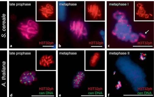
Next we investigated the cell cycle dependent distribu-tion patterns of phosphorylated threonine 32 of histone H3 in both A. thaliana and S. cereale. In both species phosphor-ylation of Thr 32 in somatic cells took place at prophase ( Fig. 3 a, d) and persisted through metaphase ( Fig. 3 b, e). In S.
ce-reale dephosphorylation occurs at late anaphase, whereas in
A. thaliana the immunolabelling was absent as early as ana-phase (not shown).
In both species we found that histone H3 phosphorylated at threonine 32 is distributed throughout the entire lengths of somatic chromosomes ( Fig. 3 a, b, d, e). At meiosis, in the
Fig. 2. Distribution pattern of Thr 3 phosphorylation of histone H3 (H3T3ph) in mitotic ( a–c ) and meiotic cells of A. thaliana ( d–f ). DAPI stained chromosomes are blue, H3T3ph-specific signals are red, and the FISH signals corresponding to the A. thaliana 180-bp centromeric repeat are green. ( a ) Early prophase stage: the immunosignals (red) are adjacent to FISH signals (green). When centromeric domains of sister chromatids are visualized as individual, H3T3ph dots are located in-tercalary to the centromeric domains in each sister chromatid (arrows). No superimposition between signals is observed. ( b ) Late prophase stage: H3T3ph is dispersed along the chromosome arms and superim-poses with centromeric FISH signal. ( c ) Anaphase: no H3T3ph label-ling is seen, whereas the FISH signals are detected. ( d ) Diakinesis: phosphorylation is initiated all over entire chromosome lengths except
a b c d e f
early steps of chromatin condensation (leptotene to pachy-tene) H3T32 phosphorylation is absent, as determined for Thr 3. In both A. thaliana and S. cereale, Thr 32 phosphor-ylation is initiated at diakinesis, and all bivalent chromo-somes were immunolabelled along their entire lengths in-cluding the supernumerary B chromosomes ( Fig. 3 c), and went through metaphase I. The immunostaining disap-peared at anaphase I and reapdisap-peared at metaphase II ( Fig. 3 f) however, and contrasting with the observations of H3T3ph the entire chromosome arms were labelled at metaphase II.
Discussion
Although the components of the chromatin code are fairly well conserved throughout eukaryotes, evidence is growing that the interpretation of the code has diverged in different organisms, particularly in plants versus animals. Our study provides evidence that in addition to the chro-mosomal patterns of histone methylation (Houben et al., 2003; Fuchs et al., 2006) phosphorylation of histone H3 also partially varies between species.
Phosphorylation of histone H3 in meristematic cells and meiocytes was followed by the use of polyclonal antibodies that recognize the phosphorylated form of Thr 3 and Thr 32. The detailed distribution patterns of Thr 3 phosphoryla-tion in meiosis, and of Thr 32 phosphorylaphosphoryla-tion in mitosis and meiosis are here described for the first time. For both post-translational modifications we show that the distribu-tion patterns of phosphoryladistribu-tion are correlated with chro-mosome condensation, although presenting some differ-ences between different species. In mitosis of grasses, the timing of phosphorylation for Thr 3 and Thr 32 begins in prophase, progresses through metaphase when
chromo-2000; Manzanero et al., 2000), H3S28 (Gernand et al., 2003) and H3T11 (Houben et al., 2005). However, in A. thaliana dephosphorylation in both residues occurs prior to ana-phase. In meiosis both threonine residues are undergoing phosphorylation at diakinesis in a late state of chromosome condensation and progress through metaphase I as it hap-pened for Thr 11 phosphorylation in plants (Houben et al., 2005). The staining of both phosphorylated threonine resi-dues of histone H3 is detected at metaphase II. Again, slight differences in dephosphorylation timing occur between different species. While in the large genome species dephos-phorylation is seen at late anaphase I or anaphase II, in A.
thaliana, it is found during first and second anaphase.
Whether the immunostaining pattern differences between the different species are caused by the different genome siz-es remains to be tsiz-ested on a larger number of specisiz-es.
Additional differences were found between the distribu-tion patterns of H3T3ph and H3T32ph in chromosomes at different cell division stages. In mitotic prophase of S.
cere-ale H3T3ph labelling is discontinuous along entire lengths
of chromosome arms suggesting a close proximity with het-erochromatic domains, namely higher density at the centro-meric pole. While in A. thaliana phosphorylation at Thr 3 and Thr 32 begins adjacent to the major centromeric hetero-chromatic domain, and spreads along the chromosome arms in late prophase, when higher chromatin condensa-tion occurs. Also in A. thaliana at early prophase some chromosomes show split signals for centromeric DNA with H3T3ph staining in a central location (between sister chro-matids), a conformation present in metaphase II chromo-somes. A comparable behaviour of centromeric regions was also reported for Arabidopsis polyploids at meiosis (Comai et al., 2003). Although those configurations are not yet ful-ly understood, the cruciform organization of pericentric
a b c
d e f
Fig. 3. Thr 32 phosphorylation of histone H3 (H3T32ph) correlates with mitotic and meiotic condensation in A. thaliana an S.
ce-reale. DAPI stained chromosomes are blue,
H3T32ph-signals are red, and the FISH sig-nals corresponding to the A. thaliana 180-bp centromeric repeat are green, inset shows H3T32ph signal only. ( a , d ) At late prophase H3T32ph-signals coincide with entire lengths of chromosomes and the same is observed at metaphase. ( c ) In diakinesis phosphorylated Thr 32 is dispersed along bivalent arms. B chromosomes are also stained (arrow). ( f ) In the second metaphase of meiosis, the immu-nosignals are spread along chromosome arms. Bars = 10 m.
In metaphase chromosomes of S. cereale Thr 3 phos-phorylation presents a punctuated distribution pattern along entire lengths of chromosome arms and the pericen-tromeric domains are hyperphosphorylated, as found for phosphorylated Thr 11 of H3 in plants (Houben et al., 2005). Nevertheless, absence of immunolabelling is observed in centromeric domains in both species. In fact, the signal gap colocalizing with the centromeres may be related to the presence of CENH3 at these regions where no phosphoryla-tion of H3 at Ser 10 has been described in plants (Schroeder-Reiter et al., 2003; Houben et al., 2007b). Collectively, these results contrast with studies in mammalian cells on mitotic chromosomes where the cell cycle dependent phosphoryla-tion at threonines 3 and 11 are restricted to the centromer-ic regions (Preuss et al., 2003; Dai et al., 2005). Moreover, there are also differences in distribution patterns between the two division phases of meiosis for both H3T3ph and H3T32ph in plants. At diakinesis, the staining spreads uni-formly all over the bivalent chromosomes except for the centromeric regions. In mitosis and first meiotic division of species with large genomes, costaining with H3T3ph and
H3S28ph antibodies revealed minor superimposition be-tween signals. A similar pattern was detected in metaphase II chromosomes which show hyperphosphorylation of Thr 3 adjacent to phosphorylated serine 28 at the pericentro-meric regions and its absence along chromosome arms. These results contrast with patterns here observed for H3T32ph and described for H3T11ph (Houben et al., 2005) which present a uniform distribution along the entire length of condensed chromosomes. Although phosphorylated threonine residues of histone H3 have been associated with chromatin condensation at mitosis and meiosis I, this dif-ferent phosphorylation dynamics of threonines 3 and 11 at metaphase II stage suggest possible different roles for these post-translational modifications.
Acknowledgements
We thank Augusta Barão and Katrin Kumke for excellent technical assistance. The antibody against H3 phosphorylated at serine 28 was kindly provided by Dr. M. Inagaki.
References
Caperta A, Neves N, Morais-Cecílio L, Malhó R, Viegas W: Genome restructuring in rye affects the expression, organization and disposition of homologous rDNA loci. J Cell Sci 115: 2839– 2846 (2002).
Comai L, Tyagi A, Lysak M: FISH analysis of meio-sis in Arabidopmeio-sis allopolyploids. Chromosome Res 11: 217–226 (2003).
Copenhaver GP, Nickel K, Kuromori T, Benito MI, Kaul S, et al: Genetic definition and sequence analysis of Arabidopsis centromeres. Science 286:2468–2474 (1999).
Dai J, Sultan S, Taylor S, Higgins JMG: The kinase haspin is required for mitotic histone H3 Thr 3 phosphorylation and normal metaphase chro-mosome alignment. Genes Dev 19: 472–488 (2005).
Demidov D, Van Damme D, Geelen D, Blattner FR, Houben A: Identification and dynamics of two classes of aurora-like kinases in
Arabidop-sis and other plants. Plant Cell 17: 836–848 (2005).
Fuchs J, Demidov D, Houben A, Schubert I: Chro-mosomal histone modification patterns – from conservation to diversity. Trends Plant Sci 11: 199–208 (2006).
Gernand D, Demidov D, Houben A: The temporal and spatial pattern of histone H3 phosphoryla-tion at serine 28 and serine 10 is similar in plants but differs between mono- and polycen-tric chromosomes. Cytogenet Genome Res 101: 172–176 (2003).
Goto H, Tomono Y, Ajiro K, Kosako H, Fujita M, et al: Identification of a novel phosphorylation site on histone H3 coupled with mitotic chro-mosome condensation. J Biol Chem 274: 25543– 25549 (1999).
Goto H, Yasui Y, Nigg EA, Inaki M: Aurora B phos-phorylates histone H3 at serine 28 with regard to the mitotic chromosome condensation. Genes Cells 7: 11–17 (2002).
Gurtley LR, Walters RA, Tobey RA: Sequential phosphorylation of histone subfractions in the Chinese hamster cell cycle. J Biol Chem 250: 3936–3944 (1975).
Hendzel MJ, Wei Y, Mancini A, Van Hooser A, Ra-nalli T, et al: Mitosis-specific phosphorylation of histone H3 initiates primarily within peri-centromeric heterochromatin during G2 and spreads in an ordered fashion coincident with mitotic chromosome condensation. Chromo-soma 106: 348–360 (1997).
Houben A, Wako T, Furushima-Shimogawara R, Presting G, Kunzel G, et al: The cell cycle de-pendent phosphorylation of histone H3 is cor-related with the condensation of plant mitotic chromosomes. Plant J 18: 675–679 (1999). Houben A, Demidov D, Gernand D, Meister A,
Leach CR, Schubert I: Methylation of histone H3 in euchromatin of plant chromosomes de-pends on basic nuclear DNA content. Plant J 33: 967–973 (2003).
Houben A, Demidov D, Rutten T, Scheidtmann KH: Novel phosphorylation of histone H3 at threo-nine 11 that temporally correlates with conden-sation of mitotic and meiotic chromosomes in plant cells. Cytogenet Genome Res 109: 148–155 (2005).
Houben A, Demidov D, Caperta AD, Karimi R, Agueci F, Vlasenko L: Phosphorylation of his-tone H3 in plants – a dynamic affair. Biochim Biophys Acta 1769: 308–315 (2007a).
Houben A, Schroeder-Reiter E, Nagaki K, Nasud S, Wanner G, et al: CENH3 interacts with the cen-tromeric retrotransposon cereba and GC-rich satellites and locates to centromeric substruc-tures in barley. Chromosoma 116: 275–283 (2007b).
Kaszás E, Cande WZ: Phosphorylation of histone H3 is correlated with changes in the mainte-nance of sister chromatid cohesion during mei-osis in maize, rather than the condensation of the chromatin. J Cell Sci 113: 3217–3226 (2000).
Manzanero S, Arana P, Puertas MJ, Houben A: The chromosomal distribution of phosphorylated histone H3 differs between plants and animals at meiosis. Chromosoma 109: 308–317 (2000). Moehs C, Mcelwain E, Spiker S: Chromosomal
pro-teins of Arabidospis thaliana. Plant Mol Biol 11: 507–515 (1988).
Pedrosa A, Jantsch MF, Moscone EA, Ambros PF, Schweizer D: Characterisation of pericentro-meric and sticky intercalary heterochromatin in Ornithogalum longibracteatum (Hyacintha-ceae). Chromosoma 110: 203–213 (2001). Pontes O, Lawrence RJ, Neves N, Silva M, Lee JH, et
al: Natural variation in nucleolar dominance reveals the relationship between nucleolus or-ganizer chromatin topology and rRNA gene transcription in Arabidopsis. Proc Natl Acad Sci USA 100: 11418–11423 (2003).
Preuss U, Landsberg G, Scheidtmann KH: Novel mitosis-specific phosphorylation of histone H3 at Thr11 mediated by Dlk/ZIP kinase. Nucleic Acids Res 31: 878–885 (2003).
Schagger H, von Jagow G: Tricine-sodium dodecyl sulfate-polyacrylamide gel electrophoresis for the separation of proteins in the range from 1 to 100 kDa. Anal Biochem 166: 368–379 (1987). Schroeder-Reiter E, Houben A, Wanner G:
Immu-nogold labeling of chromosomes for scanning electron microscopy: a closer look at phosphor-ylated histone H3 in mitotic metaphase chro-mosomes of Hordeum vulgare . Chromosome Res 11: 585–596 (2003).
Tamada H, Thuan NV, Reed P, Nelson D, Katoku-Kikyo N, et al: Chromatin decondensation and nuclear reprogramming by nucleoplasmin. Mol Cell Biol 26: 1259–1271 (2006).
Wei Y, Allis CD: A new marker for mitosis. Trends Cell Biol 8: 266 (1998).
Wolffe A: Chromatin Structure and Function, pp 447 (Academic Press, San Diego 1998). Yeh E, Haase J, Paliulis LV, Joglekar A, Bond L, et
al: Pericentric chromatin is organized into an intramolecular loop in mitosis. Curr Biol 18: 81–90 (2008).