HEMATOLOGICAL AND BIOCHEMICAL
PARAMETERS IN HEREDITARY
SPHEROCYTOSIS
UNDER
OXIDATIVE STRESS
. Elisa Granjo, Alice Santos-Silva, Irene Rebelo, Ana N6voa,
Elfsio Costa, Jose Barbot, Leticia Ribeiro, and Alexandre Quintanilha
Departamento de Hematologia do Hospital SJoao do Porto, Porto the Instituto de Biologia Molecular e Celular, Univ. Porto, Porto theiCBAS
the Departamento de Bioqufmica Faculdade Fanmicia Univ. Porto, Porto
the Servi~o de Hematologia Hospital Maria Pia, Porto
and the Centro Hospitalar de Coimbra, Coimbra, Portugal
A broad spectrum of clinical manifestations in hereditary spherocytosis (HS) has been documented.
The aim of this study 'Yas to investigate a correlation be!Ween the development of an oxidative stress, during inflammatory or infectious stress, and the development of a hemolytic event in HS patients. We evaluated hematological and biochemical changes i) imposed by infection in 2 HS patients and in 2 HS patients who had undergone splenectomy (more than six months before) ii) in 5 HS patients immediately after splenectomy and iii) in 4 HS patients, more than six months after splenectomy. In both inflammatory and infectious stresses we found a reduction in
RBCs
concentration and in MCHC, ~ rise in M CV, and neutrophilic monocytic leukocytosis. The changes in RBCs seemed to correlate with neutrophils and monocytes values. To clarify this, we studied the same parameters and a few others in iiii) a group of 6 HS patients who had undergone splenectomy (more than six months) and compared the results with those presented by a healthy control, including 29 individuals with no HS and no infection.Address reprint requests to: Alice Santos-Silva. PhD. Faculdade de Fanruicia. Departamento de Bioqufmica, RuaAnibal Cunba.164. 4050-Porto. Portugal. Tel.:+ 3512 2002564;Fax;+ 3512 2003977; e-mail: [email protected]
Molecular Biology of Hematopoiesis 6. edited by Abraham et al.
338 E. Granjo et aL
Our data suggest that inflammatory and infectious stress by inducing leukocyto-sis and leukocyte activation, may induce oxidative and proteolytic modifications upon RBCs, leading to an accelerated aging process, to a premature removal or even to a hemolytic event.
In Europe, Hereditary Spherocytosis (HS) is the most common congenital hemolytic anemia. A broad spectrum of clinical manifestations has been documented, which can range from mild to extremely severe. A set of mutations, namely in spectrin, ankirin and, band 3, may underlie HS.1
-3The mode of transmission is dominant or
reces-sive, with intermediate situations.
During its life span any normal red blood cell (RBC) is exposed to various insults and undergoes physical and chemical changes. .c-7 These modifications become more
pronounced with cell age, and with the development of unusual physiologic circum-. stances, such as increased mridative and proteolytic stresscircum-. In these circumstances, the
accelerated and premature development of senescent modifications may trigger the premature removal of RBCs or even a hemolytic event. Cells, such as spherocytes, which develop intracellular defects early during their life span, are also removed from circulation at an earlier stage.
RBC senescence includes several modifications, namely a reduction in its meta-bolic activity, resulting from a general decreased enzymatic activity. The pathway for the removal of senescent or damaged RBCs involves the development of a senescent cell antigen immunologically related to band 3 protein, 8.9 which marks the RBC for death, by triggering the binding of specific auto-antiband 3 antibodies10
•11 and
comple-ment activation.12 The modified antigenicity of band 3 may result from proteolytic
cleavage, clustering or even by exposure of unusual epitopes.
Inflammatory and infectious diseases often present a rise in leuk:ocytes, namely in neutrophils, and a reduction in total RBCs. 13.14 The exposure of circulating RBCs to
oxygen metabolites and proteases, which are known to be produced by activated neu-trophils, may account for some oxidative and proteolytic RBC damage, leading to an accelerated aging process and premature removal.7.1S-19 In the case of spherocytes
pre-senting a destabilized membrane structure, with loss of membrane components, and with an abnormal reduced life span, the impact of an oxida!ive and proteolytic stress is probably enhanced, triggering a hemolytic event. _
The aim of this study was to evaluate whether a correlation
can
be drawn between an inflammatory or an infectious stress and the development of a hemolytic event in HS patients. Splenectomy (SPL) was used as the model of inflammatory stress, and bac-terial infection as the model of infectious stress. In searchingfor correlation we studied several hematological and biochemical parameters, that included: concentration of total WBC and of the several WBC types; concentration of RBCs; hematocrit (Ht); hemo-globin (Hb) concentration; hematimetric indexes; direct bilirrubin (DB) and total bilir-rubin (TB). The same parameters and a few others were studied in HS patients who had undergone splenectomy several months before (more than 6 months) and corn-. pared to a healthy control, to investigate the impact of splenectomy in the spherocyteaging process and to compare it to the normal RBC aging. The other studied param-eters included: membrane ·bound hemoglobin (%MBH); the band 3 profile (high molecular weight aggregates, monomer, and total proteolytic fragments) as a cumu-lative marker of RBC aging; the RBC glucose-6-phosphate dehydrogenase (G6PD) activity as an index of RBC age; lactoferrin (Lactof) concentration, a product of neutrophil activatio~ as a marker of leukocvte activation.
..
(~. ,.
MATERIALS
AND METHODS
Subjects. We evaluated the referred hematological and biochemical parameters· imposed by infection in 2 HS patients and in other 2 HS patients who had undergone splenectomy several months before (more than 6 months). The changes unposed by inflammatory stress induced immediately after surgical spleen removal were evaluated
in 5 HS patients. The same parameters were studied in 4 HS patients, to evaluate the modifications imposed by the removal of the spleen (not less than 6 months before). To clarify the results obtained with the previous groups, we studied the same parame-ters and a few others in 6 HS patients, who had undergone splenectomy more than 6
months before and in 29 healthy individuals (with no HS and no infection).
Assays. Whole blood and plasma (EDTA as anticoagulant) and serum were used
for hematological and biochemical procedures.
WBC and RBC count,Ht, Hb concentratio~ MCV, MCH, MCHC, RDW, and the
WBC differential count were evaluated by a Ortho Counter. The morphology of the blood cells was evaluated in a Wright stained blood film.
To evaluate erythrocyte G6PD activity, RBCs were isolated after centrifugation on a double density gradient (Histopaque 1119 and
tern,
Sigma), washed with phos-phate buffered solution (PBS) pH 7.4 and lysed by thermic shock. The activity was then evaluated using a commercially available kit (Test-Combination G6PDH, Boehringer Mannheim}, with modifications. 20 The G6PD activity was expressed as a function ofhemolysate hemoglobin content (UI/g Hb ), which was evaluated by a calorimetric method (Hb-Boehringer Mannheim). .
RBC membrane ghosts were prepared by hypotonic lysjs21 using phenylmethyl-sulfonyl fluoride· as a protease inhibitor (final concentration of 0.1 mM). Protein concentration of the prepared membranes was determined 22
%MBH was measured spectrophotometrically, 15 after dissociation of membrane
oomponents with Triton X-100 (5% in Dodge buffer) at 415nm, and compared with membrane protein concentration.
To study band 3 profile RBC membranes were treated with an equal volume of a solubilization buffer (0.125M 1iis HQ pH 6.8, 4% sodium dodecyl sulfate (SDS), 20% glycerol, 10% 2-mercaptoethanol} heat denatured and subjected to SDS PAGE (20 Jig of protein/lane) using the discontinuous LaelDlilli system23 (9% separating gel;
4,5% stacking gel). Membrane proteins were then electrophoretically transferred from SDS gels to nitrocellulose24 (22 J1lD. porosity). Additional reactive sites. were blocked (3% gelatin and 0.1% 'lliton-X 100 in PBS pH 7.0/lh}. Monoclonal antibodies anti-human band 3, produced in mouse, recognizing an epitope located in the cytoplasmatic pole of the band 3 molecule (Sigma) were then added (dilution 1: 3,000} and incubated for 4 h; the washing of the nitrocellulose was 'followed by the addition and incubation with anti-mouse lgG peroxidase-linked (Amersham) for 1 h (dilution 1: 15,000). Both incubations were carried out at room temperature in PBS pH 7.0 containing 0.1% detergent and 0.5% gelatin. The washes rised the same. buffer without gelatin. The immunoblot was developed by the addition of hydrogen peroxide and horseradish per-oxidase color developer reagent. The band 3 profile was quantified by densitometry (Cybertech CS1).
Plasma lactoferrin was evaluated by an enzyme immunoassay (Bioxytech lacto f enzyme immunoassay, Oxis International, Inc.) .
. . Statistics. All measurements are expressed as mean value ± standard deviation.
340 E. Granjo et aL
differences between each value presented by two groups was evaluated by the Student t-test and P
<
0.05 was considered statistically significant. To draw graphs we used the Excel Microsoft..RESULTS
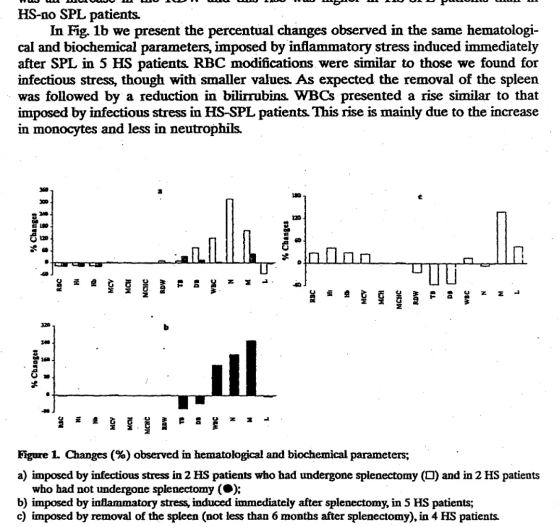
Figure la shows the percentual changes· in some hematological and biochemical parameters, imposed by infectious stress in 2 HS-SPL patients and in 2 HS-no SPL patients.
In
both cases we observed .a reduction in RBC concentration, which is accom-panied by an increase in bilirrubins. These modifications are followed by an increase in WBC concentration. This rise is Specially evident in HS-SPL patients and results mainly from arise
in neutrophils and less in monocytes. In HS-no SPL patients the WBC concentration rise is minor and results mainly from a rise in monocytes and less in neutropbils. Along with these changes the MCV rises and the MCHC reduces. There was an increase in the RDW and this rise was higher in HS-SPL patients than in HS-no SPL patients.In
Fig. lb we present the percentual changes observed in the samehematologi-cal and biochemical parameters, imposed by ·inft.ammatory stress induced immediately after SPL in
5
HS patients. RBC modifications were similar to those we found for infectious stress, though with smaller values. As expected the removal of the spleen was followed by· a reduction in bilirrubins. WBCs presented a rise similar to that imposed by infectious stress in HS-SPL patients. This rise is mainly due to the increase in monocytes and less in neutrophils.--
•
IlD c:; •
:.
•
tj12D
I2D
=
•
..
.:•
'$.• u
•
;!! D.QI 1!C
..
i! te a 6
ill: ~ •~· z; :Ill .;a.
..
..
..
:Ill :Ill
..
=
i! !
e
6a
I
• • u z z ..:a:11! 1-
..
..
:Ill :Ill ;J:
:E
-
b-:
:•
•...
u . '#
•
..
Figure L Olanges (%) obseived in hematological and biochemical parametexs;
a) imposed by infectious stress in 2 HS patients who had undergone splenectomy (D) and in 2 HS patients
who had not undergone splenectomy (e);
b) imposed by inflammatory stress. induced immediately after splenectomy, in 5 HS patients;
c) imposed by removal of the spleen (not less than 6 months after splenectomy), in 4 HS patients.
·-,.
Figure le shows the impact of SPL (more than six months after splenectomy) in
the same hematological and biochemical parameters, observed in 4 HS patients. We found a rise
in RBC concentration and
in the hematimetric indexes; the RDW and the
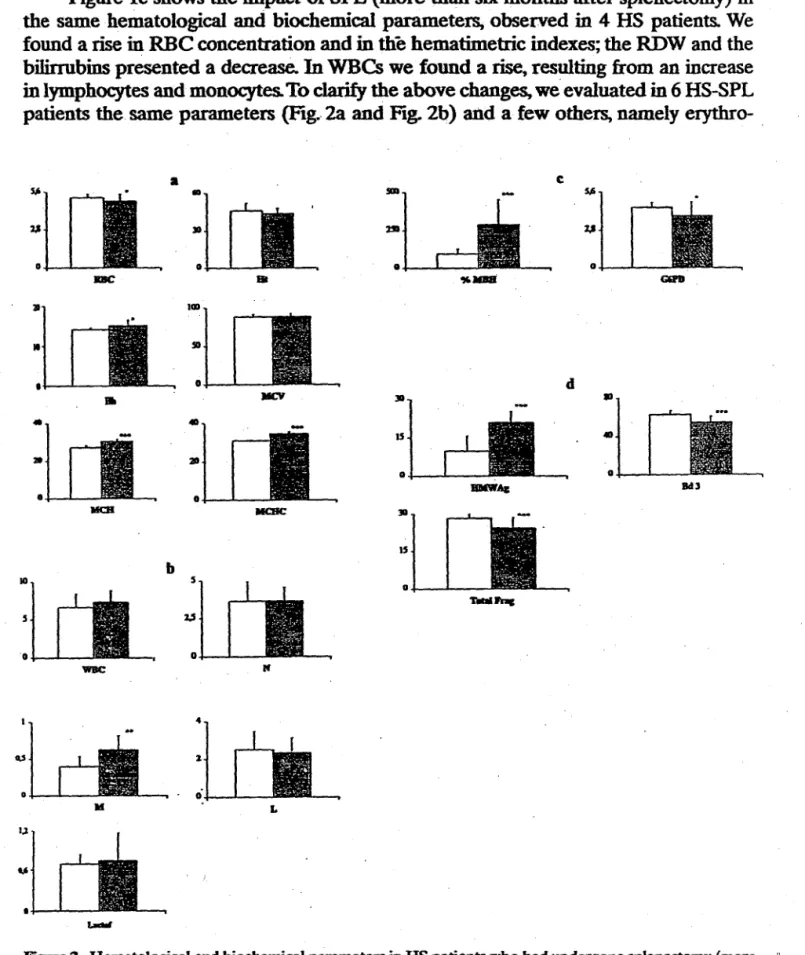
bilirrubins presented a decrease.. In WBCs we found a rise, resulting from an increase in lym.phocytes and monocytes. To clarify the above changes, we evaluated in 6 HS-SPL patients the. same parameters (Ftg .. 2a and Fig. 2b) and a few others, namelyerythro-a c
5,6
•
Slit 5,62,1 ~ 2,1
0 0 0
DC
..
"JIIIIi1111
SI
0 d
..
JIC'I Jll.,
•
•
IS
•
210 0
BlllWA& Bell
•
0MCll IICBC Jll
15
b
10 5
0
T.aiJ'nc
5 2,:1
D 0
M
4
"
%0
..
0L
1,1
u
I
L-dll
Figure 2. Hematological and biochemical parameters in HS patients who bad undergone splenectomy (more
than 6 months) and in a healthy controL
0 Controls (n = 29) • Patients HS-SPL (n
=
6)*P<O.OS -P<O.Ol-*P<O.OOl
a) RBC (xl012/l); Ht (% ); Hb. MCHC (gldl); MCV (fl); MCH (pg);
b) WBC. N. M. L (xl0'/1); Lactof (J.Lgfml);
c) %MBH (xlQ-4}; G6PD (UI/g Hb);
342 E. Granjo et aL
cyte G6PD activity and the %MBH (Fig. 2c ); band 3 profile (Fig. 2d); and plasma lacto-ferrin concentration (Fig. 2b ). The results were compared with those presented by a control including 29 healthy individuals {with no HS and no infection). We observed an evident improvement in RBC population though some of the characteristics of HS . . were still observed, namely a high M CH C. A significant reduction in erythrocyte G6PD, a significantly higher %MBH, and a different. band 3 profile were~, also observed. Actually, HS-SPL patients presented a significant increase in high molecular weight aggregates (HMW Ag) and a significant decrease in band 3 monomer (Bd 3) and in its proteolytic fragments (fotal frag). Concerning leukocytes we found increased values for WBC concentration and a significantly higher value in monocyte concentration. Plasma lactoferrin presented no statistical significance, although presenting a higher
value than that presented by the control. ·
DISCUSSION
Hereditary spherocytes, on account of its membrane modifications, are more sen-sitive to oxidative and proteolytic stresses. During an inflammatory or an infectious process, activated leukocytes may be an important source of products, namely oxygen metabolites, and .proteases. Either· of these leukocyte activation products can damage the neighbouring RBCs, imposing oxidative and proteolytic lesions to RBCs, which may lead to an accelerated aging and removal process. Therefore, the inlpact of inflamma-tory or infectious stress in spherocytes is probably enhanced and may impose an even more accelerated aging and removal process, leading to a hemolytic event.
We showed (Fig. la) that the changes in the hematological and biochemical para-meters studied, imposed by infectious stress in both HS-SPL and HS-no SPL patients, were similar. The reduction in RBC concentration was accompanied by a rise in bill-robins, revealing an increased RBC removal Along with these changes we observed a rise in WBC concentration, which was specially evident in HS-SPL patients and resulted mainly from a rise in neutrophils and less in monocytes. Hs-no SPL patients presented a minor rise in WBC concentration and resulted mainly from a rise in mono-cytes and less in neutropbils. These results suggest the involvement of these leukomono-cytes
in accelerated RBC aging, and premature removal. In addition, we showed that the MCV presented a rise and the MCHC a reduction in both HS patients, suggesting that molecular modifications occurred As expected, there was an increase in the RDW and the rise was higher in ·HS-SPL patients. This is probably the result of the increase in RBCs presenting the referred molecular changes. The change in the RDW value of HS-no SPL patients was lower since the spleen will filter the RBCs presenting those mol-ecular changes. In factT the reduction in RBC concentration and the rise in TB was higher in HS-no SPL, denoting an enhanced splenic RBCs removal It seems that the presence of the spleen, acting as a filter of damaged or senescent RBCs, renders the HS patients more susceptible to develop a hemolytic event during an inflammatory or an infectious stress.
The percentual changes (Hg.lb) in the same hematological and biochemical para-meters, imposed by inflammatory stress induced immediately after splenectomy in HS patients, were similar to those found for infectious stress, though with smaller"values. As expected after the removal of the spleen, a reduction in bilirubin was observed denoting reduced RBC destruction.
before) presented the expected changes for RBC concentration and bilirrubins. Actu-ally, the rise in RBC concentration and the reduction in bilirubin was followed by
clin-ical and hematologclin-ical improvement of the patients. The removal of the spleen, the filter of the damaged or senescent RBCs, seems to underlie these changes. However, we must expect that the absence of that filter may account for the accumulation of damaged RBCs. Contrary to the previous stressfull situations, in this case we observed a rise in·WBes, resulting from an increase in lymphocytes and monocytes. This suggests that the absence of the spleen may trigger a different mechanism to command the granulo-monocytopoiesis or even the immune system.
To clarify the above changes in RBCs, denoting accelerated RBC damage or senescence and the involvement of activated leukocytes in its development, we evalu-ated in HS-SPL patients and in a healthy control the same parameters and a few others, which could give us some information about the age of RBCs, about the amount of oxidative and proteolytic lesions suffered by RBCs during their life span and about leukocyte activation. Several months after splenectomy the HS patients presented an evident improvement in RBC concentration, showing that the absence of the spleen reduced the removal of damaged or senescent RBCs. We must note, however, that both MCH and MCHC maintained higher values when compared to the control, as it is characteristic of spherocytes. The reduction of RBC removal seems to be followed by the accumulation of older circulating RBCs, as shown by the significant decrease in erythrocyte G6PD activity (Fig. 2c). Strengthening this hypothesis we found also a
significantly higher %MBH and a different band
3
profile (Fig. 2d). HS-SPL presented a significant increase in HMWAg and a significant decrease in band 3 monomer and inits proteolytic fragments. When compared this profile with that presented by the control, it appears that HMWAg might result from oxidation and crosslinking of band
3 monomer and its proteolytic fragments or even from a further proteolysis of band 3
followed by oxidation and crosslinking .of proteolytic fragments. These results provide further evidenee of the premature senescence of RBCs in these patients. We must emphasize that these changes for band 3 profile are similar to those found in patients under chronic stressfull situations, such as cardiovascular disease, 15 and similar to those found for RBC aging in vitro, i.e. exposed to aCtivated neutrophils or neutrophi!ic elastase.16 Concerning. leukocytes, we found higher values for HS-SPL patients for
all leukocyte types, tbough only for monocyte8 we did observe a signifiCantly higher value when compared to the control (Fig. 2b ). We must emphasize that mon~cytes
!,. • are the WBC which pr~nt a higher rise in both inflammatory and infectious stress in
!' . HS-no
SPL
,.,
The destabilized membrane structure of spherocytes seems to underlie membrane
:i. vesiculation, premature aging and premature removal of the cells. Whenever an
~,
inftammatory or an infectious stress develops, a rise in leukocytes, mainly in neutrophilsF
and monocytesoCcurs.
It is known that leukocytes present a reduced deformability andr:.
that its activation may produce and release oxygen metabolites and proteases. The rise· in leukocytes of reduced deformability may siow the blood flow and favour the inter-action of the released leukocyte activation products with the surrounding cells, namely · with the spherocytes. It seems, therefore, reasonable to assume that the change in blood
5 tlow and the easier interaction of leukocyte activation products with the circulating ·\. RBCs, is even more enhanced within the splenic microvasculature, favouring oxidative, · and proteolytic damage of spherocytes. The o.xidative and proteolytic lesions, by \ inducing splenic sequestration, may lead to premature removal of a higher number of
E. Granjo et aL
In conclusion, our data show that inflammatory and infectious stress by inducing leukocytosis and eventually leukocyte activation, may underlie and trigger hemolytic events in HS patients. In addition, o/oMBH aDd band 3 profile may provide further evi-. dence of the oxidative and proteolytic modifications imposed upon RBCs by leukocyte
activation products.
ACKNOWLEDGMENT
Supported in part by research grants from the "Funda~o Gomes Tei"(eira", Universidade do Porto, and PRAXIS XXI nOUl.l/SAU/1240/95 (JNICf).
REFERENCES
1. Delaunay J. Alloisio N. Morle L. Cane G: La spherocytose hereditaire en 1995: l'apport de la
gene-tique moleculaire. Hematologie 2:115. 1995.
2. Cynober T. Mohandas N. Tchernia G: Red cell abnormalities in hereditary spherocytosis: relevance
to diagnosis and uriderstanding of the variable expression of clinical severity. J Lab Clin Med 128:259,
1996.
3. Delaunay J: Hereditary hemolytic anemias due to defects in membrane proteins. Curr Opinion Hematol59,1993. ·
4. Lutz HU: ~rocyte clearance. In: Harris JR, editor. Blood cell biochemistry. Vol 1 Erythroid cells.
New York: Plenum Press, 1981. p 81.
5. Brovelli A. Castellana MA. Minetti G. Piccinini G. Seppi C. Renzis MR. Balduini C: COnformational
changes and oxidation of membrane proteins in senescent human erythrocyte. In: Magnani M and De
Flora A, editors. Red Blood Cell Aging. New York: Plenum Press. 1991, p 59.
6. Castellana MA, Piccinini G. Minetti G. Seppi C. Balduini C. Brovelli A: Oxidation of membrane
pro-teins and functional activity of band 3 in human red cell senescence. Arch Gerontal Geriatr Suppl
3:101, 1992.
7. Bosch FH. Were JM, Schipper L. Roerdinkholder-Stoelwinder B. Huls T. Wdlekens. FLA. Wichers G,
Halie Mf: Determinants of red blood cell deformability in relation to cell age. Eur J Haematol52:35,
1994.
8. Kay MMB. Goodman S. Wbitfield C. Wong P. Zaki L. Rudloff V: Senescent cell antigen in
immuno-logically related to band 3. Proc Nad Acad Sci 80:1631, 1984.
9. Kay MMB: Localization of senescent cell antigen on band 3. Proc Natl Acad Sci 81:57~3. 1984.
10. Lutz HU: Naturally occurring anti-band 3 anb.Dodies. 'Thmsf Med Rev 7:201. 1992.
11. Kay MMB: Role of physiologic autoantibody in the removal of senescent human red cells. J Supra
Mol Struct 9-.555, 1978. •
12. Lutz HU. Bussolino S. Flepp R. Fasler S. Stammler P. Kazatchkine MD, Arese P: Naturally occurring
anti-band 3 antibodies and complement together mediate phagocytosis of oxidatively stressed human
erythrocytes. Proc Natl Acad Sci USA 84:7368. 1987.
13. Weiss DJ, Klausner JS: Neutrophil-induced erythrocyte injury: a potential cause of erythrocyte
destruction in anaemift associated with inflammatory disease. Vet Pathol 25:450. 1988.
14. Rebelo I, Guerra FC. Leite LP. Quintanilha A: Lactoferrin as a sensitive blood marker of neutrophil
activation in normal pregnancies. Eur J Obstet Gynccol Reprod Bioi 62:189. 1995.
15. Santos-Silva A. Castro EMB. Teixeira NA, Guerra FC, Quintanilha A: Altered erythrocyte membrane
band 3 profile as a marker in patients at risk for cardiovascular disease Atherosclerosis 116:199,1995.
16. Santos-Silva A, Castro EMB, Teixeira NA, Guerra FC, Quilitanilha A: Erythrocyte membrane band
3 profile imposed by cellular aging, by activated neutrophils and by neutrophilic elastase. Clin Cbim
Acta 275(2):185. 1998.
17. MucCurdy PR. Sherman S: Irreversibly sickled cells and red cell survival in sickle cell anemia. Am J
Med 64:253. 1978.
18. Rachmilewitz EA. S~ E. Shalev O. Galili
u.
Schrier SI.: Eiythrocyte membrane alterations inP..
19. l(aneko JJ: Comparative erythrocyte metabolism. Adv Vet Sci Comp Med 18:117, 1974.
20.. Santos-Silva A, Guerra FC, Ri'beiro MM. Teixeira N: Deficiencia em glucose-6-fosfato desidrogenase
(G6PD)~ Estabelecimento de valores de referencia dA sua actividade no eritr6cito. Estudo de dois
hemizigotos. Rev Port Farm 39:84.
1~-21. Dodge IT, MitcheU C, Hanaha.B J: The preparation and chemical characteristics of hemoglobin-free
ghosts of human erythrocytes. Arch Biochem Biophys 100:119, 1963.
22. Bradford MM: A rapid and sensitive method for the quantitation of microgram quantities of protein
utilizing the principle of protein dye binding. Anal Biochem 72:. 255, 1976.
23. Laemmli UK: Oeavage of structural proteins during the assembly of the head of bacteriophage T4.
Nature 227:680, 1970.
24. Towbin H, Stahelin T, Gordon J: Electrophoretic transfer of proteins from polyacrylamide gels to