Erythropoietin levels in the different clinical forms of hereditary
spherocytosis
Hereditary spherocytosis (HS) is the most common red cell membrane disorder affecting one in 2500 individuals of northern European ancestry (Bolton-Maggs et al, 2004). It is a congenital haemolytic anaemia that is dominantly inherited in 75% of cases, the remaining cases show a recessive or non-dominant pattern of inheritance, such as spontaneousde novo
mutations, which are often caused by family-specific private mutations (Agre et al, 1986; Eber et al, 1990; Miraglia del Giudiceet al, 1998).
Hereditary spherocytosis is highly heterogeneous in terms of clinical and laboratory presentation, ranging from an asymp-tomatic condition to a severe life-threatening transfusion-dependent anaemia (Bolton-Maggs et al, 2004). According to haematological and clinical presentation, HS is usually classi-fied as mild, typical or moderate, and severe (Bolton-Maggs
et al, 2004). The presence of spherocytes in the peripheral
blood smear, the increase in red blood cell (RBC) osmotic fragility and reticulocytosis are hallmarks of HS (Iolasconet al, 1998).
The erythrocytes in HS, by loosing membrane surface area, undergo a spherocytic shape change and a reduction in their deformability. These changes are because of defects in RBC membrane proteins, including ankyrin, band 3, spectrin and protein 4Æ2. The increased rigidity of the cell membrane leads to premature spleen trapping and destruction of spherocytes, causing the haemolysis experienced by these patients. The clinical manifestations of HS are, therefore, usually abrogated by splenectomy (Tse & Lux, 1999).
In healthy humans, erythropoiesis counterbalances the continuous removal of aged RBCs. About 1% of circulating RBCs are destroyed daily and replaced by reticulocytes. Erythropoietin (EPO) is the main growth factor responsible
S. Rocha,1E. Costa,2C. Catarino,1,3
L. Belo,1,3E. M. B. Castro,1,3J. Barbot,2
A Quintanilha1,4and A. Santos-Silva1,3
1Instituto de Biologia Molecular e Celular, Universidade do Porto,2Hospital Crianc¸as Maria Pia, Servic¸o Hematologia,3Faculdade de Farma´cia, Servic¸o Bioquı´mica, Universidade do Porto, and4Instituto Cieˆncias Biome´dicas Abel Salazar, Universidade do Porto, Porto, Portugal
Received 2 June 2005; accepted for publication 2 August 2005
Correspondence: Alice Santos Silva, Servic¸o de Bioquı´mica, Faculdade de Farma´cia, Rua Anı´bal Cunha, 164, 4099-030 Porto, Portugal. E-mail: assilva@ff.up.pt
Summary
Erythropoietin (EPO), the main growth factor responsible for the regulation of red blood cell production, may be overproduced when blood loss or haemolysis occurs. Patients with mild hereditary spherocytosis (HS) are able to maintain normal haemoglobin concentration, whereas typical and severe HS patients develop an anaemic state. Splenectomy usually reverses anaemia. We aimed to clarify the role of EPO in the response to enhanced spherocyte destruction, and to look for a linkage with the broad clinical spectra of HS. EPO levels, reticulocyte count and production index (RPI), other parameters used to classify HS and the protein deficiencies underlying HS were evaluated
in previously diagnosed unsplenectomised (n¼24) and splenectomised
(n¼10) patients presenting mild, typical or severe HS. A significant increase
in EPO was observed in all unsplenectomised HS patients. In the mild form, a significant correlation of EPO with reticulocyte count and RPI was observed; however, this correlation disappeared in typical HS patients. Splenectomised HS patients presented a correction in EPO levels in all forms of HS, although the reticulocyte count and RPI sustained slightly higher values. Our data show HS as a disease linked to an overproduction of EPO, according to the severity of the disease; however, a disturbance in erythropoiesis seems to occur in typical HS. Moreover, splenectomy leads to a correction in the EPO levels.
Keywords: hereditary spherocytosis, erythropoietin, erythropoietin response,
for the regulation of RBC production in steady-state condi-tions and for enhancing the production rate of RBCs, whenever blood loss or haemolysis occurs (Krantz, 1991; Jelkmann, 1992). Besides the kidney, which is the main site of production in the adult, additional organs and cells, including liver, uterus, endothelial cells, vascular smooth muscle cells and insulin-producing cells have been found to secrete EPO (Fisher, 2003; Maieseet al, 2005).
The primary stimulus for increasing EPO synthesis is tissue hypoxia resulting from reduced blood oxygen availability. By inducing EPO gene expression and synthesis, hypoxia leads to a rise in the plasmatic levels of EPO (Ebert & Bunn, 1999). In the bone marrow, the erythroid progenitor cells, erythroid burst-forming units (BFU-E) and erythroid colony-forming units (CFU-E), present specific receptors for EPO on their surface. When EPO binds to the receptors on these cells, they proliferate and develop into erythroblasts and, finally, to reticulocytes that enter the peripheral circulation and will mature into RBC (Lappin, 2003).
When the reticulocytes are released normally from the bone marrow, remaining in circulation for the normal period of time for maturation into erythrocytes (about 24 h), the number of circulating reticulocytes is an accurate reflection of the erythropoietic activity. However, under an erythro-poietic stimulus, reticulocytes are prematurely released and the maturation time of the ‘stressed’ reticulocytes in the peripheral blood may be as long as 3 days. In this case, the reticulocyte count needs to be corrected for both changes in haematocrit (degree of anaemia) and in the effect of EPO on premature reticulocyte release from the bone marrow. Considering the common features in HS patients (anaemia and reticulocytosis), we have calculated the reticulocyte production index (RPI) as an appropriate way to measure the effective RBC production (Hillman & Ault, 2002).
Patients with mild HS, despite the increased RBC destruc-tion, are able to maintain a normal haemoglobin concentra-tion, probably on account of the physiologically increased EPO synthesis and erythroid expansion. However, in moderate and severe HS an anaemic state develops, suggesting a disturbance in that physiological mechanism.
Splenectomy of HS patients is usually effective in reducing haemolysis and improving the anaemia by leading to a significant prolongation of the RBC life span, though not always to a normal value (Chapman & McDonald, 1968; Baird
et al, 1971). Since splenectomy abrogated the clinical mani-festations, the diagnosis of HS was likely to lead to splenec-tomy irrespective of the severity of HS, for several years, and explains why some mild and typical HS patients were splenectomised (Zinkham, 1969). Nowadays, patients are selected for splenectomy on the basis of their age, clinical symptoms and presence of complications, such as gallstones (Bolton-Maggset al, 2004).
The erythrocyte membrane protein deficiencies underlying HS are usually identified by sodium dodecyl sulphate-polyacrylamide gel electrophoresis (SDS-PAGE) of membrane
proteins (Bolton-Maggs et al, 2004). The same protein defi-ciency may account for a mild, typical or severe presentation of the disease. It is accepted that the most commonly affected proteins are ankyrin and band 3 protein, and less frequently spectrin and protein 4Æ2 (Miraglia del Giudice et al, 1994; Lanciottiet al, 1997). We recently reported that band 3 and ankyrin deficiencies are the major causes for HS in a Portuguese studied sample (Rochaet al, 2005).
In this study, the plasmatic EPO levels of unsplenectomised HS patients presenting a mild or a typical form of HS, and splenectomised HS patients presenting a mild, typical or severe HS were evaluated, in order to clarify the role of EPO in the physiological response to enhanced spherocyte destruction and to look for a connection to the broad haematological and clinical spectra of the disease and to the underlying erythrocyte membrane protein deficiencies. Moreover, we also evaluated the EPO response after splenectomy according to severity of HS.
Materials and methods
Subjects
We studied 56 individuals from the north of Portugal: 22 controls, 18 patients with mild HS (three were splenectom-ised), 13 patients presenting typical HS (four were splenec-tomised) and three splenectomised patients with severe HS. The control group included three children, 15 adults and four elderly; the mild HS group included 10 children and eight adults; the typical HS group included seven children and six adults and the severe HS patients included two children and one adult. We used children in this study because it is in childhood that the disease is usually diagnosed, with the severe cases recommended for splenectomy at an appropriate age. We felt, therefore, that children should be a good target group to study, in order to assemble all forms of the disease. Actually, many HS adult patients with typical and severe HS are splenectomised and, therefore, could not meet all of our proposed aims. We included in all groups of patients a similar number of children and adults, and no elderly were included. Patients who had undergone splenectomy had been sub-mitted to surgery more than 1 year before this study.
The 34 HS patients were previously diagnosed and classified as presenting mild, typical or severe HS, according to haematological and biochemical data, and clinical follow-up. The membrane protein deficiency underlying HS was identi-fied and quantiidenti-fied for all patients by electrophoretic analysis of RBC membrane proteins.
shown by the individuals at time of blood collection for the present studies.
In the case of severe HS patients, there was no contamin-ation with transfused blood, as the last transfusion took place at least 4 months before the collection of blood for our studies. All severe HS patients were splenectomised. Three HS patients classified as mild had been splenectomised in adulthood, as they developed a symptomatic vesicular litiasis. None of the controls and patients presented chronic inflammatory diseases, abnormal renal function or iron and folic acid deficiencies, at time of blood collection.
Assays
Blood samples [ethylenediaminetetraacetic acid (EDTA) and heparin as anticoagulants] were collected and processed for the haematological and biochemical analysis. To exclude immune haemolysis a direct antiglobulin test was performed for all studied cases (Reganet al, 2001).
We evaluated the RBC count, haemoglobin concentration, the red cell distribution width (RDW), mean cell haemoglobin (MCH) and mean cell haemoglobin concentration (MCHC), by using an automatic blood cell counter (Sysmex K1000, Sysmex, Hamburg, Germany).
The search for spherocytic cells was performed in Wright stained blood films (International Committee for Standardi-sation in Haematology, 1984). Osmotic fragility test was carried out on fresh blood and after 24 h of incubation at 37C (Roperet al, 2001). Serum concentration of total bilirubin was measured by routine biochemical procedures.
Reticulocyte count was performed by microscopic counting on blood smears after vital staining with new methylene blue (Reticulocyte stain; Sigma, St Louis, MO, USA). Considering the common features in HS patients – anaemia and reticul-ocytosis – we have calculated the reticulocyte production index (RPI) as an appropriate way to measure the effective RBC production (Hillman & Ault, 2002). Plasma was separated after centrifugation and EPO plasma levels were evaluated by an enzyme-linked immunosorbent assay (Human EPO Quanti-kine IVD ELISA Kit; R&D Systems Inc, Minneapolis, MN, USA).
To prepare the RBC membranes for electrophoretic analysis, plasma and leucocytes were previously isolated from RBCs and discarded after centrifugation on a double density gradient (Histopaque 1Æ077 and 1Æ119; Sigma). RBCs were washed in saline solution. The erythrocytes were lysed by hypotonic lyses according to Dodge et al (1963). The obtained membranes were washed in Dodge buffer, with phenylmethylsulphonyl fluoride added as a protease inhibitor, in the first two washes, with a final concentration of 0Æ1 mmol/l. The protein concen-tration of the RBC membrane suspensions was determined by the Bradford method (Bradford, 1976).
The electrophoretic analysis of the red cell membrane proteins was carried out on a discontinuous SDS-PAGE, using a 5–15% linear acrylamide gradient gel and a 3Æ5–17%
exponential acrylamide gradient gel, according to Laemmli (1970) and Fairbankset al (1971), respectively. The proteins were stained with Coomassie brilliant blue, and scanned (Darkroom CN UV/wl; BioCaptMW, version 99; Vilber Lourmat, Marne-La-Valle´e, France). The relative amount of each major protein (as a percentage of total), was quantified by densitometry (Bio1D++, version 99; Vilber Lourmat).
The electrophoretic analysis for each RBC membrane sample was performed in duplicate gels, and, in each gel, duplicates of each sample were loaded alongside with six control samples. The mean values presented by the studied subjects were compared with those presented by the control samples. A protein deficiency was considered significant whenever the mean value of that protein was lower than the mean value minus 2·SD presented by the control samples for the same protein (subject mean protein % value < control mean protein % value)2·SD).
Statistical analysis
Measurements are presented as mean ± SD or as median values (inter-quartile range). For statistical analysis, we used the Statistical Package for Social Sciences (spss, version 12.0, SPSS Inc, Chicago, IL, USA) for Windows. To evaluate the differences between groups, we used the Student’s t-test whenever the parameters presented a Gaussian distribution and the Mann–Whitney test in the case of a non-Gaussian distribution. Spearman’s rank correlation coefficient was used to evaluate relationships between sets of data. AP-value lower than 0Æ05 was considered as statistically significant.
Results
In Table I, we present the haematological values for controls and HS patients, according to the severity of the disease. For each form of the disease, we present the results for unsple-nectomised and spleunsple-nectomised patients. We observed that, in unsplenectomised patients, worsening of the disease was associated with a reduction in RBC count, Hb concentration and haematocrit, and a rise in MCHC, RDW, and in osmotic fragility. In splenectomised HS patients, we found a correction for all those parameters, except for MCHC and the osmotic fragility, which sustained a higher value when compared with controls, and, actually, presented a trend to rise with severity of the underlying disease.
The bilirubin levels (Table II) increased with worsening of the disease and reached significantly higher values in typical HS unsplenectomised patients. Splenectomised HS patients showed a reduction in these values, though sustaining a trend to higher values, when compared with controls.
compared with the control values. In splenectomised HS patients the values had been corrected by splenectomy.
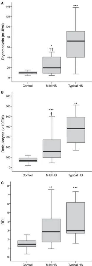
Considering the reticulocyte count and RPI, reflecting the rate of RBC production, we found that in HS unsplenectom-ised patients (Table II; Fig 1B and C) the reticulocyte count increased significantly with the severity of the disease and the same occurred for RPI, though the rise observed for typical HS patients in RPI was not as striking as that observed in mild HS, in which the values were double those of the control. When comparing these EPO levels, reticulocyte count and RPI presented by unsplenectomised patients with mild and typical HS, we found significantly different values for EPO (P< 0Æ01) (Fig 1A) and for reticulocyte count (P< 0Æ05) (Fig 1B), but not for RPI (Fig 1C).
The 34 studied HS patients showed different erythrocyte membrane protein deficiencies underlying the disease (Table III). In 26 patients an isolated band 3 or combined with protein 4Æ2 deficiency was observed; 17 had mild HS, eight typical HS and one severe HS. An ankyrin and/or combined spectrin and protein 4Æ2 deficiencies was found in one case of mild HS and two of typical HS. In four patients with spectrin deficiency, two of them presented as typical HS and the others as severe HS. A case of protein 4Æ2 deficiency was also observed, presenting as typical HS.
Concerning splenectomised patients, a correction for EPO was observed in all forms of the disease; a higher reticulocyte count and RPI than controls was observed in mild, typical and severe patients.
Looking for the relationship between the erythropoietic stimulus and the effective RBC production in accordance with the severity of the disease, we studied the correlation of EPO levels with reticulocyte count and RPI in unsplenectomised mild and typical HS patients. Mild HS patients (Fig 2) showed a significant positive correlation between EPO and the reticulocyte count (r¼0Æ825;P< 0Æ001), as well as with RPI (r¼0Æ757; P< 0Æ01). However, no significant correlations were observed between EPO and reticulocyte count (r¼0Æ200), as well as with RPI (r¼0Æ117) for typical HS patients (Fig 3).
We also looked for a connection between the type of protein deficiency and EPO levels in mild and typical HS unsplenec-tomised patients (Fig 4). We found that in typical HS patients presenting a primary ankyrin deficiency, the EPO levels were higher than the median values observed in controls and in mild unsplenectomised HS patients. The same occurred for the two studied cases of spectrin deficiency presenting a typical HS. However, for primary band 3 deficiency with typical HS, we found that in three of the five studied cases, the EPO levels were within the values observed for controls (one case) and mild (two cases) unsplenectomised HS patients.
Discussion
The membrane pathology in HS is effected by a change in cell shape from the deformable biconcave disc into a spherocytic
poorly deformable cell, seen on the blood smears as sphero-cytes. The vertical and horizontal interactions between mem-brane constituents account for the integrity, strength and deformability of the cell. Disruptions of vertical interactions, because of membrane protein deficiencies, favour membrane vesiculation and, therefore, the development of spherocytes.
Hereditary spherocytosis occurs in most racial groups, is the commonest cause of inherited chronic haemolysis in the Northern Europe and usually presents an autosomal inherit-ance pattern. It was described in 1871 and, since then, much has been clarified about the structure of the red cell membrane and the genetic defects causing HS. However, some contro-versies still exist about the clinical outcome of the disease. It presents a clinical broad spectrum, being usually classified as mild, typical or moderate and severe, according to clinical and analytical follow-up. Although the clinical severity of HS differs among families, it is quite similar within a family presenting an autosomal pattern of inheritance (Eber et al, 1990; Iolascon et al, 1998). The differences in HS clinical expression within a family have been ascribed to a heterozy-gous combination of interacting alleles, a common allele and a weak allele and to a coinheritance of another erythrocyte disorder, able to affect HS phenotype (Alloisio et al, 1996, 1997). The mechanism underlying the heterogeneous clinical outcome of the disease among families is, also, still poorly clarified. The same type and amount of the primary protein deficiency underlying HS may present a different clinical expression. Recently, we reported that similar values of protein deficiencies in the deficient proteins (primary and secondary) was observed in mild HS patients, while in typical and severe HS a trend to an unbalance was found, suggesting the importance of a balanced interaction between proteins in the clinical outcome of the disease (Rocha et al, 2005). Some authors have reported a relationship between clinical severity of HS with specific membrane protein defects (Miraglia del Giudice et al, 1994), and others with the quantitative defici-ency in spectrin, even when it is secondary to a primary deficiency in another membrane protein (Agre et al, 1986).
Moreover, the severity of the disease is closely associated with the severity of haemolysis and spleen size.
In accordance with the literature (Iolascon et al, 1998; Bolton-Maggset al, 2004), we observed that in unsplenectom-ised HS patients, worsening of the disease was associated with a reduction in RBC count, Hb concentration and haematocrit, and a rise in MCHC, RDW, and in osmotic fragility (Table I). These changes were associated with an increased haemolysis, as suggested by the rise in bilirubin levels (Table II). The haemolytic reduction in the number of circulating RBCs, leading to a reduction in tissue oxygen delivery, triggers the physiological mechanism to compensate this condition, by enhancing EPO production to stimulate erythropoiesis. Actu-ally, we observed an erythropoietic stimulus, as shown by the increase in the EPO levels, in reticulocyte count and in the RPI (Table II; Fig 1A–C). In unsplenectomised mild HS patients the median levels of EPO were double those of the control values and the same occurred for reticulocyte count and for RPI. In typical unsplenectomised HS patients, the significantly higher haemolysis, as shown by the significant increase in bilirubin concentration, was associated to a rise in EPO levels that was almost quadruple those presented by mild HS patients. However, this erythropoietic stimulus was not paralleled by a similar effective reticulocyte production. Typical unsplenectomised HS patients presented a median reticulocyte count value, which was only 2Æ5-fold the value of the mild unsplenectomised HS patients, and the median value of RPI was similar to those presented by the mild unsplenec-tomised HS patients. Considering the overlap between the error bars for reticulocyte count and for RPI between the mild and typical groups (Fig 1B and C), as well as the small number of typical unsplenetomised HS patients, we removed the outliers in this group, and found no substantial effect in the statistical results, nor in the subsequent conclusions.
To clarify the results, we studied the correlations between EPO levels and the effective reticulocyte production. We found that in mild HS a significant positive correlation was observed between stimulus and effective erythropoietic response (Fig 2);
Table II. Biochemical and haematological data for controls and HS patients according to severity of HS.
Group Bilirubin (lmol/l) Erythropoietin (mUI/ml) Reticulocytes (109/l) RPI
Controls (n¼22) 8Æ80 (5Æ83–13Æ4) 9Æ68 (7Æ58–11Æ6) 67Æ4 (55Æ8–96Æ3) 1Æ43 (1Æ18–1Æ86) Mild HS (n¼18)
15 unspl. 9Æ00 (6Æ66–36Æ0) 19Æ7 (7Æ58–42Æ0)* 156 (91Æ6–287)*** 2Æ84 (1Æ54–4Æ69)** 3 spl. 19Æ0 (10Æ2–27Æ6) 8Æ70 (5Æ29–12Æ9) 91Æ1 (90Æ0–108) 1Æ85 (1Æ80–2Æ04)
Typical HS (n¼13)
9 unspl. 34Æ7 (14Æ0–106)** 72Æ2 (34Æ3–108)*** 380 (233–509)*** 2Æ96 (2Æ62–6Æ20)***
4 spl. 17Æ0 (11Æ0–24Æ5) 8Æ27 (6Æ18–10Æ0) 141 (108–158)** 2Æ92 (2Æ00–2Æ99)**
Severe HS (n¼3)
3 spl. 13Æ1 (7Æ42–18Æ8) 11Æ2 (8Æ40–14Æ8) 127 (66Æ2–156) 1Æ83 (1Æ12–2Æ58)
Data are presented as median (IQR) values. no spl., unsplenectomised; spl., splenectomised. ***P< 0Æ001; **P< 0Æ01; *P< 0Æ05 (HSversuscontrol).
however, in typical HS the same was not observed, suggesting a failure or a disturbance in the response mechanism, despite the significantly higher erythropoietic stimulus (Fig 3).
As far as we know, only Guarnoneet al(1996) have reported an over production of EPO in compensated and anaemic HS patients (some of them were splenectomised), and suggested this feature as a biological characteristic of the disease. Our data, in agreement with this report, shows high levels of EPO in all unsplenectomised HS patients, whatever the severity of the disease. This overproduction of EPO reflects the adaptive response to tissue hypoxia resulting from enhanced and premature RBC removal.
Moreover, we found that the balance between the erythro-poietic stimulus and the effective production of reticulocytes observed in mild patients was no longer observed in typical HS patients. The significantly higher levels of EPO in typical HS patients were not able to induce the expected reticulocyte production.
All the major events involved in RBC development, survival, growth and maturation are maintained by EPO binding to the receptors of the maturating erythroid cells (Lappin, 2003). These receptors appear gradually along the maturation of progenitor cells, and begin to disappear with the onset of haemoglobin synthesis in the early erythroblast (D’Andrea & Zon, 1990; Remyet al, 1999). Afterwards, the rate of prolif-eration will be mainly determined by the availability of the erythropoietic nutrients, iron, vitamin B12 and folic acid.
It is known that in chronic diseases associated with inflammatory processes, the release of cytokines (tumour necrosis-factor, interferon-gamma) may affect erythropoiesis, inducing usually a mild to moderate anaemia. The more likely explanation for the development of this anaemia is a decreased production and/or action of EPO. Cytokines involved in the inflammatory response may inhibit both EPO synthesis and erythroid colony formation (Faquin et al, 1992; Means & Krantz, 1992), as well as downregulate the EPO receptor at the surface of the progenitor erythroid cells (Taniguchiet al, 1997). 140
120
100
80
60
40
Er
ythropoietin (m
Ul/ml)
A
B
C
Reticulocytes (× 10E9/l)
RPI
20
0
700
600
500
400
300
200
100
0
8
7
6
5
4
3
2
1
0
Control
* ††
***
**
** *** †
***
Mild HS Typical HS
Control Mild HS Typical HS
Control Mild HS Typical HS
Fig 1. Median values (IQR) for erythropoietin levels (A), reticulocyte
count (B) and reticulocyte production index (C) in control, mild and typical unsplenectomised HS patients. ***P< 0Æ001; **P< 0Æ01; *P< 0Æ05 (HSversuscontrol); P< 0Æ05; P< 0Æ01 (mildversustypical).
Table III.Protein deficiency and clinical classification.
Spectrin deficiency
Ankyrin and/ or combined spectrin and protein 4Æ2 deficiencies
Isolated band 3 or combined with protein 4Æ2 deficiency
Protein 4Æ2 deficiency
Mild HS
spl. – – 3 –
unspl. – 1 14 –
Typical HS
spl. – – 3 1
unspl. 2 2 5 –
Severe HS
spl. 2 – 1 –
Total 4 3 26 1
In HS, the reduced deformability of the RBCs imposes an enhanced spherocyte sequestration and removal by the macro-phages of the reticulo-endothelial system. We speculated if in
HS, as occurs in chronic diseases, this enhanced macrophage activation could be involved in the inhibition of EPO synthesis and/or of the EPO response, as seems to occur with worsening of the disease. Another possible explanation for the inhibition of EPO response could involve the EPO receptors, which may be lacking or presenting an impaired transduction signal, possibly also because of the inflammatory cytokines and breakdown products of dead or dying cells (Erslev, 2001).
Strengthening the hypothesis of the involvement of inflam-matory cytokines in the inhibition of EPO response with worsening of HS, we found that the EPO levels were corrected after splenectomy, although the reticulocyte count and the RPI could still sustain higher values. Splenectomy, by reducing RBC removal and, therefore, macrophage activation seems to allow, thereafter, an appropriate EPO response.
The electrophoretic analysis of the erythrocyte membranes (Table III) showed that isolated band 3 or combined with protein 4Æ2 deficiency was the major cause for HS, and that spectrin deficiency and ankyrin and/or combined spectrin and protein 4Æ2 deficiencies presented a similar prevalence in the studied group (n¼4 andn¼3, respectively). Only one case of protein 4Æ2 deficiency was observed. These results are slightly different from those, we recently reported for a larger sample of Portuguese HS patients (Rochaet al, 2005), in which the major cause for HS was a primary band 3 deficiency, followed by primary ankyrin deficiency and spectrin defici-ency. The differences are probably the result of the different size of the studied groups. The search for a connection between EPO levels and the type of protein deficiency underlying HS (Fig 4), showed that in typical HS patients both with a primary ankyrin deficiency and with a spectrin deficiency, the EPO levels were higher than the values observed in controls and in mild unsplenectomised HS patients. However, for primary band 3 deficiency with typical HS, we found that in three of the 500
A
B 400
300
200
100
0
8
6
4
RPI
2
0
0 10 20
Erythropoietin (m30 40UI/ml)
r=0·825
P < 0·01
r = 0·757
P < 0·01 50 60
0 10 20
Erythropoietin (mUI/ml) 30 40 50 60
Reticulocytes (10E9/l)
Fig 2. Correlation of erythropoietin levels with reticulocyte count and
reticulocyte production index (RPI) in mild unsplenectomised HS patients.
800
600
400
200
0
0 20 40 60 80 100 120 140 Erythropoietin (mUI/ml)
0 20 40 60 80 100 120 140 Erythropoietin (mUI/ml)
RPI
8
6 7
4 5
3
2
1
r=0·200
r = 0·117
Reticulocytes (10E9/l)
A
B
Fig 3. Correlation of erythropoietin levels with reticulocyte count and
reticulocyte production index (RPI) in typical unsplenectomised HS patients.
140
120
100
80
60
40
20
0
Spectrin Ankyrin
mild HS typical HS
Band 3
Er
ythropoietin (m
Ul/ml)
Fig 4. Erythropoietin values according to erythrocyte protein
five studied cases, the EPO levels were within the values observed for controls (one case) and for mild (two cases) unsplenectomised HS patients. We may speculate if, in this type of protein deficiency, an inhibition of EPO synthesis may occur instead of an inhibition of the EPO response, as seems to occur in the other types of protein deficiencies. However, this hypothesis warrants further studies considering the number of the studied cases. Anyway, this hypothesis seems to be reasonable, as, in the case of protein band 3 deficiency, fewer vertical protein interactions may coexist with a normal cytoskeleton, and, in the case of skeletal protein deficiency, a fewer number of vertical interactions coexist with an almost normal number of integral membrane proteins. It is known that the pathway for the removal of senescent RBCs involves a senescent neoantigen, which has been related to band 3 protein (Kayet al, 1983). Clustering, proteolysis or even exposure of unusual epitopes, trigger the binding of specific autoanti-band 3 antibodies and complement activation (Lutz et al, 1987; Lutz, 1992), marking the cell for death.
While spectrin- and ankyrin-deficient RBCs escape opsonisation by releasing band 3-containing vesicles with increasing cell age, in the case of band 3-deficient RBCs they do not lose band 3 with cell aging (Reliene et al, 2002). Therefore, band 3 clusters required for the bivalent binding of immunoglobulin (Ig)G naturally occurring anti-band 3 antibodies (Kayet al, 1991; Lutz, 1992) may further enhance macrophage activation, which may condition erythropoiesis in a different way.
Since EPO was cloned in 1985, recombinant human erythropoietin (rHu-EPO) has been used for the treatment of anaemia associated with renal failure, human immunode-ficiency virus (HIV) infection in az(ido)t(hymidine) (AZT)-treated patients, and chemotherapy (Cazzola et al, 1998; Lacombe & Mayeux, 1998). Recently, it has also been administered to infants with HS presenting a severe anaemia. Most of them improved their response in achieving a compensated haematological state (Tcherniaet al, 2000; Schiff
et al, 2003); however, few of them did not reach sustained transfusion-independence despite EPO therapy. Recently, it was reported that chronic kidney disease patients may also present resistance to EPO therapy (Macdougall, 2004) and that patients who persistently failed to respond expressed abnor-mally raised levels of the pro-inflammatory cytokines tumour necrosis factor and interferon-gamma, which are known to inhibit erythropoiesis. Further studies are warranted to clarify the mechanism that may disturb the erythropoietic response in these pathologies and in HS, as it may lead to the improvement of new therapeutic strategies.
In conclusion, our data shows HS as a disease linked to an overproduction of EPO in accordance with the severity of the disease. Moreover, it suggests that further studies are warran-ted to clarify the inhibition of the EPO response observed with worsening of HS, and to clarify the erythropoietic response according to the type of protein deficiency underlying the disease.
Acknowledgements
This study was in part supported by a grant (POCTI- PL 122204 – BIC) from the ‘Fundac¸a˜o para a Cieˆncia e Tecnologia’.
References
Agre, P., Asimos, A., Casella, J.F. & McMillan, C. (1986) Inheritance pattern and clinical response to splenectomy as a reflection of ery-throcyte spectrin deficiency in hereditary spherocytosis.New Eng-land Journal of Medicine,315,1579–1583.
Alloisio, N., Maillet, P. & Carre´, G. (1996) Hereditary spherocytosis with band 3 deficiency. Association with a non-sense mutation of the band 3 gene (Allele Lyon), and aggravation by a low expression allele occurring in trans (Allele Genas). Blood, 88,
1062–1069.
Alloisio, N., Texier, P. & Valliet, A. (1997) Modulation of clinical expression and band 3 deficiency in hereditary spherocytosis.Blood,
90,414–420.
Baird, R.N., Macpherson, A.I.S. & Richmond, J. (1971) Red-blood cell survival after splenectomy in congenital spherocytosis.Lancet, ii,
1060–1061.
Bolton-Maggs, P.H.B., Stevens, R.F., Dodd, N.J., Lamont, G., Titten-sor, P. & King, M.-J. (2004) Guidelines for the diagnosis and management of Hereditary Spherocytosis.British Journal of Hae-matology,126,455–474.
Bradford, M.M. (1976) A rapid and sensitive method for the quanti-fication of microgram quantities of protein utilizing the principle of the protein dye binding.Analytical Biochemistry,72,248–254. Cazzola, M., Guarnone, R., Cerani, P., Centenara, E., Rovati, A. &
Beguin, Y. (1998) Red blood cell precursor mass as an independent determinant of serum erythropoietin level.Blood,91,2139–2145. Chapman, R.G. & McDonald, L.L. (1968) Red cell life span after
splenectomy in hereditary spherocytosis. Journal of Clinical Investigation,47,2263–2267.
D’Andrea, A.D. & Zon, L.I. (1990) Erythropoietin receptor. Subunit structure and activation.Journal of Clinical Investigation,86,681–687. Dodge, J.T., Mitchell, C. & Hanahan, D.J. (1963) The preparation and chemical characteristics of hemoglobin-free ghosts of human erythrocytes. Archives of Biochemistry and Biophysics, 100, 119– 130.
Eber, S.W., Armbrust, R. & Schro¨ter, W. (1990) Variable clinical severity of hereditary spherocytosis: relation to erythrocyte spectrin concentration, osmotic fragility and autohemolysis. Journal of Pediatrics,117,409–416.
Ebert, B.L. & Bunn, H.F. (1999) Regulation of the erythropoietin gene. Blood,94,1864–1877.
Erslev, A.J. (2001) Anemia of chronic disease. In:Williams Hematology, 6th edn (ed. by E. Beutler, M.A. Lichtman, B.S. Coller, T.J. Kipps & U. Seligsohn), pp. 481–487. McGraw-Hill, New York.
Fairbanks, G., Steck, T.L. & Wallach, D.F.H. (1971) Electrophoresis of the major polypeptides of the human erythocyte membrane. Bio-chemistry,10,2606–2616.
Faquin, W.C., Schneider, T.J. & Goldberg, M.A. (1992) Effect of in-flammatory cytokines on hypoxia-induced erythropoietin produc-tion.Blood,79,1987–1994.
Guarnone, R., Centenara, E., Zappa, M., Zanella, A. & Barosi, G. (1996) Erythropoietin production and erythropoiesis in compen-sated and anaemic states of hereditary spherocytosis.British Journal of Haematology,92,150–154.
Hillman, R.S. & Ault, K.A. (2002) Clinical approach to anemia. In: Hematology in Clinical Practice, 3rd edn (ed. by R.S. Hillman & K.A. Ault), pp. 12–26. McGraw-Hill, New York, NY.
International Committee for Standardization in Haematology (1984) ICSH reference method for staining of blood and bone marrow films by azure B and eosin Y (Romanowsky stain). British Journal of Haematology,57,707–710.
Iolascon, A., del Giudice, E.M., Perrotta, S., Alloisio, N., Morle´, L. & Delaunay, J. (1998) Hereditary Spherocytosis: from clinical to molecular defects.Haematologica,83,240–257.
Jelkmann, W. (1992) Erythropoietin: structure, control of production, and function.Physiological Reviews,72,449–489.
Kay, M.M.B., Goodman, S.R., Sorensen, K., Whitfield, C.F., Wong, P., Zaki, L. & Rudloff, V. (1983) Senescent cell antigen is immunologically related to band 3. Proceedings of the National Academy of Sciences United States of America,80,1631–1635. Kay, M.M.B., Marchalonis, J.J., Schluter, S.F. & Bosman, G. (1991)
Human erythrocyte aging: cellular and molecular biology. Transfu-sion Medical Review,3,173–195.
Krantz, S.B. (1991) Erythropoietin.Blood,77,419–434.
Lacombe, C. & Mayeux, P. (1998) Biology of erythropoietin. Haema-tologica,83,724–732.
Laemmli, U.K. (1970) Cleavage of structural proteins during the assembly of the head of the bacteriophage T.Nature,227,680–685. Lanciotti, M., Perutelli, P., Valetto, A., Di Martino, D. & Mori, P.G. (1997) Ankyrin deficiency is the most common defect in dominant and non dominant hereditary spherocytosis. Haematologica, 82,
460–462.
Lappin, T. (2003) The cellular biology of erythropoietin receptors.The Oncologist,8,15–18.
Lutz, A. (1992) Naturally occurring anti-band 3 antibodies. Transfu-sion Medical Review,6,201–211.
Lutz, H.U., Bussolino, F., Flepp, R., Fasler, S., Stammler, P., Kazatchkin, M.D. & Arese, P. (1987) Naturally occurring anti-band 3 antibodies and complement together mediate together mediate pha-gocytosis of oxidatively stressed human erythrocytes.Proceedings of the National Academy of Sciences USA,84,7368–7372.
Macdougall, I.C. (2004) Could anti-inflammatory cytokine therapy improve poor treatment outcomes in dialysis patients?Archives de Pediatrie,19,V73–V78.
Maiese, K., Li, F. & Chong, Z.Z. (2005) New avenues of exploration for of erythropoietin.Journal of the American Medical Association,293,
90–95.
Means, R.T., Jr & Krantz, S.B. (1992) Progress in understanding the pathogenesis of the anemia of chronic disease.Blood,80,1639–1647.
Miraglia del Giudice, E., Iolascon, A., Pinto, L., Nobili, B. & Perrotta, S. (1994) Erythrocyte membrane protein alterations underlying clinical heteregoneity in hereditary spherocytosis. British Journal of Hae-matology,93,828–834.
Miraglia del Giudice, E., Francese, M., Nobili, B., Morle´, L., Cutillo, S., Delaunay, J. & Perrotta, S. (1998) High frequency of de novo mutations in ankyrin gene (ANK1) in children with hereditary spherocytosis.Journal of Pediatrics,132,117–120.
Regan, F., Newlands, M. & Bain, B.J. (2001) Acquired haemolytic anaemias. In:Dacie and Lewis Practical Haematology(ed. by S.M. Lewis, B.J. Bain & I. Bates), pp. 199–229. Churchill Livingstone, London.
Reliene, R., Mariani, M., Zanella, A., Reinhart, W.H., Ribeiro, M.L., Miraglia del Giudice, E., Perrotta, S., Iolascon, A., Eber, S. & Lutz, H.U. (2002) Splenectomy prolongs in vivo survival of erythrocytes differently in spectrin/ankyrin- and band 3-deficient herediatry spherocytosis.Blood,100,2208–2215.
Remy, I., Wilson, I.A. & Michnick, S.W. (1999) Erythropoietin receptor activation by ligand-induced conformation change.Science,
283,990–993.
Rocha, S., Rebelo, I., Costa, E., Catarino, C., Belo, L., Castro, E.M.B., Cabeda, J.M., Barbot, J., Quintanilha, A. & Santos-Silva, A. (2005) Protein deficiency balance as a predictor of clinical outcome in hereditary spherocytosis.European Journal of Haematology,74,374– 380.
Roper, D., Layton, M. & Mitchell Lewis, S. (2001) Investigation of the hereditary hemolytic anemias: membrane and enzyme abnormalities. In: Dacie and Lewis Practical Haematology(ed. by S.M. Lewis, B.J. Bain & I. Bates), pp. 167–198. Churchill Liv-ingstone, London.
Schiff, M., Hays, S., Sann, L. & Putet, G. (2003) Recombinant human erythropoietin (r-HuEPO) therapy in a newborn with hereditary spherocytosis.Archives de Pediatrie,10,333–336.
Taniguchi, S., Dai, C.H., Price, J.O. & Krantz, S.B. (1997) Interferon gamma downregulates stem cell factor and erythropoietin receptors but not insulin-like growth factor-I receptors in human erythroid colony-forming cells.Blood,90,2244–2252.
Tchernia, G., Delhommeau, F., Perrotta, S., Cynober, T., Bader-Meu-nier, B., Nobili, B., Rohrlich, P., Salomon, J.L., Sagot-Benevot, S., Miraglia del Giudice, E., Delaunay, J., DeMattia, D., Schischmanoff, P.O., Mohandas, N. & Iolascon, A. (2000) Recombinant ery-thropoietin therapy as an alternative to blood transfusions in infants with hereditary spherocytosis.The Hematology Journal,1,146–152. Tse, W.T. & Lux, S.E. (1999) Red blood cell membrane disorders.
British Journal of Haematology,104,2–13.