Birth Weight and Polycystic Ovary Syndrome
in Adult Life: Is There a Causal Link?
Stavroula A. Paschou1, Dimitrios Ioannidis2, Evangeline Vassilatou3, Maria Mizamtsidi1, Maria Panagou2, Dimitrios Lilis2, Ioanna Tzavara2, Andromachi Vryonidou1*
1Department of Endocrinology and Diabetes, Hellenic Red Cross Hospital, Athens, Greece,2Department of Endocrinology and Diabetes,“Amalia Fleming”Hospital, Athens, Greece,3Endocrine Unit, Second Department of Medicine,“Attikon”University Hospital, Athens, Greece
*mahi_vr@hotmail.com
Abstract
Objectives
Several studies have demonstrated associations of birth weight with metabolic and repro-ductive abnormalities in adults. The aim of this study was to investigate the birth weight in women with PCOS and its correlation with clinical and biochemical characteristics of the syndrome.
Materials and Methods
We studied 288 women with PCOS according to the NIH criteria and 166 women with nor-mal cycle and without clinical hyperandrogenism. Birth weight and anthropometric charac-teristics were recorded, and levels of serum androgens, SHBG, insulin and fasting glucose were measured.
Results
Birth weight data were available for 243/288 women with PCOS and age- and BMI-matched 101/166 controls. No differences were found (p>0.05) in birth weight among women with PCOS and normal controls. Birth weight of PCOS women was negatively correlated with DHEAS levels (p = 0.031, r = -0.143) and positively correlated with waist circumference (p<0.001, r = 0.297) and body mass index (BMI) (p = 0.040, r = 0.132). Birth weight of con-trols was negatively correlated with SHBG levels (p = 0.021, r = -0.234). Women from both groups were further divided in 6 categories according to birth weight (A.<2.500 gr, B. 2.501-3.000 gr, C. 3.001-3.500 gr, D. 3.501-4.000 gr, E. 4.001-4.500 gr, F.>4.500 gr). No statistically significant differences were observed in the distribution percentages be-tween PCOS women and controls. (A. 7% vs 7.9%, B. 26.8% vs 20.8%, C. 39.1% vs 48.5%, D. 21.4% vs 20.8%, E. 4.9% vs 2%, F. 0.8% vs 0%), (in all comparisons, p>0.05).
OPEN ACCESS
Citation:Paschou SA, Ioannidis D, Vassilatou E, Mizamtsidi M, Panagou M, Lilis D, et al. (2015) Birth Weight and Polycystic Ovary Syndrome in Adult Life: Is There a Causal Link?. PLoS ONE 10(3): e0122050. doi:10.1371/journal.pone.0122050
Academic Editor:Harpal Singh Randeva, University
of Warwick–Medical School, UNITED KINGDOM
Received:December 1, 2014
Accepted:February 11, 2015
Published:March 19, 2015
Copyright:© 2015 Paschou et al. This is an open access article distributed under the terms of the Creative Commons Attribution License, which permits unrestricted use, distribution, and reproduction in any medium, provided the original author and source are credited.
Data Availability Statement:All relevant data are within the paper.
Funding:These authors have no support or funding to report.
Conclusions
Women with PCOS do not differ from controls in birth weight distribution. However, birth weight may contribute to subtypes of the syndrome that are characterized by adrenal hyper-androgenism and central obesity.
Introduction
Birth weight can be considered as the“reflection”of endometrial life, representing in a large
scale the maternal environment. It has been hypothesized that Polycystic ovary syndrome
(PCOS) may have early origins in intrauterine life [1]. Evidence from experimental and clinical
studies suggest that the endometrial environment may induce permanent changes in tissue structure or function favoring the development of PCOS in adult life. Prenatal androgen excess or deranged nutritional conditions during pregnancy may result in homeostatic adaptations of female fetuses through complicated mechanisms of fetal programming, which can lead to a
more masculine and obese phenotype [2–3].
A few studies have been conducted to investigate a causative link between women’s birth
weight and PCOS and results were inconclusive [4–8]. Some of them have demonstrated an
as-sociation of low birth weight with hyperandrogenism and PCOS phenotype in adult life [4–6].
Furthermore, there are data showing that not only low but also increased birth weight may be
associated with the presence of PCOS [5–6]. On the contrary, other large epidemiological
stud-ies did not show any association of birth weight with reproductive and metabolic abnormalitstud-ies
in PCOS patients, their female or their male relatives [7–8].
The aim of this study was to investigate birth weight in women with PCOS and its possible association with clinical and biochemical characteristics of the syndrome.
Materials and Methods
Patients and Controls
We studied 288 women with PCOS (25 ± 6.1 years) and 166 women (25.7 ± 6.4 years) as nor-mal controls. Patients with PCOS were selected from the outpatient clinics of two centers
(Hel-lenic Red Cross Hospital and“Amalia Fleming”Hospital). Control women were medical or
dietology students or hospitals0personnel.
Research has been approved by the Institutional Review Boards of the Hellenic Red Cross Hospital and "Amalia Fleming" Hospital in Athens, Greece and all clinical investigations have been conducted according to the principles expressed in the Declaration of Helsinki. Written informed consent was obtained from all participants.
We used the National Institute of Health (NIH) diagnostic criteria for PCOS patients; less than eight menses per year and a free androgen index (FAI) greater than 5 and/or clinical
hyperandrogenism (presence of acne and/or hirsutism) [9]. Other causes of anovulation and
hyperandrogenism were excluded with appropriate tests. Control women reported regular menstrual cycles and no clinical evidence of hyperandrogenemia.
Study protocol
Medical and reproductive history of PCOS patients and controls was recorded and physical ex-amination was undertaken by endocrinologists. Birth weight and gestational age were self-reported for the majority of participants, while for the rest information was obtained from medical records. Nevertheless, in a previous study, it has been shown that there is a high degree
of concordance between self-reported and actual birth weight [8].
Anthropometric measurements including weight, height and waist circumference were per-formed. Clinical hyperandrogenism was assessed by the presence of acne and/or hirsutism
(modified Ferriman—Gallwey score>8) [10].
Morning blood samples were drawn from all participants, after an overnight fast. Plasma
glucose, insulin, total testosterone (TT),Δ4-androstenedione (Δ4A), dehydroepiandrosterone
sulfate (DHEA-S) and sex hormone binding globulin (SHBG) were determined in the early fol-licular phase of the menstrual cycle. HOMA-IR (homeostatic model assessment-insulin resis-tance) was calculated by using the mathematic model HOMA-IR = glucose x insulin/405
(glucose in mg/dl) for the evaluation of insulin resistance [11].
Assays
Plasma Glucose levels were measured by an enzymatic, colorimetric method in a Cobas Integra/400/700/800 autoanalyzer (Roche Laboratory Systems).
Serum insulin was measured by an immunoradiometric assay (IRMA, DIASource
Immuno-assays S.A.) with a sensitivity of 1μIU/ml and intra- and interassay coefficients of variation of
2.1% and 6.5%, respectively. Serum SHBG levels were measured by an immunoradiometric assay (IRMA, Immunotech s.r.o.) with a sensitivity of 0.4 nmol/l and intra- and interassay coef-ficients of variation of 6.1 and 8.3%, respectively. TT was measured by radioimmunoassay (RIA, Cisbio Bioassays) with a sensitivity of 0.086 ng/ml and intra- and interassay coefficients
of variation of 6% and 8.5%, respectively.Δ4A levels were measured by radioimmunoassay
(RIA, DIASource Immunoassays S.A.) with a sensitivity of 0.03 ng/ml and intra- and interassay coefficients of variation of 4.5% and 9%, respectively. DHEA-S levels were measured by
radio-immunoassay (RIA, Coat-A-Count DPC-Siemens) with a sensitivity of 1.1μg/dl and
intra-and interassay coefficients of variation of 5.3% intra-and 8.1%, respectively.
Statistical Analysis
Results are presented as mean ± SD for continuous variables. Results are presented as absolute numbers or percentages for categorical variables. Differences in continuous variables between groups were tested using the independent T-Test or Mann-Whitney U test, as appropriate.
Dif-ferences in categorical variables between groups were tested usingχ2test with Yates
Correc-tion. Pearson’s or Spearman’s Correlation was used to explore the association between pairs of
continuous variables, as appropriate. All statistical analyses were performed using the
Statisti-cal Package for Social Sciences (SPSS 16.0, Inc, Chicago, IL, USA). A p value of<0.05 was
con-sidered statistically significant.
Results
The clinical and biochemical characteristics of the study participants are shown inTable 1.
Pa-tients and controls were matched for age and BMI. Women with PCOS had significantly
in-creased waist circumference (p<0.001), and HOMA-IR (p<0.001) compared to controls. They
p<0.001 and DHEA-S p<0.001) and significantly reduced levels of SHBG (p<0.001)
com-pared to controls, as expected (Table 1).
Birth weight data were available for 243/288 PCOS women and for 101/166 controls. No
dif-ferences in birth weight were found (p>0.05) between PCOS women (3243 ± 522 gr) and
nor-mal controls (3212 ± 451 gr) (Table 2).
Table 1. Comparison of important characteristics between patients with PCOS and controls.
PCOS Patients (n = 288) Controls (n = 174) p value
Age (years) 25±6.1 25.7±6.4 ns
BMI (kg/m2) 28.4
±7.3 27.2±7.5 ns
Waist Circumference (cm) 89.7±17.8 83±16.3 <0.001
HOMA-IR 3.27±2.1 2.2±1.5 <0.001
TTesto (ng/ml) 0.89±0.24 0.41±0.14 <0.001
Δ4A (ng/ml) 3.48±1.3 1.8±0.5 <0.001
DHEAS (μg/dl) 295.5±122.6 218.8±87.7 <0.001
SHBG (nmol/lt) 31.6±13.3 51.5±21.5 <0.001
Birth Weight (gr) 3243±522 (*n = 243) 3212±451 (*n = 101) ns
BMI: body mass index; HOMA-IR: homeostatic model assessment-insulin resistance; TTesto: Total Testosterone;Δ4A: androstenedione; DHEAS: dehydroepiandrosterone sulfate; SHBG: sex hormone binding globulin.
doi:10.1371/journal.pone.0122050.t001
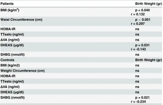
Table 2. Correlations of birth weight with important clinical and biochemical parameters of PCOS.
Patients Birth Weight (gr)
BMI (kg/m2) p = 0.040
r = 0.132
Waist Circumference (cm) p<0.001
r = 0.297
HOMA-IR ns
TTesto (ng/ml) ns
Δ4A (ng/ml) ns
DHEAS (μg/dl) p = 0.031
r = -0.143
SHBG (nmol/lt) ns
Controls Birth Weight (gr)
BMI (kg/m2) ns
Weight Circumference (cm) ns
HOMA-IR ns
TTesto (ng/ml) ns
Δ4A (ng/ml) ns
DHEAS (μg/dl) ns
SHBG (nmol/lt) p = 0.021
r = -0.234
BMI: body mass index; HOMA-IR: homeostatic model assessment-insulin resistance; TTesto: Total Testosterone;Δ4A: androstenedione; DHEAS: dehydroepiandrosterone sulfate; SHBG: sex hormone binding globulin.
In PCOS women birth weight was negatively correlated with DHEA-S levels (p = 0.031, r =
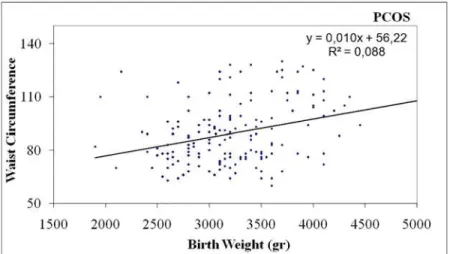
-0.143) (Table 2,Fig. 1), positively correlated with waist circumference (p<0.001, r = 0.297)
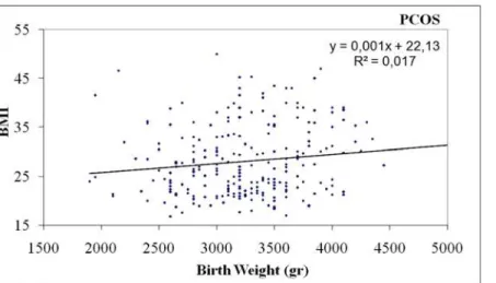
(Table 2,Fig. 2) and body mass index (BMI) (p = 0.040, r = 0.132) (Table 2,Fig. 3). In controls,
birth weight was negatively correlated with SHBG levels (p = 0.021, r = -0.234) (Table 2,
Fig. 4).
The negative correlation of birth weight with DHEA-S levels in PCOS women was con-firmed when we separated them in quartiles according to DHEA-S levels. Women from the highest quartile had statistically significant lower birth weight compared to the women from
the lowest quartile (3118 ± 563 vs 3326 ± 497 gr, p = 0.033) (Table 3).
Women from both groups were further divided in 6 categories according to birth weight
(A.<2.500 gr, B. 2.501–3.000 gr, C. 3.001–3.500 gr, D. 3.501–4.000 gr, E. 4.001–4.500 gr, F.>
4.500 gr) and percentage of women for each category was calculated. The distribution
percent-ages in the above categories for both PCOS patients and controls are shown inTable 4. There
was no statistically significant difference in the distribution percentages for each category be-tween the two groups (A. 7% vs 7.9%, B. 26.8% vs 20.8%, C. 39.1% vs 48.5%, D. 21.4% vs
20.8%, E. 4.9% vs 2%, F. 0.8% vs 0%), (in all comparisons, p>0.05) (Table 4).
Discussion
In the present study we investigated the birth weight in women with PCOS and its possible cor-relation with clinical and biochemical characteristics of the syndrome. We did not find any dif-ferences neither in the mean birth weight nor in the distribution percentages of birth weight categories between PCOS women and normal controls.
Based on Barker’s intrauterine programming theory for chronic diseases [12] and the fact
that insulin resistance is a cardinal feature of PCOS, a few studies have been conducted to in-vestigate whether a causative link exists between birth weight and PCOS in adult life. An asso-ciation between low or high birth weight and PCOS was found in some of them, but their
results are constrained by the small number of participants [4,13–14], investigation of specific
group of patients, i.e. with precocious pubarche [13] and a deficient reproductive and
metabol-ic phenotyping [5,15–16].
Our findings are similar to the results of two large epidemiological studies. The first one, a well-designed and family-based study from the United States of America, showed that birth weight in families with PCOS defined by NIH criteria did not differ from the general popula-tion, even when it was corrected for gestational age. Furthermore, the investigators did not find any association of birth weight with reproductive and metabolic abnormalities both in women
with PCOS and their relatives, males or females [8]. The second, a Finnish birth cohort study
of 2007 women, showed no relationship between birth weight and self-reported PCOS
symp-toms of oligomenorrhea and hirsutism at the age of 31 years [7].
At variance, two recent studies with a large number of participants have shown that the presence of PCOS is not only associated with low but with increased birth weight as well. The first one was a retrospective birth cohort study of 948 singleton female babies from Australia. The results after adjusting for gestational age, suggested that two discrete fetal programming pathways, one related to high birth weight and the other to thinness at birth, are operating
to-wards the manifestation of PCOS in adult life [5]. The second, a study from Denmark with the
huge number of 523.757 participants had similar findings. Data were extracted from the Dan-ish Civil Registration System and the DanDan-ish National Patient Register with the ICD codes and
showed that the risk of PCOS was increased only in women born4,500 g. Moreover, women
born from mothers diagnosed with pre-gestational or gestational diabetes were at increased
Similar results yielded from two other studies which investigated the link between birth weight and PCOS from another aspect i.e. by examining the birth weight of newborns of moth-ers with PCOS. The first one from Chile, found a significantly higher prevalence of babies small for gestational age in mothers with PCOS compared to newborns of normal women,
matched for age and weight at the beginning of pregnancy [17]. The second one was a study
from the Swedish Medical Birth Record comprising 1.195.123 singleton births between 1995 and 2007. A higher risk for being large for gestational age (1.39, 1.19 to 1.62) was documented
among 3787 births from mothers with a PCOS diagnosis [18].
A possible explanation for these conflicting data could be that PCOS encompasses various phenotypic subtypes that are dictated by the parental genetic traits of the individual, the mater-nal contribution during fetal life and the adult environment. In large epidemiological studies, small subtypes (with low or high birth weight) probably yield no significant differences. Anoth-er explanation may be that the relation of birth weight and PCOS risk is not linear but a
U-shaped, as it has been shown for insulin resistance and type 2 diabetes [19]. If this is the case
then, a much larger number of patients is needed to prove this hypothesis.
Fig 1. Negative correlation of birth weight with DHEAS levels in women with PCOS.
doi:10.1371/journal.pone.0122050.g001
Fig 2. Positive correlation of birth weight with waist circumference in women with PCOS
An important finding of our study was that birth weight was negatively correlated with lev-els of DHEA-S and positively correlated with waist circumference and body mass index only in women with PCOS. To our knowledge, this association has not been found before, and it may be relevant to the fact that previous studies had not examined these parameters or included them in statistical analysis.
DHEA-S, produced by the adrenal glands, is an important pro-hormone of sex steroids from in utero and throughout life. Adrenal glands are vital organs for survival. Adverse endo-metrial conditions, which are reflected by low birth weight, may modulate the function of such organs, by changes in blood flow redistribution or in metabolic rates, a phenomenon known as
developmental plasticity. This programming of“hyper-function”of the adrenal glands, which
may be crucial for fetus survival in an adverse intrauterine environment, could lead to obesity
and adrenal hyperandrogenism in childhood and possibly in adult life [20–21]. Several studies
have shown that low birth weight is positively correlated with high DHEA-S levels in girls just
before adrenarche until early adolescence [22–24]. Furthermore, a longitudinal study of a small
group of lean girls with low birth weight and precocious pubarche from Spain showed that
Fig 3. Positive correlation of birth weight with BMI in women with PCOS.
doi:10.1371/journal.pone.0122050.g003
Fig 4. Negative correlation of birth weight with SHBG levels in Controls.
more than one third of them developed functional adrenal hyperandrogenism in adolescence [25].
Approximately 20–30% of women with PCOS have elevated levels of DHEA-S and this may
be the only abnormality in circulating androgens in almost 10% of these women. Furthermore,
an augmented adrenal response to exogenous ACTH stimulation [26–27] as well as an
en-hanced adrenal steroid production capacity up to menopause was documented in some
women with PCOS [28–29]. However, taking into consideration that low birth weight (<2500
gr) infants represent only 3–10% of total births in relevant studies [30] we can assume that
other factors apart from birth weight such as heritability, insulin resistance or ovarian
hyperan-drogenemia could be responsible for high DHEA-S levels in women with PCOS [31]. Thus,
those with low birth weight may constitute a subtype of the syndrome.
Obesity mainly central, is found in almost 50% of PCOS women and constitutes a character-istic feature of PCOS that can be encountered even in normal-weight patients. It can exacerbate insulin resistance as well as hyperandrogenemia and lead to an unfavorable metabolic profile
[32–33]. Obesity and insulin resistance are strong heritable traits in PCOS families including
male relatives [34–35]. We found that high birth weight in PCOS women was positively
associ-ated with BMI and waist circumference, a surrogate marker of central adiposity.
Epidemiological studies have documented that both low and high birth weight confer an
in-creased risk for childhood and adult obesity [36–37]. Fetal under- or over-nutrition can have
long term consequences for descendants’health and continue the vicious cycle of obesity
epi-demic [38]. Thus, high birth weight in women with PCOS could have an additive effect on the
development of obesity, insulin resistance and hyperandrogenism in adulthood [34].
In control women, the above correlations did not exist. However, birth weight in these women was negatively correlated with the levels of SHBG. SHBG has an important role in the
regulation of bioavailable sex steroids, throughout human life (prenatal & antenatal) [39].
Ge-netic, hormonal and metabolic factors modulate SHBG production from the liver [21].
More-over, SHBG was proved to have an inverse relation with obesity and insulin resistance and low
SHBG levels are an independent risk factor for type 2 diabetes development [40]. In PCOS
women SHBG levels are often low, independent of obesity and insulin resistance and may
Table 3. Birth weight in women with PCOS according to DHEAS levels
DHEAS Q1* DHEAS Q2 DHEAS Q3 DHEAS Q4*
Birth Weight (gr) 3326±497 3298±550 3230±467 3118±563
Q: Quartile;
*Q1 vs Q4 p = 0.033, in all other comparisons p>0.05.
doi:10.1371/journal.pone.0122050.t003
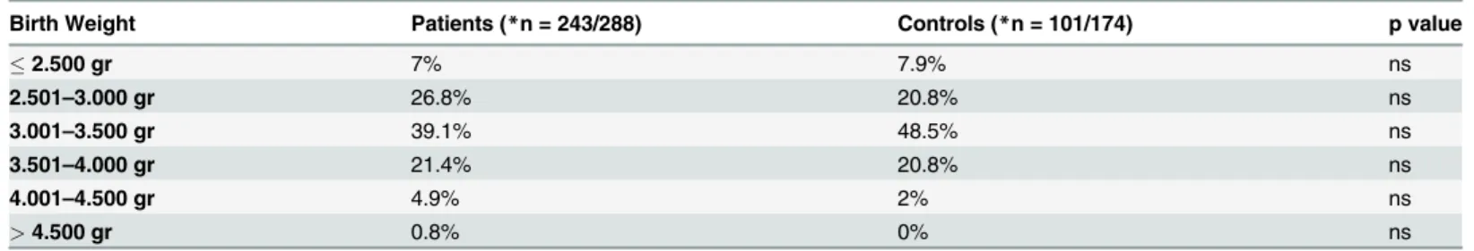
Table 4. Percentages of patients and controls according to birth weight categories.
Birth Weight Patients (*n = 243/288) Controls (*n = 101/174) p value
2.500 gr 7% 7.9% ns
2.501–3.000 gr 26.8% 20.8% ns
3.001–3.500 gr 39.1% 48.5% ns
3.501–4.000 gr 21.4% 20.8% ns
4.001–4.500 gr 4.9% 2% ns
>4.500 gr 0.8% 0% ns
contribute to hyperandrogenic phenotype of the syndrome [34]. However, data upon birth weight correlation with SHBG in adult non PCOS women are very limited. In 592
premeno-pausal women from the Nurses’Health Study no correlation was found between birth weight
and SHBG levels [41]. In a study of young women from France, intrauterine growth restriction
had no effect on SHBG levels [42]. A possible explanation for this finding is that SHBG levels
may be the consequence of fetal programming for obesity and/or insulin resistance in adult women, not genetically determined to develop PCOS.
One limitation of our study could be the sample size. While it is large enough for a clinical study, it is relatively small from a general epidemiological point of view. Strengths of this study is the well-defined population of Caucasian women with PCOS and controls, that were all full-term born, with a similar socio-economic status.
In conclusion, in this cohort of women with PCOS birth weight was similar to age- and BMI-matched control women, probably due to a neutral net-effect of combined genetic, epige-netic and maternal factors upon birth weight. However, birth weight was associated with clini-cal and biochemiclini-cal parameters of PCOS and may contribute to the phenotypic subtypes of the syndrome. Low birth weight was associated with adrenal hyperandrogenism, while high birth weight was associated with central obesity, both of which are features of the syndrome.
Author Contributions
Conceived and designed the experiments: SAP DI EV MM MP DL IT AV. Performed the ex-periments: SAP DI EV MM MP DL IT AV. Analyzed the data: SAP DI EV MM MP DL IT AV. Contributed reagents/materials/analysis tools: SAP DI EV MM MP DL IT AV. Wrote the paper: SAP DI EV MM MP DL IT AV.
References
1. Conway G, Dewailly D, Diamanti-Kandarakis E, Escobar-Morreale HF, Franks S, Gambineri A, et al. The polycystic ovary syndrome: a position statement from the European Society of Endocrinology. Eur J Endocrinol. 2014; 17: P1–29.
2. Xita N, Tsatsoulis A. Fetal origins of the metabolic syndrome. Ann N Y Acad Sci. 2010; 1205: 148–55. doi:10.1111/j.1749-6632.2010.05658.xPMID:20840267
3. de Zegher F, Ibáñez L. Early Origins of polycystic ovary syndrome: hypotheses may change without
no-tice. J Clin Endocrinol Metab. 2009; 94: 3682–5. doi:10.1210/jc.2009-1608PMID:19808859
4. Melo AS, Vieira CS, Barbieri MA, Rosa-E-Silva AC, Silva AA, Cardoso VC, et al. High prevalence of polycystic ovary syndrome in women born small for gestational age. Hum Reprod. 2010; 25: 2124–31. doi:10.1093/humrep/deq162PMID:20573680
5. Davies MJ, March WA, Willson KJ, Giles LC, Moore VM. Birthweight and thinness at birth independent-ly predict symptoms of poindependent-lycystic ovary syndrome in adulthood. Hum Reprod. 2012; 27: 1475–80. doi: 10.1093/humrep/des027PMID:22373955
6. Mumm H, Kamper-Jørgensen M, Nybo Andersen AM, Glintborg D, Andersen M. Birth weight and
poly-cystic ovary syndrome in adult life: a register-based study on 523,757 Danish women born1973–1991. Fertil Steril. 2013; 99: 777–82. doi:10.1016/j.fertnstert.2012.11.004PMID:23200688
7. Laitinen J, Taponen S, Martikainen H, Pouta A, Millwood I, Hartikainen AL, et al. Body size from birth to adulthood as a predictor of self-reported polycystic ovary syndrome symptoms. Int J Obes Relat Metab Disord. 2003; 27: 710–5. PMID:12833115
8. Legro RS, Roller RL, Dodson WC, Stetter CM, Kunselman AR, Dunaif A. Associations of birth weight and gestational age with reproductive and metabolic phenotypes in women with polycystic ovarian syn-drome and their first-degree relatives. J Clin Endocrinol Metab. 2010; 95: 789–99. doi: 10.1210/jc.2009-1849PMID:19965924
9. Zawadzki JK, Dunaif A. Diagnostic criteria for polycystic ovary syndrome; towards a rational approach. In: Dunaif A, Givens JR, Haseltine F, Merriam G, eds. Polycystic ovary syndrome. Boston: Blackwell Scientific. 1992;377–384.
11. Matthews DR, Hosker JP, Rudenski AS, Naylor BA, Treacher DF, Turner RC. Homeostasis model as-sessment: insulin resistance and beta-cell function from fasting plasma glucose and insulin concentra-tions in man. Diabetologia. 1985; 28: 412–9. PMID:3899825
12. Barker DJ. In utero programming of chronic disease. Clin Sci (Lond). 1998; 95: 115–28. PMID: 9680492
13. Ibáñez L, Potau N, Francois I, de Zegher F. Precocious pubarche, hyperinsulinism, and ovarian hyper-androgenism in girls: relation to reduced fetal growth. J Clin Endocrinol Metab. 1998; 83: 3558–3562. PMID:9768664
14. Pandolfi C, Zugaro A, Lattanzio F, Necozione S, Barbonetti A, Colangeli MS, et al. Low birth weight and later development of insulin resistance and biochemical/clinical features of polycystic ovary syndrome. Metabolism. 2008; 57: 999–1004. doi:10.1016/j.metabol.2008.02.018PMID:18555843
15. Michelmore K, Ong K, Mason S, Bennett S, Perry L, Vessey M, et al. Clinical features in women with polycystic ovaries: relationships to insulin sensitivity, insulin gene VNTR and birth weight. Clin Endocri-nol (Oxf). 2001; 55: 439–46. PMID:11678825
16. Cresswell JL, Barker DJ, Osmond C, Egger P, Phillips DI, Fraser RB. Fetal growth, length of gestation, and polycystic ovaries in adult life. Lancet. 1997; 350: 1131–1135. PMID:9343501
17. Sir-Petermann T, Hitchsfeld C, Maliqueo M, Codner E, Echiburú B, Gazitúa R, et al. Birth weight in off-spring of mothers with polycystic ovarian syndrome. Hum Reprod. 2005; 20: 2122–6. PMID:15802312
18. Roos N, Kieler H, Sahlin L, Ekman-Ordeberg G, Falconer H, Stephansson O. Risk of adverse pregnan-cy outcomes in women with polypregnan-cystic ovary syndrome: population based cohort study. BMJ 343: d6309. BMJ. 2011;343: d6309. doi:10.1136/bmj.d6309PMID:21998337
19. Harder T, Rodekamp E, Schellong K, Dudenhausen JW, Plagemann A. Birth Weight and Subsequent Risk of Type 2 Diabetes: A Meta-Analysis. Am J Epidemiol. 2007; 165: 849–857. PMID:17215379
20. Pasquali R. The hypothalamic-pituitary-adrenal axis and sex hormones in chronic stress and obesity: pathophysiological and clinical aspects. Ann N Y Acad Sci. 2012; 1264: 20–35. doi: 10.1111/j.1749-6632.2012.06569.xPMID:22612409
21. Xita N, Tsatsoulis A. Genetic variants of sex hormone-binding globulin and their biological conse-quences. Mol Cell Endocrinol. 2010; 316: 60–5. doi:10.1016/j.mce.2009.08.025PMID:19733622
22. Ruder EH, Hartman TJ, Rovine MJ, Dorgan JF. Birth characteristics and female sex hormone concen-trations during adolescence: results from the Dietary Intervention Study in Children. Cancer Causes Control. 2011; 22: 611–21. doi:10.1007/s10552-011-9734-7PMID:21327460
23. Tenhola S, Martikainen A, Rahiala E, Parviainen M, Halonen P, Voutilainen R. Increased adrenocortical and adrenomedullary hormonal activity in 12-year-old children born small for gestational age. J Pediatr. 2002; 141: 477–82. PMID:12378185
24. Opdahl S, Nilsen TI, Romundstad PR, Vanky E, Carlsen SM, Vatten LJ. Association of size at birth with adolescent hormone levels, body size and age at menarche: relevance for breast cancer risk. Br J Can-cer. 2008; 99: 201–6. doi:10.1038/sj.bjc.6604449PMID:18594544
25. Ibáñez L, Potau N, Marcos MV, De Zegher F. Adrenal hyperandrogenism in adolescent girls with a his-tory of low birthweight and precocious pubarche. Clin Endocrinol (Oxf). 2000; 53: 523–527. PMID: 11012579
26. Azziz R, Black V, Hines GA, Fox LM, Boots LR. Adrenal androgen excess in the polycystic ovary syn-drome: sensitivity and responsivity of the hypothalamic-pituitary-adrenal axis. J Clin Endocrinol Metab. 1998; 83: 2317–23. PMID:9661602
27. Gennarelli G, Holte J, Stridsberg M, Lundqvist U, Massobrio M, Bäckström T, et al. Response of the pi-tuitary-adrenal axis to hypoglycemic stress in women with the polycystic ovary syndrome. J Clin Endo-crinol Metab. 1999; 84: 76–81. PMID:9920065
28. Puurunen J, Piltonen T, Jaakkola P, Ruokonen A, Morin-Papunen L, Tapanainen JS. Adrenal androgen production capacity remains high up to menopause in women with polycystic ovary syndrome. J Clin Endocrinol Metab. 2009; 94: 1973–8. doi:10.1210/jc.2008-2583PMID:19318449
29. Markopoulos MC, Rizos D, Valsamakis G, Deligeoroglou E, Grigoriou O, Chrousos GP, et al. Hyperan-drogenism in women with polycystic ovary syndrome persists after menopause. J Clin Endocrinol Metab. 2011; 96: 623–31. doi:10.1210/jc.2010-0130PMID:21177795
30. Hediger ML, Overpeck MD, Maurer KR, Kuczmarski RJ, McGlynn A, Davis WW. Growth of infants and young children born small or large for gestational age: findings from the Third National Health and Nutri-tion ExaminaNutri-tion Survey. Arch Pediatr Adolesc Med. 1998; 152: 1225–1231. PMID:9856434
31. Goodarzi MO, Carmina E, Azziz R. DHEA, DHEAS and PCOS. J Steroid Biochem Mol Biol. 2014; pii: S0960-0760(14)00117–4.
33. Villa J, Pratley RE. Adipose tissue dysfunction in polycystic ovary syndrome. Curr Diab Rep. 2011; 11: 179–84. doi:10.1007/s11892-011-0189-8PMID:21424395
34. Diamanti-Kandarakis E, Dunaif A. Insulin resistance and the polycystic ovary syndrome revisited: an update on mechanisms and implications. Endocr Rev. 2012; 33: 981–1030. doi:10.1210/er.2011-1034 PMID:23065822
35. Azziz RJ. Polycystic ovary syndrome is a family affair. J Clin Endocrinol Metab. 2008; 93: 1579–81. doi: 10.1210/jc.2008-0477PMID:18463352
36. Gluckman PD, Hanson MA, Morton SM, Pinal CS. Life-long echoes–a critical analysis of the develop-mental origins of adult disease model. Biol Neonate. 2005; 87: 127–139. PMID:15564779
37. Ross MG, Desai M. Developmental programming of offspring obesity, adipogenesis, and appetite. Clin Obstet Gynecol. 2013; 56: 529–36. doi:10.1097/GRF.0b013e318299c39dPMID:23751877
38. Cnattingius S, Villamor E, Lagerros YT, Wikström AK, Granath F. High birth weight and obesity—a vi-cious circle across generations. Int J Obes (Lond). 2012; 36: 1320–4. doi:10.1038/ijo.2011.248PMID: 22158263
39. Rosner W. Sex steroids and the free hormone hypothesis. Cell. 124: 455–6. PMID:16469688
40. Ding EL, Song Y, Manson JE, Hunte DJ, Lee CC, Rifai N, et al. Sex Hormone–Binding Globulin and Risk of Type 2 Diabetes in Women and Men. N Engl J Med. 2009; 361: 1152–63. doi:10.1056/ NEJMoa0804381PMID:19657112
41. Tworoger SS, Eliassen AH, Missmer SA, Baer H, Rich-Edwards J, Michels KB, et al. Birthweight and body size throughout life in relation to sex hormones and prolactin concentrations in premenopausal women. Cancer Epidemiol Biomarkers Prev. 2006; 15: 2494–501. PMID:17164375