BRUNA DA CONCEIÇÃO CORREIA PARREIRA
Genetic variants associated with ectopic calcifications
Doutoramento em Ciências Biotecnológicas (Especialidade em Biotecnologia Molecular)
Trabalho efetuado sob a orientação de: Doutor Jácome Bruges Armas Doutora Ana Rita Couto Rendeiro Professora Doutora Deborah Power
UNIVERSIDADE DO ALGARVE
Faculdade de Ciências e Tecnologia
iii
Genetic variants associated with ectopic calcifications
Declaração de autoria de trabalho
Declaro ser a autora deste trabalho, que é original e inédito. Autores e trabalhos consultados estão devidamente citados no texto e constam da listagem de referências incluída.
iv
Copyright: Bruna da Conceição Correia Parreira. Universidade do Algarve. Departamento de Ciências e Tecnologia.
A Universidade do Algarve reserva para si o direito, em conformidade com o disposto no Código do Direito de Autor e dos Direitos Conexos, de arquivar, reproduzir e publicar a obra, independentemente do meio utilizado, bem como de a divulgar através de repositórios científicos e de admitir a sua cópia e distribuição para fins meramente educacionais ou de investigação e não comerciais, conquanto seja dado o devido crédito au autor e editor respetivos.
v
O trabalho aqui apresentado foi realizado no Serviço Especializado em Epidemiologia e Biologia Molecular (SEEBMO) do Hospital de Santo Espirito de Angra do Heroísmo, liderado pelo Doutor Jácome Bruges Armas e no Centro de Ciências do Mar (CCMAR) da Universidade do Algarve, liderado pelo Professor Doutor Adelino Canário.
O trabalho foi co-financiado pelo Fundo Regional da Ciência e Tecnologia dos Açores através do projeto M3.1.2/F/023/2011 e pela Fundação para a Ciência e Tecnologia (FCT-Portugal) através do projeto UID/Multi/04326/2013.
vii
AGRADECIMENTOS
Abro esta dissertação de doutoramento agradecendo a todos aqueles que de forma direta ou indireta estiveram comigo ao longo de todo este percurso e que sem os quais não se teria tornado numa realidade. Agora, que alcanço os meus objetivos não poderia deixar de reconhece-las.
No plano institucional, agradeço à Direcção Regional da Ciência e Tecnologia pela concessão de uma bolsa de doutoramento (M3.1.2/F/023/2011), e ao Hospital de Santo Espirito da Ilha Terceira e ao SEEBMO, pelo apoio ao meu projeto e pelas excelentes condições de acolhimento que me proporcionaram para o desenvolvimento desta investigação. Muito obrigada, pois sem o vosso apoio este doutoramento não teria sido viável. Quero ainda agradecer à Junta de Freguesia dos Altares pelo apoio na impressão desta tese.
Ao meu orientador Doutor Jácome Bruges Armas por me ter colocado o desafio de fazer um doutoramento, pela competência científica e pela orientação dada com toda a dedicação e compreensão. Para mim, foi uma imensa honra e orgulho tê-lo como orientador por mais de 6 anos (desde o mestrado).
A minha profunda gratidão à minha co-orientadora Doutora Ana Rita Couto Rendeiro, pelos seus preciosos conselhos e discussões de ideias que em muito me ajudaram a percorrer este caminho. Obrigada pela sua amizade, por todas as palavras de encorajamento e acima de tudo por ter sempre acreditado em mim.
À minha co-orientadora Doutora Deborah Mary Power da Universidade do Algarve pela sua orientação sempre sábia, generosa e paciente relativamente às duvidas que me iam surgindo e aos problemas que tive de enfrentar. Apesar do Atlântico nos separar a sua voz do outro lado do telefone foi sempre jovial e encorajadora.
À equipa do CCMAR, a qual tive a oportunidade de conhecer graças a uma temporada de um mês que passei na Universidade do Algarve. Quero agradecer especialmente à Doutora Rute Felix e ao meu amigo Doutor João Cardoso que tanto admiro pela forma humilde e paciente como compartilha o saber. Muito obrigada pelo convite e principalmente por todos os ensinamentos dados. Um agradecimento também à Rita Costa pelo trabalho de expressão nos peixes.
A todos os colegas e amigos do SEEBMO, com quem convivi diariamente, partilhando receios e ambições e dos quais sempre tive o maior apoio. Em particular, quero agradecer à Marta, Fabiana, Bruno e Vanessa por toda a colaboração que tiveram neste trabalho.
viii
Um agradecimento muito especial à Madalena que para além de ter colaborado neste trabalho me ajudou imenso na formatação desta tese. Também um agradecimento especial à Fernanda e Diana pela ajuda na recolha da informação das amostras e à enfermeira Vânia do HSEIT pela ajuda na elaboração das árvores das famílias.
A toda a minha família, que seguramente são os que mais compartilham da minha alegria; aos meus queridos pais que sempre estiveram do meu lado me incentivando e que não mediram esforços para que eu chegasse até ao fim desta etapa. A todos os meus irmãos pelos momentos de alegria e distração, em particular à minha querida irmã Cidália que se mostrou sempre disponível para me ajudar em qualquer coisa que precisasse. Aos meus sogros, por amarem e protegerem o meu filho quando não pude estar presente e pela sua generosa ajuda. Ao meu marido Rui pelo seu companheirismo e acima de tudo por ter muitas vezes acolhido as minhas inseguranças e as ter revertido em força. E por ultimo, ao meu bem mais precioso, ao meu filho Lucas, a quem dedico este trabalho; pelo seu lindo sorriso, pela sua ternura, enfim… pelo simples facto de existir. Foram muitas horas que lhe roubei, mas espero poder lhe retribuir este tempo que não vivemos, embora esteja certa que a minha valorização pessoal terá um contributo para a sua educação.
E por fim, agradeço a Deus por me ter conduzido até aqui e por nunca me ter faltado!
ix
ABSTRACT
Diffuse idiopathic skeletal hyperostosis (DISH) is a common skeletal disorder characterized by the presence of new bone formation in ligaments and entheses. DISH can co-exist with Chondrocalcinosis (CC) and it has been suggested that both diseases share the same pathogenic mechanism. To date, two genes, COL6A1 and FGF2, have been shown to have a weak positive association with DISH susceptibility. The main objective of this thesis was to investigate the genetic basis of the DISH/CC disease, making use of Next Generation Sequencing technology, association and expression studies, in a group of DISH/CC samples from the Azores biobank. Two regulatory variants in the RSPO4 gene were significantly more frequent in controls than in DISH/CC patients. These may protect against the DISH/CC phenotype, possibly by altering gene expression of the RSPO4 gene. Using whole exome sequencing we identified a significant association between the DISH/CC disease and a genetic variant in BMP4 (rs17563), a gene involved in endochondral bone formation. Another of the candidate genes associated with DISH/CC was ABCC6 that is of relevance in ectopic calcification disorders. Although inconclusive, the expression studies performed in human cartilage tissue indicated overexpression of ABCC6 in DISH and CC patients relative to the controls, raising the hypothesis that this gene may be involved in calcium pyrophosphate formation in DISH and CC. A comparative approach using teleosts revealed that the abcc6 gene is expressed in skin but was not associated with ectopic calcification of the scales. Furthermore, comparative genomics revealed the abcc6 has only been retained in the genome of bony vertebrates. In summary, I identify for the first time potential gene variants that protect (RSPO4) or predispose (BMP4) to DISH/CC. The relevance of the
ABCC6 gene in this phenotype remains to be proven. It is unlikely that one major gene is
responsible for DISH/CC and instead it appears to be a polygenic disease.
xi
RESUMO
A hiperostose idiopática difusa do esqueleto (DISH) é uma doença musculoesquelética comum caracterizada pela formação óssea de novo em ligamentos e enteses. A DISH pode coexistir com a condrocalcinose e por isso tem sido sugerido que ambas partilham o mesmo mecanismo patogénico. COL6A1 e FGF2 são os dois genes de suscetibilidade conhecidos com uma ligação genética fraca à DISH.
O objetivo principal desta tese foi investigar a genética da DISH/CC, utilizando a sequenciação de nova geração e estudos de associação e expressão, num grupo de amostras de doentes com DISH/CC do AZORBIO.
Duas variantes na região reguladora do gene RSPO4 são significativamente mais frequentes nos controlos do que nos doentes DISH/CC. Estas variantes podem afetar a expressão do gene, conferindo proteção à doença. Utilizando a sequenciação exómica identificamos uma associação significativa entre a DISH/CC e a variante genética rs17563 no gene BMP4, um gene diretamente envolvido na formação óssea endocondral. Outro gene candidato estudado foi o ABCC6, que parece ser relevante em doenças caracterizadas por calcificações ectópicas. Embora inconclusivos, os estudos de expressão em tecidos de cartilagem humana mostraram que o gene ABCC6 apresenta expressão superior nos doentes DISH e CC em relação a um doente controlo, levantando a hipótese de que este aumento de expressão poderá estar envolvido com a deposição de cristais de pirofosfato de cálcio nestes doentes. Uma abordagem comparativa utilizando teleósteos revelou que o gene abcc6 está expresso na pele dos peixes, mas não está associado com a calcificação ectópica das escamas. Além disso, nos resultados da genómica comparativa o gene abcc6 só foi encontrado no genoma de vertebrados ósseos, indicando que este gene poderá estar envolvido em inovações específicas dos vertebrados.
Concluindo, foi identificado pela primeira vez potenciais variantes genéticas que protegem (RSPO4) ou predispõem (BMP4) à DISH/CC. A relevância do gene ABCC6 neste fenótipo necessita de ser provada. É pouco provável que um único gene esteja envolvido no aparecimento de DISH/CC, e por isso a doença parece ser poligénica. Palavras-chave: Condrocalcinose, DISH, WES, BMP4, ABCC6, RSPO4.
xiii
RESUMO ALARGADO
A hiperostose idiopática difusa do esqueleto (DISH, MIM 106400) é uma doença musculoesquelética comum caracterizada pela calcificação progressiva e ossificação de tecidos moles, em particular ligamentos e enteses [1, 2]. Em alguns casos a deposição óssea pode levar a alterações biomecânicas do sistema músculo-esquelético e/ou a formação de massas cervicais obstrutivas [3, 4]. Desconhece-se a prevalência e a incidência exata da DISH, porém sabe-se que é mais frequente no sexo masculino e que a sua prevalência aumenta com a idade, afetando principalmente os indivíduos com mais de 40 anos [5]. Várias evidências sugerem que fatores genéticos estão envolvidos na etiologia da DISH, como a existência de casos familiares com início precoce (na terceira ou quarta década de vida) [29], e a maior frequência da DISH em uma raça específica de cães, a raça boxer [30, 31]. Variantes de suscetibilidade para a DISH foram encontradas nos genes COL6A1 e FGF2, no entanto estas estão localizadas em regiões não codificantes da proteína e são consideradas variantes comuns na população em geral, o que sugere um efeito menor ou não patogénico. Embora os genes COL6A1 e FGF2 pareçam estar envolvidos na suscetibilidade da DISH, a genética da DISH é ainda desconhecida.
A DISH pode coexistir com um grande número de outras doenças reumáticas, sendo exemplos destas a ossificação do ligamento lateral posterior (OPLL, MIM 602475) [6], Ossificação do ligamento flavum [8], Espondilite anquilosante (AS) (MIM 106300) [7-21] e Condrocalcinose (CC) [22, 23]. A coexistência da DISH com CC é muito comum na ilha Terceira (Açores) e parece ser uma manifestação endémica. Estudos anteriores, conduzidos pelo nosso grupo, levaram à identificação e caracterização de doze famílias com início precoce de DISH e/ou CC levando o grupo a sugerir que ambas as doenças, designadas como fenótipo DISH/CC, poderiam partilhar o mesmo mecanismo patogénico. Estas famílias parecem apresentar um tipo familiar precoce, autossómica dominante, com um fenótipo que inclui calcificações entesopáticas periféricas e axiais [24]. Um fenótipo similar foi relatado em outros estudos no passado [25, 26]. Estes doentes têm a sua qualidade de vida gravemente comprometida devido ao início precoce e ao fenótipo exuberante.
Este estudo foi realizado nos Açores, um arquipelago português localizado no meio do Oceano Atlântico, numa pequena ilha com apenas 56.467 habitantes, Ilha Terceira (Census, 2011). Em populações isoladas ou com mobilidade reduzida, como é o caso da
xiv
Ilha Terceira, existe uma elevada taxa de casamentos consanguíneos e uma alta probabilidade de ancestralidade comum que é particularmente valiosa no mapeamento de genes envolvidos em doenças monogénicas mendelianas e, portanto, investigar o fenótipo DISH/CC nessa população aumenta a probabilidade de identificar o gene causador desta patologia. Na ilha Terceira Açores foi estabelecido um biobanco com produtos biológicos e dados associados para a população da Ilha Terceira. Os Biobancos são essenciais na investigação, por conterem coleções de amostras e dados armazenados de forma organizada. Actualmente, o biobanco dos Açores (AZORBIO) do Serviço Especializado de Epidemiologia e Biologia Molecular (SEEBMO) tem uma colecção de material biológico e dados associados de doentes açorianos de diferentes patologias [27].
O objetivo principal desta tese foi investigar doentes e famílias afetadas com o fenótipo DISH/CC para determinar a sua base genética. Para atingir esse objetivo, foram utilizados diferentes estudos que se encontram divididos ao longo dos capítulos desta tese.
No capítulo 2 foi realizada uma revisão da literatura sobre a genética da ossificação dos ligamentos da coluna vertebral (OSL). Com base na sua análise verifica-se que a contribuição genética para o desenvolvimento destas ossificações parece ser inegável. A existência de casos familiares com início precoce (terceira/quarta década de vida) [1], a existência de modelos animais [2, 3], e a existência de uma grande variedade de genes com associação positiva, essencialmente na OPLL, são evidências que reforçam ainda mais esta predisposição genética. É provável que esta contribuição genética não seja causada por um único gene com grande efeito (padrão de herança mendeliana), mas sim por uma variedade de variantes em diversos genes. Para além disso existem também múltiplos fatores exógenos que podem estar envolvidos na patogénese da doença, mas provavelmente em indivíduos suscetíveis. Portanto, a OSL parece ser genética e multifatorial. A presença de OSL tem também sido associada com inúmeros distúrbios metabólicos de diferentes etiologias. A coexistência de ossificação dos ligamentos da coluna espinhal com alguns transtornos monogénicos tem sido relatada na literatura e estes normalmente estão associados a uma perturbação na homeostasia do cálcio e fosfato, levando-nos acreditar que os genes envolvidos nestes processos são bons candidatos para a etiologia da OSL.
No capítulo 3 são apresentados os materiais e métodos utilizados ao longo de todo o trabalho. No capítulo 4, encontra-se descrito um caso de CC associado à síndrome de Gitelman. A CC é caracterizada pela deposição de sais de cálcio na cartilagem articular, membranas sinoviais e, em alguns casos, nos tecidos moles periarticulares [28]. Os sais
xv
depositados são normalmente compostos por pirofosfato de cálcio desidratado (CPP), embora outros sais de cálcio, como a hidroxiapatite, também possam ser encontrados [28]. Sabe-se que mutações no gene ANKH, em algumas famílias, são a causa monogénica de CC articular familiar (CCAL2, MIM 118600). Esse gene está relacionado com o metabolismo do pirofosfato inorgânico (PPi) sendo responsável pelo transporte deste pela membrana. A CC pode ocorrer sob 3 formas: 1) hereditária, 2) esporádica, e 3) associada a doenças metabólicas como hiperparatiroidismo, hemacromatose, doença de Wilson, Síndrome de Gitelman entre outras. A síndrome de Gitelman é uma doença genética renal autossómica recessiva, causada por uma mutação no gene SLC12A3, o qual codifica o transportador NCCT (thiazide-sensitive sodium-chloride cotransporter) expresso no túbulo contornado distal do rim. A síndrome é caracterizada por alcalose metabólica, hipocalemia, hipocalciúria e hipomagnesiémia. A hipomagnesiémia está associada a uma redução na concentração de magnésio celular e pertence à lista de causas de CC secundária associada a síndromes metabólicos. A hipomagnesiémia parece favorecer a CC através da elevação intra-articular dos níveis de PPi extracelular e/ou através da redução da solubilidade dos cristais de CPP [4]. Neste estudo foi identificado um caso de CC associada a hipomagnesiémia causada por uma mutação patogénica homozigótica no gene SLC12A3. Alguns familiares deste doente são portadores da mutação heterozigótica e apresentavam CC.
No capítulo 5 foi realizado um estudo de sequenciação de dois genes candidatos ao fenótipo DISH/CC, o gene LEMD3 e o gene RSPO4 para verificar uma possível associação com o fenótipo em estudo. Foram identificadas várias variantes no gene
RSPO4, no entanto não foram observadas diferenças estatisticamente significativas na
ocorrência destas variantes genéticas no fenótipo DISH/CC relativamente ao grupo controlo. Duas variantes na região reguladora do gene RSPO4 (rs146447064 e rs14915407) são significativamente mais frequentes em controlos do que em doentes DISH/CC, indicando que estas podem eventualmente afetar a expressão do gene, conferindo proteção à doença. A variante estudada no gene LEMD3 é extremamente rara e parece não estar envolvida com o fenótipo em estudo.
No capítulo 6 desta tese descreve-se o estudo de sequenciação exómica realizado em quatro indivíduos das 12 famílias anteriormente referidas com o objetivo de determinar o possivel gene envolvido no fenótipo DISH/CC, aparentemente autossómico dominante. A sequenciação completa do exoma é uma técnica capaz de identificar rapidamente todas as variantes codificantes no genoma de um individuo. Esta técnica tem sido uma ferramenta
xvi
essencial para a deteção de variantes patogénicas causadoras de doenças. Das filtragens efetuadas surgiram vinte e uma variantes genéticas relevantes, em dezassete genes que estão direta ou indiretamente relacionados com a mineralização e/ou ossificação. Identificamos uma associação significativa entre a doença DISH/CC e a variante genética rs17563 no gene BMP4, um gene diretamente envolvido na formação óssea endocondral (p = 0,009, OR = 2,331).
No capítulo 7 descreve-se o estudo do gene ABCC6, um gene que surgiu das filtragens da sequenciação exómica. Este gene contém 31 exões e está localizado no cromossoma 16, assim como os seus dois pequenos pseudogenes ABCC6P1 e ABCC6P2. O gene codifica o transportador MRP6 que pertence à família de transportadores da membrana dependentes de ATP. Pensa-se que está envolvido no transporte de adenosinas trifosfato. O MRP6 é composto por 1503 aminoácidos, três segmentos membranares, consistindo em 17 hélices hidrofóbicas e dois domínios de ligação a nucleótidos. A proteína apresenta expressão ubiquitária no entanto é principalmente no fígado e rins que esta se expressa mais. Alterações neste gene e também no gene ENPP1, já anteriormente designado como associado à OPLL, estão associadas ao Pseudoxantoma Elasticum (PXE, MIM 264800) e à calcificação arterial generalizada da infância (GACI2, MIM 614473). A PXE é caracterizada por calcificações das fibras elásticas na pele, artérias, trato gastrointestinal e retina. A PXE é geralmente autossómica recessiva podendo ser esporádica ou dominante com penetrância variável. A GACI, doença autossómica recessiva, é caracterizada por calcificações na lâmina elástica interna das artérias musculares e estenose. O gene
ABCC6 foi considerado um candidato ideal porque: 1) mutações genéticas neste gene são
a maior causa da PXE, e em alguns casos podem causar GACI, doenças que tal como a DISH/CC causam calcificações ectópicas; 2) a maioria dos casos de GACI são devidos a mutações no gene ENPP1, levando-nos a pensar que os dois genes podem ter processos fisiológicos comuns e, para além disso, o gene ENPP1 foi anteriormente estudado por nós e por outros grupos por poder estar associado à CC; 3) recentemente foi estudado a associação de um transportador de adenosinas - ENT1- com a DISH; como o gene
ABCC6 pode também ser um transportador de adenosinas, leva-nos também a pensar que
pode haver algum mecanismo fisiológico comum entre os dois transportadores. A variante rara não sinónima rs41278174 no gene ABCC6 foi encontrada num doente. A variante é altamente conservada entre mamíferos, e de acordo com os algoritmos utilizados (SIFT e PolyPhen) pode afetar a proteína. Verificámos que esta variante é frequente na ilha Terceira- Açores nomeadamente em controlos masculinos quando
xvii
comparados com doentes do sexo masculino (DISH/CC e AS). Esta variante está localizada no domínio transmembranar da proteína MRP6, um domínio essencial na especificidade do substrato de transportadores ABC [25]. Mutações no domínio transmembranar também podem afetar a integração da proteína na membrana celular levando a uma perda de função [26]. A variante rs41278174 poderá alterar a especificidade do transportador MRP6 nos homens, conferindo um efeito protetor através de um mecanismo desconhecido. Foram encontradas outras variantes neste gene mas de acordo com o estudo de associação não apresentam associação positiva com a DISH/CC. Os estudos de expressão por qPCR mostraram que os transcritos de ABCC6 são pouco expressos em tecidos de cartilagem humana, no entanto verificou-se que nos doentes DISH e CC o gene apresentava expressão superior em relação a um doente controlo. Este aumento de expressão poderá estar envolvido com a formação de cristais de CPP em doentes DISH e CC.
No capítulo 8 foi realizado uma abordagem comparativa utilizando teleósteos, que revelou que o gene abcc6 está expresso na pele mas não está associado com a calcificação ectópica das escamas. Além disso, na genómica comparativa o gene abcc6 só foi encontrado no genoma de vertebrados ósseos, indicando que este gene poderá estar envolvido com inovações específicas dos vertebrados.
Os resultados deste trabalho sugerem que vários genes parecem estar envolvidos na etiologia do fenótipo DISH/CC. A associação positiva encontrada para o gene BMP4 deverá ser reproduzida com um maior número de doentes para verificar a sua veracidade. A expressão aumentada do gene ABCC6 encontrada nos doentes DISH e CC deverá também ser confirmada com mais amostras de cartilagem a fim de esclarecer e confirmar se este gene está mesmo envolvido na formação de deposição de CPP nos doentes com DISH e CC.
Palavras-chave: Condrocalcinose, DISH, WES, BMP4, ABCC6, RSPO4, calcificações ectópicas, variantes genéticas, genes, Açores, PPi, CPP.
xix
LIST OF ABBREVIATURES
A
A- Affected A1- allele1 A2- allele 2 AA- Amino acids A-Allelic testABC- ATP binding cassete
ABCC6,1,3 - ATP-binding cassete subfamily
C, member 6, 1,3
ABCC6P1- ATP-binding cassette subfamily C,
member 6 pseudogene 1
ABCC6P2- ATP-binding cassette subfamily C,
member 6 pseudogene 2
ABCG2- ATP Binding Cassette Subfamily G
Member 2
ABI-SOLiD- Applied Biosystems Sequencing by Oligonucleotide Ligation and Detection Aca- Anolis carolinensis
ACE- Angiotensin I Converting Enzyme ACVR1- Activin A receptor, type I
AD- Autosomal dominant Aga- Anopheles gambiae
AHSG: Alpha 2-Heremans-Schmid
glycoprotein
AIP- Aryl hydrocarbon receptor-interacting
protein Ala- alanine
ALPL-Alkaline Phosphatase, Liver/Bone/Kidney
AMDH- Acromesomelic dysplasia, Hunter-Thompson
Ame- Astyanax mexicanus Amel- Apis mellifera
AMER3- APC Membrane Recruitment Protein
3
AMP- Adenosine monophosphate
ANKH- progressive ankyloses protein homolog ANO6- Anoctamin 6
ANTXR2- Anthrax Toxin-receptor 2 AOMS1,2- Abdominal obesity-metabolic
syndrome 1,2
AP2S1- Adaptor Related Protein Complex 2
Sigma 1 Subunit
AR- Autosomal recessive
ARL6IP1- ADP ribosylation factor like protein
6 interacting protein 1 AS- Ankylosing spondylitis as- altered splicing
ASARM- Acidic serine- and aspartate- rich MEPE- associated motif
ASPN- Asporin
At- Annealing temperature ATP- Adenosine triphosphate
AVH- Ankylosing Vertebral Hyperostosis AZORBIO- AZOresBIObank
B
BC- Breast cancer
Bfl- Branchiostoma floridae
BID- BH3 Interacting Domain Death Agonist
BMP- Bone Morphogenetic Proteins
BMP2- Bone morphogenetic protein 2 BMP4- Bone morphogenetic protein 4 BMP9- Bone morphogenetic protein 9 BMPR1B- Bone morphogenetic protein
receptor type 1B
BOS- Buschke ollendorff syndrome BSA- Bovine Serum Albumin
xx
Bta- Bos taurus
C
C2H2- Cys2His2-like
CASR- calcium-sensing receptor
CC- Chondrocalcinosis CCAL1- Chondrocalcinosis 1 CCAL2- Chondrocalcinosis 2
CCDC91- Coiled-coil domain containing 91
CCMAR- Centro de Ciências do Mar CD4 cells- cluster of differentiation 4 cells
CDKN1β- Cyclin-dependent kinase inhibitor
1β
cDNA- Complementary DNA Cel- Caenorhabditis elegans
CFTR- Cystic Fibrosis transmembrane
Conductance Regulator CHISQ- Chi- squared CHISQ- Chi-squared Chr- chromosome
CLCN5- Chloride voltage-gated channel 5 CLCNKB- Chloride channel clc-kb
Cluf- Canis lupus familiaris
CMDD- Craniometaphyseal dysplasia cMGP- carboxylated Matrix Gla Protein Cmi- Callorhinchus milii
COL11A2- Collagen Type XI Alpha 2 Chain COL17A1- Collagen Type XVII Alpha 1 Chain COL1A1- Collagen Type I Alpha 1 Chain COL2A1- Collagen Type II Alpha 1 Chain COL6A1- Collagen Type VI Alpha 1 Chain COL6A4P1- collagen Type VI, alpha-4,
pseudogene 1
COQ7- Coenzyme Q7
CPP- calcium pyrophosphate
CPPD- Calcium pyrophosphate deposition disease
Csa- Ciona intestinalis C-Spine- Cervical spine Ct- cycle threshold Ct- Cycle threshold CTD- Citoplasmatic domain Cys- cystein
D
Dgg- dragon fishDISH- Diffuse idiopathic skeletal hyperostosis
DKK1- Dickkopf WNT Signaling Pathway
Inhibitor 1
Dla- Dicentrarchus labrax DM- Diabetes mellitus
Dme- Drosophila melanogaster DMP- Deborah Mary Power
DMP1- Dentin matrix acidic phosphoprotein 1
DMSO- Dimethyl sulfoxide DNA- Deoxyribonucleic acid DNase- Deoxyribonuclease DNase- Deoxyribonuclease Dno- Dasypus novemcinctus Dpu- Daphnia pulex
DR- Diabetic retinopathy
DRD2- Dopamine receptor
Dre- Danio rerio
DYRK1B- Dual specificity Tyrosine
Phosphorylation regulated kinase 1β
E
E- Exon
EDIL3- EGF Like Repeats And Discoidin Domains 3
EDTA- Ethylenediamine tetraacetic acid
ENPP1 /NPPS/PC1- ectonucleotide
pyrophosphatase/phosphodiesterase 1
xxi
ER- Endoplasmic reticulum
ERAP1- endoplasmic reticulum
aminopeptidase 1
ESR1- Estrogen Receptor 1
EST- Expressed sequence tag EtOH- Ethanol
ExoSAP-IT- Exonuclease I and Shrimp Alkaline Phosphatase
Ext- extracellular
F
F- Female
FAM- Fluorescein amidite
FGF2- Fibroblast growth factor 2 FGF23- Fibroblast growth factor 23
FGFR1- Fibroblast Growth Factor Receptor 1
FHH- Familial Hypocalciuric hypercalcemia
FLNC- Filamin C
FOPNL- FOP related protein
FRZB- Frizzled-related protein 1
FZD5- Frizzled Class Receptor 5
G
Gac- Gasterosteus aculeatus
GACI- Generalized arterial calcification of infancy
GCM2- Glial cells missing Homolog 2
GDF5- Growth/differentiation factor 5
GERP- Genomic Evolutionary Rate Profiling Gga- Gallus gallus
GH- Growth hormone Gln- Glutamine
GLUT9- Glucose transporter 9
Gmo- Gadus morhua
GNA11- G Protein Subunit Alpha 11
GNAS1- Guanine nucleotide binding protein,
alpha stimulating
GPR10- G protein-coupled receptor 101
GS- Gitelman syndrome
H
HAPLN1- Hyaluronan And Proteoglycan Link
Protein 1
HDL- High density lipoprotein
HGD- Homogentisate 1,2-Dioxygenase HGMD- Human genetic mutation database HiDi formamide- highly deionized formamide His- histidine
HLA- Human Leukocyte antigen
HLA-B- Human Leukocyte antigen B HLA-DQA2- major histocompatibility
complex, class II, DQ alpha 1
HOA1- Hydroxyacid oxidase 1
Hro- Helobdella robusta Hsa- Homo sapiens
HSEIT- Hospital de Santo Espirito da Ilha Terceira
HWE- Hardy –Weinberg equilibrium
I
i- intron
IBD- Identity by descent
IBSP- Integrin binding sialoprotein
IFNG-Interferon, Gamma
IGF1- Insulin-like growth factor 1
IL-1- Interleukin-1
IL-15RA- Interleukin 15 Receptor Alpha
IL-17- Interleukin-17
IL-1β- Interleukin 1 Beta
IL-23- Interleukin-23
IL-23R- Interleukin-23 receptor
IL-6- Interleukin-6
xxii
IPD-IMGT/HLA- Immuno Polymorphism Database- ImMunoGenetics information system for Human Leucocyte Antigene
J
JAG1- Jagged 1
JBA- Jácome Bruges-Armas
K
K- Potassium Kb- Kilobases
L
LaTaq- Long and accurate taq LBP- Low back pain
Lch- Latimeria chalumnae
LEMD3- LEM domain containing 3 LEP- Leptin
LEPR- Leptin receptor
Lji- Lottia gigantea Loc- Lepisosteus oculatus
LRP6- LDL Receptor Related Protein 6 LRP6- Low density lipoprotein receptor related protein 6
L-Spine- Lumbar spine
M
M- Males
MAF- Minor allele frequency
MATN3- Matrilin-3
MCP- Metacarpophalangeal Mdo- Monodelphis domestica MEN1- Menin 1
MGP- Matrix Gla Protein
MHC- Major Histocampatibility Complex MIF- Macrophage inhibitory factor MIM- Mendelian Inheritance in Man min- minutes
miRNA- micro ribonucleic acid
miRNA SNP- micro ribonucleic acid related single nucleotide database
mRNA- messenger ribonucleic acid Mmu- Mus musculus
MODY- Maturity-Onset Diabetes of the young MRNA- Messenger ribonucleic acid
MRP1- Multidrug resistance protein 1 MRP3- Multidrug resistance protein 3 MRP6- Multidrug resistance protein 6
MTP- Microsomal triglyceride transfer protein MYH11- Myosin heavy chain 11
N
NA- Not applicable
NCBI-National Center for Biotechnology Information
NCCT- sodium chloride cotransporter NCX1- Sodium/calcium exchanger
NDE1- Neurodevelopment protein 1
NDNC4- Nonsyndromic congenital nail disorder-4
NFB1- Nucleotide Binding Fold 1 NFB2- Nucleotide Binding Fold 2 ng- nanograms
NGS- Next generation sequencing Nm: nanometers
NOMO3- Nodal modulator 3
NPT3- Sodium phosphate transporter protein 3
NSAIDS- Nonsteroidal anti-inflammatory drugs
NSCL/P- Nonsyndromic cleft lip with or without palate
NTC- No template control
O
xxiii
OA- Osteoarthritis
Oan- Ornithorhynchus anatinus Ola- Oryzias latipes
OLF- Ossification of the ligament flavum OMIM- Online Mendelian Inheritance in man Oni- Oreochromis niloticus
OPG- Osteoprotegerin
OPLL- Ossification of the Posterior longitudinal ligament
OR – Odds ratio
ORF- Open reading frame
OSL- Ossification of Spinal Ligaments
P
P- Proband
PCR- Polymerase chain reaction
PCSK1- Proprotein convertase subtilisin/kexin
1
PD- Paget disease
PDB4- Paget disease of bone 4
PHEX- Phosphate regulating endopeptidase
homolog, X-linked Pi- inorganic phosphate
PLCG2- Phospholipase C Gamma 2
Pm- p value maternal Pma- Petromyzon marinus
POH: Progressive osseous heteroplasia PolyPhen- Polymorphism Phenotyping v2
POMC: Pro-opiomelanocortin
Pp- pvalue paternal
PPi- inorganic pyrophosphate
PPP2R2D- Protein Phosphatase 2 Regulatory
Subunit B delta
PRKAR1A- Protein Kinase CAMP-Dependent
Type I Regulatory Subunit Alpha Pro- Proline
PTCH1- Patched 1
PTH- Parathyroid hormone
PTHR2- Parathyroid Hormone 2 Receptor
Ptr- Pan troglodytes
PXE- Pseudoxanthoma elasticum
Q
qPCR- Quantitative Polymerase chain reaction
R
r.p.m.- rotations per minute RA- Rheumatoid arthritis
RANKL- receptor activator of nuclear factor-kappaB ligand
RDD- RNase-free DNase set Ref- References
RIN- RNA Integrity Number RLT- RNeasy lysis buffer RNA- Ribonucleic acid RNA- Ribonucleic acid
RPS18- GRibosomal Protein S18
RR- Regulatory regions RRM- RNA recognition motif
RSPH9- radial spoke head 9 homolog RSPO2- R-spontin 2
RSPO4- R-spontin 4
RSPS15A- Ribosomal protein S15A
RT-PCR- Reverse transcription polymerase chain reaction
RUNX2- Runt-related transcription factor 2 RXRB- Retinoid X Receptor Beta
S
SEEBMO- Specialized Service of Epidemiology and Molecular Biology
SETS- Sequencing by oligonucleotide ligation and detection Experimental Tracking Systems SIFT- Sorting Intolerant From Tolerant
xxiv SLC17A3- Solute carrier family 17 member 3
SLC29A1- Solute carrier family 29 member 1 SLC2A9- Solute carrier family 2 member 9 SLC34A3- Solute carrier family 34 member 3
SNP- Single nucleotide polymorphism SNV- Single Nucleotide Variant
SPDA1,2,3- Spondyloarthropathy,
Susceptibility To, 1,2,3 SPL- Splicing
Spu- Strongylocentrotus purpuratus
SQSTM1- Sequestosome-1
STK38L: serine/threonine kinase 38 like
T
T- Trend test
T1D- Type 1 Diabetes T2D- Type 2 Diabetes Tca- Tribolium castaneum
TDT- transmission disequilibrium test
TGFBR2- Transforming Growth Factor, Beta
Receptor II
TGFβ- Transforming Growth Factor, Beta
TGFβ1- Transforming Growth factor Beta 1 TGFβ3- Transforming Growth factor Beta 3
Th17- T helper 17
THR- Tip hip replacement THR- Total hip replacement TMD- transmenbrane domain TNAP- Tissue-nonspecific alkaline phosphatase
TNF- tumor necrosis factor
TNFRSF11A- TNF Receptor Superfamily
Member 11a
TNFRSF11B- TNF Receptor Superfamily
Member 11b
Tni- Tetraodon nigroviridis T-Spine- Thoracic spine
Twy- Tiptoe Walking Yoshimura
U
ucMGP- un-carboxylated matrix Gla Protein UHM- U2Af homology motif kinase 1 UN- Unaffected
UTR- Untranslated region
V
VDR- Vitamin D Receptor
VKORC1- Vitamin K epoxide reductase
complex subunit 1 vs- versus
W
WES- Whole exome sequencing WNT- Wingless
Wt- Wild type
X
XLD- X-linked dominant XLR- X-linked recessive Xma- Xiphophorus maculatus Xtr- Xenopus tropicalis
XYLT1- Xylosyltransferase 1
Y
YWHAZ- Tyrosine
3-Monooxygenase/Tryptophan
5-Monooxygenase Activation Protein Zeta
Z
ZFR- Zucker fatty rat
xxv
LIST OF CONTENTS
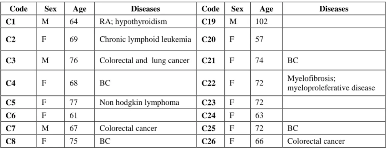
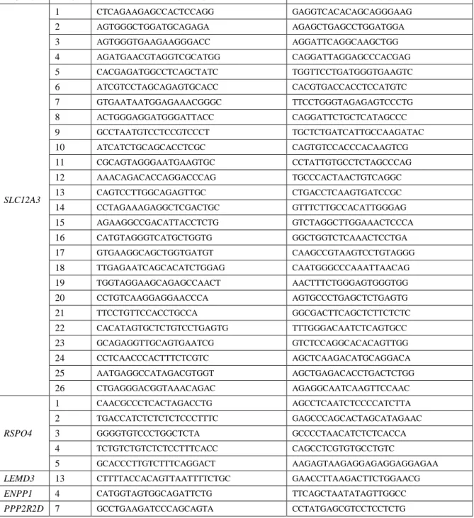
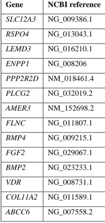
AGRADECIMENTOS ... vii ABSTRACT ... ix RESUMO ... xi RESUMO ALARGADO ... xiii LIST OF ABBREVIATURES ... xix TABLES ... xxix SUPPLEMENTARY TABLES ... xxxi FIGURES ... xxxiii SUPPLEMENTARY FIGURES ... xxxv 1. INTRODUCTION ... 3 1.1 Background of the study ... 3 1.2 Objectives of the thesis ... 4 1.3 Thesis outline ... 5 2. LITERATURE REVIEW ... 9 2.1 Introduction ... 9 2.2 Diagnosis ... 9 2.3 Epidemiology ... 11 2.4 Evidences for a genetic aetiology ... 12 2.4.1 Familial cases ... 13 2.4.2 Animal models ... 15 2.4.2.1 Animal models for DISH ... 15 2.4.2.2 Animal models for OPLL ... 17 2.4.2.3 Other animal models with OSL ... 18 2.4.3 Genetic variants associated ... 20 2.4.3.1 Genetic studies on DISH ... 20 2.4.3.2 Genetic studies on OPLL ... 22 2.4.4 Genetic studies on OLF ... 28 2.5 Association with other diseases ... 29 2.5.1 Monogenic disorders ... 29 2.5.2 Complex disorders ... 31 2.5.3 Rheumatic disorders co-existing with OSL ... 33 3. MATERIAL AND METHODS ... 43 3.1 Collections- patients/families and associated data ... 43 3.1.1. Families DISH/CC ... 43 3.1.2 DISH/CC patients not related ... 46 3.1.3 Gitelman syndrome family ... 47 3.1.4 Ankylosing Spondylitis (AS) patients ... 48 3.1.5 Control population without DISH/CC ... 49 3.1.6 Representative population of Terceira Island ... 50 3.1.6.1 Randomized cohort ... 50 3.1.6.2 Two regions – Angra do Heroísmo and Praia da Vitória ... 52 3.1.7 Total hip replacement group ... 55 3.2 Methods ... 56
xxvi
3.2.1 Gene sequencing ... 56 3.2.1.1 DNA extraction and quantification ... 56 3.2.1.2 Amplification of regions of interest ... 56 3.2.1.3 Sequencing ... 60 3.2.1.4 Sequence analysis ... 61 3.2.2 Tissue expression ... 61 3.2.2.1 Cartilage homogenization ... 61 3.2.2.2 RNA isolation ... 62 3.2.2.3 RNA purification ... 63 3.2.2.4. RNA quality assessment ... 64 3.2.2.5 Gene Expression ... 65 4. CHONDROCALCINOSIS ASSOCIATED WITH GITELMAN SYNDROME ... 67 4.1 Abstract ... 67 4.2 Introduction ... 68 4.3 Material and methods... 69 4.4 Results ... 70 4.5 Discussion ... 72 4.6 Conclusion ... 74 4.7 Future work ... 74 5. INVESTIGATING THE ROLE OF THE RSPO4 AND LEMD3 GENES WITH DISH/CC PHENOTYPE ... 77 5.1 Abstract ... 77 5.2 Introduction ... 78 5.3 Material & methods ... 80 5.3.1 Subjects ... 80 5.3.2 RSPO4 & LEMD3 sequencing ... 80 5.3.3 Statistical analysis ... 81 5.4 Results ... 81 5.4.1 RSPO4 sequencing ... 81 5.4.2 LEMD3 sequencing ... 87 5.5 Discussion ... 89 5.6. Supplementary material ... 91 6. WHOLE EXOME SEQUENCING OF PATIENTS SHOWING EXUBERANT ECTOPIC CALCIFICATIONS IN THE AXIAL AND APPENDICULAR SKELETON ... 95 6.1. Abstract ... 95 6.2 Introduction ... 96 6.3 Material & methods ... 97 6.3.1. Subjects ... 97 6.3.2. Exome capture ... 98 6.3.3. WES filtering ... 99 6.3.3.1. Filtering candidate genes provided by WES results ... 99 6.3.3.2. Filtering within candidate genes ... 100 6.3.3.3. Filtering pathogenic variants ... 101 6.3.4. Validation and Evaluation of Genes/Variants ... 102
xxvii
6.3.5. Association studies ... 103 6.3.6. Statistical analysis ... 103 6.4 Results ... 103 6.4.1. Exome capture - variants detected ... 103 6.4.2. Filtering Results ... 104 6.4.3. Association between variants and DISH/CC phenotype ... 108 6.4.3.1. Segregation of variants ... 108 6.4.3.2. Case/control studies... 109 6.5. Discussion ... 110 6.6. Conclusion ... 113 6.7. Future work ... 113 6.8. Supplementary material ... 114 7. INVESTIGATING THE ROLE OF ABCC6 GENE IN ECTOPIC CALCIFICATION ... 119 7.1 Abstract ... 119 7.2. Introduction ... 120 7.3. Material & methods ... 123 7.3.1. Subjects ... 123 7.3.2. Gene sequencing... 123 7.3.2.1. Mutation screening ... 123 7.3.2.2. Statistical analysis ... 124 7.3.3. Gene expression ... 125 7.3.3.1.RNA isolation and quality control... 125 7.3.3.2 Reverse transcription- RT-PCR ... 125 7.3.3.3.Quantitative RT-PCR (qRT-PCR) ... 125 7.3.3.4. Typing ABCC6 gene variants ... 126 7.4. Results ... 127 7.4.1. Sequencing ... 127 7.4.1.1. ABCC6 variants ... 127 7.4.1.2. Case/control study ... 130 7.4.1.3 SNP frequencies ... 134 7.4.2. Expression ... 136 7.5. Discussion ... 138 7.6. Supplementary material ... 142 8. THE ORIGIN OF ABCC6 GENE IS POTENTIALLY LINKED WITH THE EMERGENCE OF A BONY SKELETON IN VERTEBRATES ... 147 8.1 Abstract ... 147 8.2. Introduction ... 148 8.3. Methods ... 150 8.3.1. Database searches and sequence retrieval ... 150 8.3.2. Multiple sequence alignments and sequence comparisons... 152 8.3.3. Phylogenetic analysis ... 152 8.3.4. Gene organization and gene synteny ... 153 8.3.5 Transcriptome database searches ... 153
xxviii
8.3.6 Polymerase chain reaction (PCR) and quantitative-PCR (qPCR)... 154 8.4. Results ... 155 8.4.1. Members of the ABCC6 in vertebrates and invertebrates ... 155 8.4.2 Sequence conservation of the amino acids altered in PXE disease ... 157 8.4.3 Phylogeny of ABCC6 ... 158 8.4.4. Gene structure and gene linkage across vertebrate ... 161 8.4.5 Expression analysis in non-mammals ... 164 8.4.6 Expression of abcc6 and abcc1 during sea bream scale formation ... 166 8.5. Discussion ... 167 8.6 Supplementary Material ... 170 9. GENERAL DISCUSSION AND CONCLUSIONS ... 191 10. BIBLIOGRAPHY ... 201 APPENDIX ... 227
xxix
TABLES
Table 2-1. DISH diagnostic criteria. ... 10 Table 2-2. Genetic variants associated with DISH. ... 22 Table 2-3. Genetic variants associated with OPLL.. ... 23 Table 2-4. Genetic variants associated with OLF... 28 Table 2-5. Monogenic disorders previously associated with OSL.. ... 30 Table 2-6. Complex disorders previously associated with OSL.. ... 32 Table 2-7. Rheumatic disorders previously seen coexisting with OSL.. ... 34 Table 3-1. Azorean families included in the study and associated data. ... 45 Table 3-2. Patients with DISH/CC and associated data. ... 46 Table 3-3. Gitelman syndrome family and associated data. ... 48 Table 3-4. AS population and associated data. ... 49 Table 3-5. Population control without DISH and or CC and associated data. ... 49 Table 3-6. Individuals from the randomized population and associated data. ... 51 Table 3-7. Representative population of two regions of Terceira Island and associated data . ... 52 Table 3-8. Total hip replacement group. ... 56 Table 3-9. Forward and reverse oligonucleotide primers used in PCR and sequencing. ... 57 Table 3-10. PCR conditions, annealing temperature and PCR product size ... 59 Table 3-11. NCBI references for the genes studied. ... 61 Table 3-12. Forward and reverse oligonucleotide primers used in PCR of β-Actin gene. ... 64 Table 4-1. Characteristics and SLC12A3 gene variants and blood chemistry levels in the proband and the thirteen selected individuals from their family pedigree. ... 72 Table 5-1. Genetic variants identified in the RSPO4 gene and functional information. ... 82 Table 5-2. Family based association test results for variants of RSPO4 gene. ... 84 Table 5-3. Association study results of genetic variants found in RSPO4 gene ... 85 Table 5-4. Statistical results using the Cochran-Armitage trend and allelic tests. ... 86 Table 5-5. Genetic variants identified in LEMD3 gene ... 87 Table 6-1. Radiology results from four selected patients for WES ... 98 Table 6-2. Number of candidate genes per sample. ... 99 Table 6-3. Number of candidate genes shared by the investigated DISH/CC patients. ... 100 Table 6-4. Known, novel and total number of SNVs and Indels for the four samples. ... 103 Table 6-5. List of variants found by WES and confirmed by Sanger Sequencing. ... 105 Table 6-6. Conservation analysis of the variants identified.. ... 107 Table 6-7. Family based association test results for variants of AMER3 gene and FLNC gene.109 Table 6-8. Association study between variants and DISH/CC phenotype.. ... 110 Table 7-1. List accession transcript numbers for ABCC6 gene, used for conservation analysis.124
xxx
Table 7-2. ABCC6 variants identified in a group of 12 DISH/CC probands.. ... 128 Table 7-3. The results of the association study of genetic variants found in the ABCC6 gene in Azorean patients with DISH/CC and AS compared to the controls.. ... 131 Table 7-4. Association study of the genetic variants in the ABCC6 gene in DISH/CC and AS patients compared to the controls... 132 Table 7-5. Frequency comparison of the genetic variants found in the coding exons of the ABCC6 gene between the DISH/CC and AS groups. ... 134 Table 7-6. Frequency in population 1 and population 2 of Terceira Island of the ABCC6 gene variant in exon 23. ... 135 Table 7-7. Details of the 13 patients undergoing hip replacement ... 136 Table 8-1. Human ABCC genes and their functions.. ... 149 Table 8-2. ABCC6; ABCC1 and ABCC3 expression analysis in human, bird, reptile, amphibian and teleost.. ... 164
xxxi
SUPPLEMENTARY TABLES
Supplementary table 5-1. Results of genetic variants found in the RSPO4 gene in Azorean patients with DISH/CC compared to the controls. ... 91 Supplementary table 6-1. List of genes and their accession transcript numbers, used for conservation analysis.. ... 114 Supplementary table 7-1. Results of genetic variants found in the ABCC6 gene in Azorean patients with DISH/CC and AS compared to the controls. ... 142 Supplementary table 7-2. RNA cartilage samples and measures of NanoVue and Agilent. ... 143 Supplementary table 8-1. Accession numbers of the ABCC6, ABCC1 and ABCC3 genes retrieved from vertebrates.. ... 170 Supplementary table 8-2. Accession numbers of the ABCC genes from invertebrates.. ... 171 Supplementary table 8-3. List of genes and their accession numbers used for linkage analysis..185 Supplementary table 8-4. Percentage of amino acid sequence identity/similarity of the ABCC6 and
ABCC1 sequences in comparison with human sequence by GeneDoc program. ... 186
Supplementary table 8-5. Size of the exons and introns for the Human and spotted gar ABCC6 orthologues genes.. ... 187 Supplementary table 8-6. CT values for the control gene rps18 and abcc6 and abcc1 genes in intact and regenerating skin at 0-96 hours. ... 188
xxxiii
FIGURES
Figure 1-1. Geographic location of the Azores. ... 4 Figure 3-1. Azorean families included in the study. ... 44 Figure 3-2. Gitelman family. ... 47 Figure 4-1. X-rays of proband showing classical chondrocalcinosis in knees ... 70 Figure 4-2. Heredogram with investigated individuals. ... 71 Figure 5-1. Typing results for RSPO4 gene in DISH/CC families. ... 83 Figure 5-2. Typing results for LEMD3 gene in DISH/CC families. ... 88 Figure 6-1. The two-level filtration approach used to analyze the WES results .. ... 101 Figure 6-2. Segregation results with DISH/CC phenotype in families AZ1-4. ... 108 Figure 7-1. Predicted transmembrane protein topology of MRP6 with locations of human variants and comparison with the amino acid in the equivalent position in other vertebrate species. .... 129 Figure 7-2. Relative expression of ABCC6 gene in cartilage tissue samples. . ... 137 Figure 7-3. Relative expression of the ABCC6 gene in normal, coxoarthrosis, DISH and CC individuals.. ... 137 Figure 8-1. Dendrogram indicating the number of ABCC6 genes and the related ABCC1 and
ABCC3 genes identified in the different vertebrate and invertebrate genomes. ... 156
Figure 8-2. Alignment of the MRP6 amino acid positions affected by 16 selected mutations previously associated with PXE disease. ... 158 Figure 8-3. Phylogenetic tree of the vertebrate and invertebrate ABCC6, 1 and 3 with the other human ABCC family members.. ... 160 Figure 8-4. Gene organization of the ABCC6 gene in human and spotted gar. ... 161 Figure 8-5. Gene synteny analysis of the ABCC6 gene environment across vertebrates.. ... 163 Figure 8-6. Expression results of abcc6 and abcc1 in sea bream skin regeneration assay.. ... 166
xxxv
SUPPLEMENTARY FIGURES
Supplementary figure 6-1. Analysis of sequence conservation among six vertebrates of variants identified in candidate genes possibly associated with DISH/CC phenotype.. ... 115 Supplementary figure 7-1. Conventional PCR amplification for β-actin gene ... 144 Supplementary figure 8-1. Sequence alignments of the MRP1 and MRP6 in vertebrates. ... 181 Supplementary figure 8-2. Phylogenetic tree of the vertebrate and invertebrate ABCC6, 1 and 3 with the other human ABCC family members. ... 183
C
3
1. INTRODUCTION
1.1 Background of the study
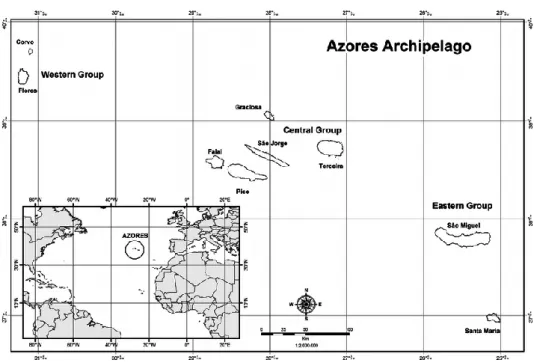
The starting point of this thesis was the identification of many families, from Terceira Island, Azores, that may represent an early onset, autosomal dominant, familial type of pyrophosphate arthropathy, with a phenotype that includes peripheral and axial enthesopathic calcifications. A detailed radiological analysis of these families showed a high level of ectopic calcification especially in elbows (82.9%) and spine (81.4%). The concurrence of spinal ossifications, resembling Diffuse Idiopathic Skeletal Hyperostosis (DISH), and CPPD Chondrocalcinosis (CC), in many of those patients, suggested a shared pathogenic mechanism [24]. For a number of years the possible major gene involved in the aethiopathogenesis, of the then designated DISH/CC phenotype, has been investigated using different strategies, including whole exome linkage and an Identity-by-state study, and two zones, in chromosomes 12 and 20, seemed relevant for further investigation [29]. For this purpose a biobank with biological products and associated data was established for the population from the Terceira Island in the Azores. Biobanks are essential in research, by having collections of samples and data stored in an organized manner. At the moment the biobank Azores (AZORBIO) of the Specialized Service of Epidemiology and Molecular Biology (SEEBMO) has a collection of biological material and associated data of Azorean patients with different pathologies [27]. The Azores archipelago (Portugal) is located in the middle of the Atlantic Ocean, 1500 km from the European mainland and is formed by nine islands of volcanic origin. The islands are grouped according to their geographic positions: Eastern (São Miguel and Santa Maria), Central (Terceira, Faial, Pico, Graciosa and São Jorge) and Western (Flores and Corvo) [24, 30] (Figure 1-1). The Azores were officially populated in 1439 and have approximately 246,746 inhabitants distributed in a very asymmetric way among islands. Terceira is a relatively small island with only 56.467 inhabitants (Census, 2011). Isolated populations or populations with reduced mobility, as is the case of Terceira Island, have proven particularly valuable for the purposes of mapping genes involved in Mendelian monogenic disorders and thus, investigating this phenotype in this population it was reasoned would increase the likelihood of identifying the causative gene mutation.
4
Figure 1-1. Geographic location of the Azores. The islands are grouped according to their geographic positions in Eastern, Central and Western. Taken from Santos et al, 2009 [30].
1.2 Objectives of the thesis
The main objective of this thesis was to proceed with the investigation of the genetic basis of the DISH/CC phenotype making use of Next Generation Sequencing technology, together with association and expression studies.
The studies presented in this thesis were guided by the following objectives:
• Investigate the association of CC with the metabolic disorder- Gitelman Syndrome;
• Investigate the candidate genes RSPO4 and LEMD3 genes, derived from a previous analysis of identity-by-descent sharing across four families with CC; • Select the best candidate genes in WES results from 4 unrelated DISH/CC
patients;
• Perform case/control and expression studies on the best candidate genes; • Characterize the candidate gene in terms of origin and evolution.
On completion of this thesis I expect to contribute to the identification of genetic factors involved in the DISH/CC phenotype.
5
1.3 Thesis outline
The Chapter 2 introduces the basic knowledge of what is known about the genetics of ossification of spinal ligaments- DISH, OPLL and OLF, and focusses on the main disorder investigated in this thesis: DISH. Chapter 3 provides a detailed presentation of the material and methods used in the thesis. The association of CC with Gitelman Syndrome through the genetic analysis of the SLC12A3 gene is presented in chapter 4. Chapter 5 covers the possible role of RSPO4 and LEMD3 genetic variants in the aetiology of DISH/CC. The whole exome sequencing study performed in order to detect genetic variants that are expected to have a potentially damaging effect on proteins with functions related to calcification and/or ossification is reported in chapter 6. In chapter 7 the candidate gene ABCC6 is investigated using case/control and expression studies to verify the association with the phenotype under study. Finally, the characterization and comparison of the ABCC6 gene in metazoans including humans using “in silico” methodologies is presented in chapter 8. The chapter 9 presents the general discussion and conclusions of this thesis. The final chapter contains the bibliography, followed by appendix with an article publication.
C
9
2. LITERATURE REVIEW
2.1 Introduction
The spine is a columnar structure in the center of the body composed by spinal bones (vertebrae) and inter-vertebral discs. It is supported by spinal ligaments (flexible band-like structures), which include the anterior and posterior longitudinal ligaments, ligament nuchae and the yellow ligament (ligamentum flavum) of the spine [31]. There is a group of diseases characterized by the ossification of spinal ligaments (OSL); the anterolateral spinal ligament [Diffuse Idiopathic Skeletal Hyperostosis (DISH; MIM 106400)], the posterior longitudinal ligament [Ossification of the Posterior Longitudinal Ligament (OPLL; MIM 602475)] and the ligament flavum [Ossification of Ligamentum Flavum (OLF)]. In some cases OPLL, DISH and OLF co-occur in the same patient [32] suggesting possible common aetiopathogenic factors. Genetic links in OPLL, DISH and OLF have been investigated and several papers cite or allude to genetic factors as playing a role in the aetiology of this diseases [33-35]. There are reports in the literature that describe familial cases of DISH and OPLL which further strengthen this genetic association. Although the evidence of a genetic predisposition in all the three diseases have been described, very few studies in DISH and OLF appear in the literature, only OPLL has been extensively investigated. However, the studies that have looked at the possible genetic links are still inconclusive and the aetiology of these diseases remains unknown. The main objective of this study is to investigate genetic variants associated with DISH susceptibility. Therefore, in this thesis, the genetic mechanisms already involved in OPLL and OLF aetiology will be explored in detail since they can give insights into the pathogenesis of DISH.
2.2 Diagnosis
DISH (DISH; MIM 106400) is the current terminology for a systemic non inflammatory disorder reported in 1925 by Knaggs [36] and later described by Forestier and Routes-Querol in 1950 [37]. This disorder has had a variety of denominations in the literature throughout the years, due to the diverse phenotypes encountered. It is a common condition amongst the elderly characterized by calcification and ossification of the anterior longitudinal ligament affecting, in particular, the right side of the spine with preservation of the intervertebral disc space. Whilst spinal involvement in DISH is nearly
10
universal, extraspinal sites, such as the elbow, shoulder, hip, knee and heel are very common [1, 5, 22, 23, 38]. The diagnosis of DISH is established using radiographies. There are two main diagnostic criteria sets to identify definite, probable or possible DISH (Table 2-1). Resnick [39] defined the first set of criteria that were, some years later, revised by Utsinger [5] for epidemiological purposes. Criteria are indicated in the following table:
Table 2-1. DISH diagnostic criteria.
Resnick Criteria [39] Utsinger Criteria [5]
De
fin
ite
1
Presence of flowing calcification and ossification along the anterolateral aspect of at least four contiguous vertebral bodies with or without associated localized pointed excrescences at the intervening vertebral body-intervertebral disc junctions.
Continuous ossification along the anterolateral aspect of at least four contiguous vertebral bodies, primarily in the thoracolumbar spine. Ossification begins as a fine ribbon-like wave of bone but commonly develops into a broad, bumpy, buttress-like band of bone.
Pro b a b le Po ss ib le 2
The presence of relative preservation of intervertebral disc height in the involved vertebral segment and the absence of extensive radiographic changes of “degenerative” disc disease, including vacuum phenomena and vertebral body marginal sclerosis.
Continuous ossification along the anterolateral aspect of at least two contiguous vertebral bodies.
Po
ss
ib
le
3
The absence of apophyseal joint bony ankylosis and sacroiliac joint erosion, sclerosis or intraarticular osseous fusion.
Symmetrical and peripheral enthesopathy involving the posterior heel, superior patella or olecranon, with the entheseal new bone having a well-defined cortical margin.
According to the criteria, the probability of DISH is as follows: ‘definite’ if criterion 1 is present, ‘probable’ if criteria 2 and 3 are present and ‘possible’ if criterion 2 or 3 is present; particularly if calcaneal spurs occur together with olecranon or patellar spurs. Exclusion criteria include: abnormal disc space height in the involved areas and/or apophyseal joint ankylosis. Resnick criteria number 3 has an exclusion factor based on the erosions, sclerosis or fusion of sacroiliac joints. This helps to exclude patients with Ankylosing Spondylitis (AS), a disease that can be confused with DISH. The third factor for exclusion of DISH diagnosis was withdrawn by Utsinger because differentiation of these two disorders should be possible with lateral and antero posterior axial x-rays. Recently efforts have been made to revise the definition of DISH in order to incorporate the current knowledge about DISH, however a new definition of DISH is still under debate [40]. Despite the need for a new definition of DISH, the criteria proposed by Utsinger are still universally accepted and widely used in the literature and will be used in this thesis.
11
OPLL (OPLL; MIM 602475) is characterized by ectopic hyperostosis and calcification of the posterior longitudinal ligament at the cervical, thoracic and lumbar spine [41]. In OPLL patients, the cervical spine (70%) is the most commonly affected, followed by the thoracic (15%) and lumbar (15%) spine [42, 43]. Some patients present myelopathy and/or radiculopathy due to chronic compression of the spinal cord and nerve roots. Symptoms of myelopathy are more severe in thoracic OPLL than in cervical OPLL due to the narrow canal, rigidity of the thoracic spine, tenuous blood supply, and inability of the spinal cord to resist much compression [42]. OPLL is diagnosed on lateral plain radiographs as an abnormal radiopacity along the posterior of the vertebral bodies [44], however because of overlying osseous structures, it is important to obtain magnetic resonance images to successfully diagnose OPLL [45]. OPLL is classified in four ossification types: continuous, segmental, mixed and localized or other [44, 46, 47]. The segmental is the most common and involves the ossification behind each vertebral body; the continuous type is an ossified mass that spans several vertebral bodies; the mixed type is a mixture of both continuous and segmental types and the localized or other type the ossification is localized to the intervertebral disk space without involvement of the vertebral body [44].
OLF, also known as ossification of the yellow ligament, is associated with serious neurologic symptoms including thoracic myelopathy and spinal stenosis [48]. The calcification is confined to the ligamentum flavum (LF) and does not extend to the closed spinal bony arch [49]. CPP and hydroxyapatite have been positively identified and are the main players in the calcification of LF [50, 51]. According to Mwaka and collaborators [52] CPP in the cervical LF seems to progress with reduction in elastic fibers, increase in collagen fibrils in the matrix, and migration of metaplastic hypertrophic chondrocytes. The lower thoracic spine is the most affected region, however several cases of cervical, upper thoracic, and lumbar areas have been reported [53, 54]. Cervical radiography, tomography, and computed tomography are useful for diagnosis, however histological examination of the calcified mass using light microscopy, scanning electron microscopy, and x-ray diffraction analysis are essential for the definitive diagnosis [49].
2.3 Epidemiology
The reported epidemiology of DISH differs in the literature. Cassin et al [55] assessed 1000 African blacks aged older than 40 years and reported that the DISH prevalence was
12
3.8% in males and 4.2% in females. Another study analysed the data from two large American Midwest metropolitan hospital populations with 1363 individuals and reported a prevalence of DISH of 25% in males and 15% in females over 50 years of age and 35% in males and 26% in females over 70 years of age [56]. Holton et al [57] postulated that the prevalence of DISH was 42% in a group of 298 males aged over 65 from the general population. A recent study of 558 Japanese found that the prevalence of DISH was 17.6% using x-ray and 27,2% using computed tomography [58]. The exact prevalence and incidence of DISH is actually unknown, and a reliable estimate of the prevalence of the disease in the general population is difficult due to the benign nature of the condition. The affected individuals do not seek medical care and normally are diagnosed during the examination of other medical conditions [57]. However, it is well known that DISH is more frequent in males and its prevalence increases with age, affecting mainly subjects over the age of 40 [5]. Furthermore DISH seems to have a higher prevalence in developed countries [59], although this predominance might be due to the more frequent use of advanced radiological examinations in developed countries than in undeveloped countries.
OPLL can be found in any population however it is more common in Asian populations, in particular amongst Japanese with a prevalence of 2 to 4% as compared with 0.01 to 2% in non-Asian populations [60]. Men are 2.5 times more likely to develop OPLL than women [41] and the age of onset may be in the fifth decade of life [61], although in some studies no association between age and the presence of OPLL was found [41]. This lack of consensus can be explained by the fact that the study which didn’t find any association probably used a group of patients with asymptomatic OPLL, while the other study only involved patients with symptomatic OPLL, already diagnosed.
OLF affects populations worldwide but there is a higher prevalence in east Asian ethnic groups, especially the Japanese, with the incidence of 12% in thoracic OLF (15% in men and 7.7% in women) [62]. OLF is common in the 6th to 7th decades [46].
2.4 Evidences for a genetic aetiology
Genetic links in OPLL, DISH and OLF have been investigated and several papers cite or allude to genetic factors as playing a role in the aetiology of these diseases [33-35]. The
13
reports in the literature that describe familial cases of DISH and OPLL and the existence of animal models further strengthen this genetic association.
2.4.1 Familial cases
The genetic predisposition to OPLL and DISH is supported by several reports of familial incidence of DISH, and by studies relating a relative recurrence risk of up to 26.1% in parents of OPLL patients and 28.9% in siblings [63].
Reports of familial DISH are uncommon in the literature. One report, dating from 1969, describes a family of Greek Cypriot ancestry with eight individuals showing signs of Ankylosing Vertebral Hyperostosis (AVH) by the third decade. Only three of them had backache as symptoms. All the individuals affected with AVH also shown tylosis (punctuate hyperkeratosis) and the mean age of individuals affected was 31 years old. Six members of this family were affected only with tylosis. Axial skeleton X-ray examination of the eight affected family members showed ossification of paraspinal ligaments, especially in lower thoracic region. There were also large osteophytes with preservation of disk space and marginal sclerosis of the sacroiliac joint. Laboratory results were all normal, the authors mention normal calcium and carbohydrate metabolism and glucose tolerance tests. Obesity was present in most individuals. Weight, in the opinion of the authors, could not account for all the x-ray changes since there was one affected individual with normal build. There was one other case of an individual with tylosis, normal spine and gross obesity. Beardwell et al suggested that the co-existence of tylosis with AVH could indicate a possible genetic link between these two disorders [64]. This link was never confirmed being this association an occasional finding. Another report of familial DISH was published in 1985 by Abiteboul et al [65], where the authors describe two families, one of French Canadian origin and another of Italian origin. French Canadian family is composed of 3 brothers and one sister definitely affected by AVH developing radiological changes by the 4th decade of age. Two other are probably affected with AVH. None of the individuals had diabetes. Italian family was first identified after a coxofemoral surgery of a 71 years old woman. Her sister was submitted to the same surgery when she was 82 years old. Former patient had five daughters, all of them observed by the authors of this study. Two of them showed radiological manifestations of the AVH. Other 2 sisters had more modest phenotypes being classified as probable AVH. None of these individuals was HLA-B*27, was obese or had diabetes. There is another report relating a family with striking cervical disease without extensive dorsal