ALTERAÇÕES DOS MARCADORES INFLAMATÓRIOS, BIOQUÍMICOS E APTIDÃO FÍSICA EM MULHERES EM RECUPERAÇÃO DO CÂNCER DE
MAMA APÓS TREINAMENTO RESISTIDO NÃO LINEAR: UM ESTUDO PILOTO
MARCO AURÉLIO FERREIRA DE JESUS LEITE
ALTERAÇÕES DOS MARCADORES INFLAMATÓRIOS, BIOQUÍMICOS E APTIDÃO FÍSICA EM MULHERES EM RECUPERAÇÃO DO CÂNCER DE
MAMA APÓS TREINAMENTO RESISTIDO NÃO LINEAR: UM ESTUDO PILOTO
Dissertação apresentada ao Programa de Pós-Graduação em Ciências da Saúde da Faculdade de Medicina da Universidade Federal de Uberlândia, como requisito parcial para a obtenção do título de Mestre em Ciências da Saúde.
Área de concentração: Ciências da Saúde.
Orientador: Guilherme Morais Puga
L533a 2017
Leite, Marco Aurélio Ferreira de Jesus, 1994
Alterações dos marcadores inflamatórios, bioquímicos e aptidão física em mulheres em recuperação do câncer de mama após treinamento resistido não linear: um estudo piloto / Marco Aurélio Ferreira de Jesus Leite. - 2017.
61 f. : il.
Orientador: Guilherme Morais Puga. Coorientador: Carlo José Freire de Oliveira.
Dissertação (mestrado) - Universidade Federal de Uberlândia, Programa de Pós- Graduação em Ciências da Saúde.
Inclui bibliografia.
FOLHA DE APROVAÇÃO
Marco Aurélio Ferreira de Jesus Leite.
Alterações dos marcadores inflamatórios, bioquímicos e aptidão física em mulheres em recuperação do câncer de mama após treinamento resistido não linear: um estudo piloto.
Presidente da banca (orientador): Prof. Dr. Guilherme Morais Puga
Dissertação apresentada ao Programa de Pós-Graduação em Ciências da Saúde da Faculdade de Medicina da Universidade Federal de Uberlândia, como requisito parcial para a obtenção do título de Mestre em Ciências da Saúde.
Área de concentração: Ciências da Saúde.
Banca Examinadora
Titular: Prof Dr. Fábio Lera Orsatti
Instituição: Universidade Federal do Triângulo Mineiro
para realização deste trabalho. Muito obrigado!
Ao Prof. Dr. Carlo José Freire de Oliveira e Prof. Nilson Penha-Silva, por sempre acreditar no meu potencial e seu imessuravel auxílio na execução da pesquisa. Sem sua expertise como co-orientador não conseguíamos alcançar estes resultados!
Aos meus colegas do Laboratório de Fisiologia Cardiorespiratória e Metabólica, Laboratório de Biofisicoquímica e Laboratório de Imunologia Aplicada pela parceria e suporte. A todos os funcionários, professores e técnicos da Faculdade de Educação Física e de Medicina da Universidade Federal de Uberlândia pelos auxílios prestados.
Aos amigos de laboratório Lucas, Franciel, Wener, Rodney, Hugo, Mário, Luciana, Igor, Juliene, Jéssica, Larissa, Brenda, Jonatas, Brenda pelo aprendizado e colaboração para elaboração desta pesquisa. A todas as voluntárias que acreditaram na minha pesquisa e reservaram seu tempo para participar de toda intervenção.
Agradeço a minha família, primeiramente minha mãe Maria pela oportunidade, amor, dedicação e por lutar dia a dia junto a mim em meus sonhos. Obrigado pela confiança. A minha tia Terezinha e ao meu primo Ernando por me darem exemplaridade e apoio. Aos meus irmãos José Victor, Natália e Carla pelo afeto e auxílios. Em especial, agradeço minha avó Arminda, a qual perdi pela luta contra o câncer. Sua simplicidade, carinho e amor me fizeram e me faz mais forte. Não fui capaz de lhe ajudar, mas espero que estes estudos possam ajudar pessoas que estão passando pela mesma situação. A vocês a minha eterna gratidão.
com câncer de mama (CM), como elevado processo inflamatório e dislipidemia. Até o momento ainda não se sabe o efeito do treinamento resistido periodizado de forma não linear (TRNL) em variáveis inflamatórias e bioquímicas em mulheres recuperando de CM. Objetivo: Verificar o efeito do TRNL sobre o perfil inflamatório, bioquímico, aptidão física e composição corporal em mulheres em recuperação do CM durante hormonioterapia. Métodos: 14 mulheres recuperando do CM durante a hormonioterapia participaram do estudo. Foram coletadas amostras de sangue e saliva para analises do perfil inflamatório, lipídico e imune e realizado avaliações antropométricas, força e resistência muscular antes e após de intervenção. O TRNL foi realizado 3x/semana em dias não consecutivos durante 3 meses. A normalidade dos dados foi verificada pelo teste Shapiro-Wilk e as comparação das variáveis foi realizada pelo teste t pareado e Wilcoxon. Ponto de corte foi ajustado para a = 5% para todas as análises. Resultados: Não houve alterações significativas na concentração plasmática das citocinas analisadas (TNF
a, IL-4, IL-6, IL-10, IL-17, IL-1RA, IFN-y). Porém houve aumento significativo de imunoglobulina A (IgA) salivar (186.29%) após intervenção. Houve também de HDL- colesterol (17.98%), massa livre de gordura (2.99%), massa magra (2.97%), força (39.16%) e resistência muscular (69.16%).Além disso houve reduções significativas de colesterol total ( 4.81%), triglicerídeos (-14.40%), LDL-colesterol (-10.23%), plaquetas (-7.90%), monócitos ( 38.89%), massa gorda (-4.53%), percentual de gordura corporal (-1.19%). Conclusão: Doze semanas de TRNL aumentou a concentração de IgA salivar, melhorou composição corporal, perfil lipídico e aptidão física em mulheres recuperando do BC durante hormonioterapia, força e resistência em mulheres em recuperação do CM.
breast cancer (BC), such as high inflammatory process and dyslipidemia. To date, the effect of nonlinear periodized resistance training (NLRT) on inflammatory and biochemical variables in women recovering from CM has not yet been known. Objective: To verify the effect of NLRT on the inflammatory, biochemical profile, physical fitness and body composition in women in CM recovery during hormone therapy. Methods: The assessments were done before and after 3 months of application of a NLRT program, consisting of three sessions of exercise per week on non-consecutive days. The normality of the data was verified by the Shapiro-Wilk test and the comparisons of results were performed by the paired t or Wilcoxon’s tests. Statistical significance was set at 5% (alpha = 0.05). Results: There were no significant changes in the plasma concentration of the analyzed cytokines (TNF-a, IL-4, IL-6, IL-10, IL-17, IL-1RA, IFN Y), but a significant increase in salivary levels of immunoglobulin A (IgA) was observed after the intervention (186.29%). Significant increases were also observed in HDL-cholesterol (17.98%), fat-free mass (2.99%), lean mass (2.97%), and muscle strength (39.16%) and endurance (65.66%). On the other hand, significant reductions were observed in the blood levels of triglycerides (-14.40%) and total (-4.81%) and LDL- cholesterol (-10.23%), in the platelets (-7.90%) and monocytes counts (-38.89%), in the amount of fat mass (-4.53%) and in the percent body fat (-1.19%). Conclusion: Twelve weeks of NPRT led to an increase in salivary IgA concentration and improvement in body composition, lipid profile and physical fitness of women under recovery from BC treatment and under hormone therapy.
1RM AC AT BC BM BMI CM CRP CT CT FCmáx FFM FM HC HDL-c HMGB1 IFN-y IL- ip IL-10 IL-15 IL-1Ra IL-2 IL-2 IL-6 IL-6 IL-8 IQR LDL-c LM LME
1 repetition maximum test Abdome circumference Aerobic training Breast cancer Body mass Body mass index Câncer de mama C-reative protein Colesterol total Total cholesterol
Frequência cardiacada máxima Fat free mass
Fat mass
Hip circumference
High density lipoprotein cholesterol High mobility group box 1
Interferon gama Interleucina-1 beta Interleucina-10 Interleucina-15
Antagonista de receptor de interleucina-1 Inteceulina-2 Interleukin-2 Interleucina-6 Interleukin-6 Interleucina-8 Interquartile range
Low density lipoprotein cholesterol Lean mass
NLRT Nonlinear periodized resistante traning NO Óxido nítrico
PCR Proteína-c reativa RT Resistant Training SEM Error of the mean
TA Treinamento aeróbio TC Triglicerídeos TGC Triglycerides
TNF-a Fator de necrose turmoral alfa TR Treinamento de resistência
TRNL Treinamentode de resistência periodizado de forma não-linear VO2máx Consumo de oxigênio máximo
1 INTRODUÇÃO...14
2 REVISÃO DE LITERATURA... 16
2.1 Processo inflamatório e efeitos adversos em pacientes oncológicos... 16
2.2 Efeitos do treinamento físico em sobreviventes de câncer...18
2.3 Efeitos dos treinamentos combinados e resistidos no perfil inflamatório em sobreviventes de câncer de mama...20
2.4 Periodização do treinamento resistido...22
3 OBJETIVOS... 23
4 A RTIGO...24
REFERÊNCIAS...46
APÊNDICES... 52
Apêndice 1. Anamnese... 52
Apêndice 2. Ficha de avaliação do recordatório alimentar... 53
Apêndice 3. Ficha de avaliação dos testes de aptidão física...54
Apêndice 4. Termo de consentimento livre e esclarecido... 55
ANEXOS... 56
Anexo 1. Parecer do comitê de ética em pesquisa...56
1. INTRODUÇÃO
Existemvários fatores de risco que podem desencadear a ocorrência de lesões proliferativas benignas da glândula mamária, sendo estes classificados em intrínsecos e extrínsecos. Os fatores de risco intrínsecos como idade, sexo e aspectos genéticos constituem parâmetros independentes e que não são modificados ao longo da vida (KAMINSKA et al., 2015). Os fatores extrínsecos são condicionados pelo estilo de vida, dieta e intervenções farmacológicas (contraceptivos hormonais orais e terapia de reposição hormonal) e suas influências sobre o processo neoplásico pode ser modificados até certo ponto (KAMINSKA et al., 2015).
Atualmente, o câncer de mama (CM) representa um grande problema na saúde mundial devido o seu alto resistro de incidência e mortalidade. No último levantamento mundial, em 2012, registrou-se 1,7 milhões de novos casos (25% de todos os cânceres) e 522.000 mortes pelo CM (WORLD HEALTH ORGANIZATION, 2013). Em países desenvolvidos como Estados Unidos, em 2016 projetou-se uma estimativa de 246.660 novos casos e 40.450 mortes por CM (SIEGEL; MILLER; JEMAL, 2016). Essas tendências não diferem em país em desenvolvimento, como no Brasil, que na estimativa de 2016, denotou a incidência de 57.960 novos casos (28,1% do total de câncer) em mulheres, sendo considerado o câncer mais frequente em mulheres no país (INCA, 2016).
Os tratamentos e intervenções oncológicos atuais asseguram a sobrevida e minimizam os riscos de mortalidade por CM (CHEN et al., 2016; STEWARD et al., 2014). Estes contemplam abordagens cirúrgicas do tipo radical ou conservadora, esvaziamento axilar de linfonodos (linfadenectomia), radioterapia, quimioterapia, imunoterapia, hormonioterapia, como outros. As aplicações dessas estatrégias oncológicas dependem da especificidade do câncer e individualidade biológica do paciente (NATIONAL CANCER INSTITUTE, 2016). Entretanto, todos tratamentos oncológicos podem oferecer riscos de efeitos adversos.
de outras comorbidades, como depressão, declínio de força, aumento de gordura (LEE et al., 2004; SCHUBERT et al., 2007), como também a reincidência do câncer nessa população (COUSSENS; WERB, 2002).
Atualmente, o treinamento físico é considerado como importante estratégia complementar para pacientes com CM, principalmente por melhorar a aptidão física e perfil inflamatório (MENESES-ECHÁVEZ et al., 2016). Porém as repostas ao treinamento físico dependem tanto da condição de saúde do paciente quanto do tipo, intensidade e protocolo de treinamento. Alguns estudos demonstraram que o treinamento aeróbio (TA) de intensidade moderada diminui a gordura corporal e melhoram significativamente o perfil inflamatório em sobreviventes de CM, como redução de proteína-c reativa (PCR), diminuição de interleucina-2 (IL-2) e aumento de interleucina-6 (IL-6) (FAIREY et al., 2005a; JANELSINS et al., 2011). Em contrapartida, todos os estudos que aplicaram intervenções isoladas de treinamento resistido (TR) em sobreviventes de CM até o presente momento, não observaram nenhuma alteração significativa na composição corporal e em marcadores inflamatórios (HAGSTROM et al., 2016; SCHMIDT et al., 2016; SIMONAVICE et al., 2014).
Intrigantemente, estes estudos que interviram e investigaram a aplicação do TR nessa população não relataram sistematização do treinamento e/ou aplicaram periodizações “tradicionais”. Nesta periodização o mesmo conjunto de exercício é realizado na mesma ordem e com intensidades constantes por longos períodos (FLECK, 2011). Em sessões de TR periodizado de forma não linear (TRNL) a intensidade e o volume do treinamento são alterados com muito mais frequência para minimizar o tédio, evitar excessos de lesões e potencializar ganhos de força (FLECK, 2011).
2. REVISÃO DE LITERATURA
2.1. Processo inflamatório e efeitos adversos em pacientes oncológicos
O ambiente pro-inflamatório desencadeado pelo câncer é devido em parte pelos fatores diretos da progressão do crescimento tumoral e incapacidade de atuação do sistema imunológico, quanto indiretos, advindos dos tratamentos oncológicos e seus efeitos adversos. Leucócitos infiltrantes de tumor, assim como as vias de sinalização relacionadas com citocinas, são componentes importantes no desenvolvimento do microambiente inflamatório tumoral (KANG et al., 2013). Os principais leucócitos infiltrantes, como macrófagos e células NK, atuam no combate de células tumorais, porém induzem a liberação extracelular de uma proteína denominada high mobility group box 1 (HMBG1) a partir da apoptose dessas células (JUBE et al., 2012).
A HMGB1 livre se liga em diferentes receptores de superfície expressos em células do sistema imunológico, como o receptor de produtos finais com glicosilação avançada (KANG et al., 2010), toll-like receptor 4 (YU et al., 2006), receptores de gatilho expressos em células mielóides-1 (EL MEZAYEN et al., 2007), e CD24+ (CHEN et al., 2009). Após a interação elas ativam vias de sinalização proteína-quinases ativadas por mitógenos (MAPKs), fator nuclear kappa beta (NF-kP), e fosfatidilinositol 3 quinases/AKT, mediando as respostas de migração,
ativação, proliferação e diferenciação celular (TANG et al., 2011), desencadeando a cascata inflamatória em suas múltiplas vias, entre elas, a ativação de mais macrófagos e liberação de mais citocinas pro-inflamatórias (IL-ip, IL-2, IL-6, IL-8, TNF-a e PCR), ativação das células endoteliais, aumento da expressão de moléculas de adesão e o aumento da expressão de inibidor-1 do ativador do plasminogénio PAI-1 e tetradecanoílo acetato de forbol (KANG et al., 2013; MENESES-ECHÁVEZ et al., 2016; TANG et al., 2011). Estas sinalizações regulam a coagulação e aumentam a permeabilidade epitelial, resultando em uma respostas inflamatórias que pode induzir graves danos teciduais e/ou até mesmo a morte (KANG et al., 2013).
Além disso, após os tratamentos oncológicos neoadjuvantes e/ou adjuvantes, o processo inflamatório ainda pode estar presente nestes sobreviventes de câncer, sendo um dos efeitos adversos apresentado por estes tratamentos (TSAVARIS et al., 2002). Embora possua efeitos anti-inflamatórios após exercício físico, a IL-6 também apresenta efeitos pro-inflamatórios quando é procedida de um patógeno e em sobreviventes de CM, é um marcador inflamatório eminente (JONES et al., 2013; LAVOY; FAGUNDES; DANTZER, 2016; PETERSEN; PEDERSEN, 2005; WIESELER-FRANK; MAIER; WATKINS, 2005). Nesta população, a IL- 6 é associada a dor (WIESELER-FRANK; MAIER; WATKINS, 2005) e também estimula o aumento de hepcidina, a qual é responsável pelo bloqueio do transporte de ferro para o meio extracelular (GRELLIER et al., 2015). Esse processo pode diminuir a eficiência de oxigenação metabólica, favorecendo diretamente a fadiga constatada em sobreviventes de CM (LAVOY; FAGUNDES; DANTZER, 2016). Por sua vez, este estado pode favorecer mudanças comportamentais pós-tratamento que também pode influenciar indiretamento o estado inflamatório.
É constato que os sobreviventes de CM aumetam o tempo de comportamento sedentário e diminuem os níveis de atividade física (queda de até 11%) após o tratamento oncológico, possivelmente pela fadiga incidida pelo tratamento (DIELI-CONWRIGHT; OROZCO, 2015; LAVOY; FAGUNDES; DANTZER, 2016; PHILLIPS et al., 2015). Além da imobilização e da inatividade física favorecer o estado sarcopenico, contribui também para redução da expressão de citocinas anti-inflamatórias e aumenta citocinas pro-inflamatórias, principalmente em no tecido adiposo (OST et al., 2016).
O menor gasto energético capacita o aumento da estocagem de gordura nos tecido adiposos, resultando em hipertrofia adipocitária. O excesso deste processo pode ocasionar a ruptura das membranas das gotículas lipídicas e a promoção da infiltração de macrófagos M1 para localidade, que por sua vez, exacerba a expressão de fator de necrose tumoral alfa (TNF a), interceulina-1 beta (IL-1P), IL-6 e interleucina-8 (IL-8) (JUNG; CHOI, 2014), na medida em que favorecem outros resultados deletérios para a saúde desta população (BARTON BURKE, 2006; CHRISTIANSEN et al., 2010). , o aumento da produção de citocinas pró- inflamatórias e hipertrofia do tecido adiposo leva ao aumento da produção de PCR no fígado e diminuição da produção de adipocinas anti-inflamatórias, como a interleucina-10 (IL-10) e a adiponectina, que podem contrabalançar positivamente a inflamação sistêmica (JUNG; CHOI, 2014).
positivos à progesterona e/ou receptor-2 do factor de crescimento epidérmico humano devem fazer o uso de tamoxifeno ou inibidores da aromatase, o que favorece uma provável diminuição da taxa metabólica basal devido a inibição da ação hormonal (BURSTEIN et al., 2016; JOHNSTON; DOWSETT, 2003).
Assim, uma vez que, a debilitação e efeitos adversos conduzida pelos tratamentos elevam a expressão de marcadores inflamatórios como IL-6, CRP, IL-8 e TNF-a (BARTON BURKE, 2006; KOZLOWSKI et al., 2003; LAMBIASE et al., 2013), torna-se evidente a existência de um ciclo progressivo e continuo do perfil pró-inflamatório desde o diagnóstico, crescimento tumoral, tratamento e desenvolvimento de efeitos adversos em sobreviventes de CM. Dentro dessa lógica, parece previsível que o treinamento físico teria efeitos benéficos associados à desaceleração e/ou inibição em algumas destas vias do ciclo inflamatórios.
2.2. Efeitos do treinamento físico em sobreviventes de câncer
O treinamento físico mostrou ser capaz de combater efeitos adversos dos tratamentos oncológicos do CM (CASLA et al., 2015; DIELI-CONWRIGHT; OROZCO, 2015; MENESES-ECHÁVEZ et al., 2016). Os possiveis mecanismos que conduzem melhora nestes parâmetros adversos nos tratamentos são o aumento da capacidade respiratória (GOLDHAMMER et al., 2005), força muscular (CASLA et al., 2015), aumento da massa magra e redução da gordura corporal (OST et al., 2016). Como as alterações na composição corporal são um fator determinante da inflamação sistêmica (OST et al., 2016), faz sentido que esse benefício também possa aparecer em sobreviventes de CM.
A IL-6 produzida pelo tecido muscular também estimula a expressão de citocinas anti- inflamatórias, tais como o antagonista do receptor de IL-1 (IL-1ra) no músculo e a IL-10 em adipócitos (PEDERSEN; FEBBRAIO, 2012). Estas citocinas, por sua vez, são responsáveis pela inibição da produção de IL-1B, TNF-a e IL-8 mediada pela atividade de monócitos humanos (PETERSEN; PEDERSEN, 2005). O aumento da produção de IL-6 a partir do tecido muscular também leva ao aumento da translocação do transportador insulino-sensível 4 para o sarcolema, e à ativação da via de sinalização AMPK (precursor da biogênese mitocondrial e biossíntese proteica) e lipólise no tecido adiposo (PEDERSEN; FEBBRAIO, 2012).
O treinamento físico também é capaz de aumentar a expressão de citoquinas menos conhecidas nestas adaptações benéficas, tais como a interleucina-15 (IL-15). Esta citocina é expressa com concentrações mais elevadas pelo músculo especificamente após TR (NIELSEN; PEDERSEN, 2007). Sua atividade provoca um declínio na degradação protéica, sendo considerada uma citocina "anabólica". A IL-15 também é capaz de aumentar a vida útil das células CD4+, CD8+ e naive que sustentam atividade imuno-protetora (NIELSEN; PEDERSEN, 2007).
Além disso, a redução das reservas de gordura no tecido adiposo pelo exercício físico diminui a infiltração de macrófagos de tipo M1 no tecido adiposo e, conseqüentemente, a expressão de citocinas pró-inflamatórias neste local (JUNG; CHOI, 2014) e melhora da resistência a insulina (SURMI; HASTY, 2008). A diminuição das reservas de gordura no tecido adiposo também leva ao aumento da expressão de adipocinas anti-inflamatórias pela infiltração de macrófagos de tipo M2 no próprio tecido, levando a um aumento das adiposinas, tal como adiponectina e IL-10, potenciando a metabolização dos ácidos graxos (JUNG; CHOI, 2014; SURMI; HASTY, 2008).
Assim, o treinamento físico é uma estratégia que deve ser encorajada no tratamento complementar em sobreviventes de CM, uma vez que, o ambiente fisiológico com menores marcadores inflamatórios reduz o risco de recorrência do câncer e até mesmo proporciona maior proteção contra outras doenças aportunas advindas do tratamento oncológico (CASLA et al., 2015; FAIREY et al., 2005a, 2005b; JANELSINS et al., 2011).
2.3. Efeitos do treinamentos combinados e resistidos no perfil inflamatório em sobreviventes de câncer de mama
combinado com exercícios aeróbios seriam mais eficientes na redução de marcadores pró- inflamatórios em sobreviventes com CM, uma vez que, estes tipos de treinamentos levam a redução de peso e aumento da massa muscular em uma magnitude maior do que a aplicação exclusiva de TA em indivíduos saudáveis com sobrepeso (HO et al., 2012). Até o presente momento, existem poucos estudos que envolveram avaliação do perfil inflamatório e aplicação de treinamento de força em sobreviventes do CM. Alguns estudos existentes envolveram o treinamento combinado (GÓMEZ et al., 2011; HUTNICK et al., 2005; ROGERS et al., 2013), ou TR isolado (HAGSTROM et al., 2016; SCHMIDT et al., 2016; SIMONAVICE et al., 2014) como intervenção nesta população. No entanto, nenhum desses estudos demonstrou a ocorrência de melhora significativa do perfil inflamatório plasmático (citocinas séricas) em sobreviventes CM submetidos ao treinamento.
A possível explicação para esta conclusão seria a multiplicidade de características individuais de cada paciente, devido às diferenças no tempo de tratamentos adjuvantes (número de sessões de quimioterapia / radioterapia aplicada / tempo de hormonioterapia) e ao período de recuperação em que foram recrutadas para a pesquisa. O estudo de Schmidt et al. (2016) foi o único estudo desenvolvido com sobreviventes CM que usaram TR de intensidade moderada a alta (60-80% de 1RM) durante a radioterapia. Após 8 semanas de TR (2x / semana) em máquinas guiadas, não foi observada melhora nos marcadores inflamatórios, mas foi evidenciado aumento na produção de IL-6 (p = 0,010) e relação IL-6 / IL-1ra (p = 0,018) no grupo controle (relaxamento muscular). Neste sentido, embora o tratamento tenha levado a um aumento nos níveis de citocinas pró-inflamatórias no grupo controle, a TR foi capaz de inibir a progressão destes marcadores no grupo intervido.
Outra hipótese para explicar a ausência de melhora no perfil inflamatório seria que as intervenções de treinamento não teriam atingido níveis suficientes de exercício (tempo de treinamento, intensidade, volume e estímulos) e/ou tempo suficiente para adaptações imunes para alterar os marcadores inflamatórios. Dois dos estudos considerados neste levantamento, interviram com dois treinos semanais durante seis meses de TR moderado a intenso (60-80% de 1RM) (SIMONAVICE et al., 2014) e combinado com intensidades moderadas de exercícios aeróbicos (60-75% do VO2max) (HUTNICK et al., 2005) em sobreviventes de CM após o
2003; SIMONAVICE et al., 2014). No estudo de Hutnick et al. (2005), a intensidade da TR não pôde ser medida devido ao uso de bandas elásticas na aplicação dos exercícios de resistência. A baixa intensidade de exercício neste estudo associada à ausência de progressão de intensidade devido ao uso de bandas elásticas pode justificar a ausência de melhora nos marcadores inflamatórios e na força dos membros inferiores (HUTNICK et al., 2005).
Um estudo recente que interveio com TR isolado também não mostrou diminuição nas citocinas em soro sanguíneo após 16 semanas (3x / semana) de treinamento progressivo de alta intensidade (80% de 1RM), mas houve redução na expressão de TNF-a produzida pelas células NK e T natural killer (NKT) (HAGSTROM et al., 2016). Os pesquisadores vinculam a diminuição deste marcador devido ao aumento dos níveis de lactato sanguíneo durante a TR. O acúmulo de lactato devido às sessões de exercício está associado a concentrações aumentadas de adenosina cíclica monofosfato (cAMP), que, por sua vez, é capaz de suprimir a expressão de TNF-a em células NK e NKT (KAST; ALTSCHULER, 2005). Além disso, a diminuição da expressão de TNF-a em células NK foi associada a um aumento na força dos membros inferiores, sugerindo que as intervenções de TR com progressão de carga e envolvendo trabalho de alta intensidade (80% de 1RM) podem promover maior sensibilização na redução de TNF a expressas por células NK em conjunto com ganho de força e resistência muscular.
Outras intervenções que adotaram treinamento combinado de baixa intensidade, envolvendo 150-160 minutos de caminhada em 48-52% da freqüência cardíaca máxima (FCmáx) em conjunto com 8 exercícios para membros superiores e inferiores usando banda elástica, não encontraram mudança nos marcadores quando comparados ao grupo controle ou em relação ao tempo de intervenção (ROGERS et al., 2013). Assim, os métodos de treinamento e suas variáveis (exercícios, cargas, amplitude e tempo de recuperação entre outros) parecem estar intimamente relacionadas com as alterações das citocinas e otimização de celulas imunológica não só em populações saudáveis, mas também em sobreviventes de CM.
Embora estes estudos não terem encontrado uma redução significativa nos marcadores inflamatórios séricos após intervenção com treinamento combinado e/ou TR em sobreviventes CM, há benefícios que devem ser destacados, já que podem aumentar a sobrevida dessa população. Esses benefícios incluem aumento do consumo máximo de oxigênio (VO2 máx)
progressão dessas citocinas. Esta premissa é justificada pela progressão de marcadores pro- inflamatórios nos grupos controles de alguns estudos (BRADLEY et al., 2011; SCHMIDT et al., 2016).
Por fim, também é importante destacar a ausência do controle de progressão de carga associada à similaridade das periodizações utilizadas nos estudos citados, que foram unicamente do tipo tradicional e/ou ausentes de periodização. A periodização do tipo ondulatório ou não-linear é bastante difundido no esporte e no TR para atletas de alto rendimento (FLECK, 2011), porém pouco se sabe da sua aplicação em populações com doenças crônicas, principalmente sobreviventes de CM. Devido a ausência de informações, se faz necessário a realização de estudos de intervenções com treinamento resistido periodizado de forma não-linear (TRNL) em mulheres com CM.
2.4. Periodização do treinamento resistido
A periodização incide em alterações programadas das variáveis agudas e crônicas de um programa de treinamento (BRADLEY-POPOVICH, 2001). Estas variáveis nas modalidades esportivas contemplam em intensidade, volume, tempo de recuperação (BRADLEY- POPOVICH, 2001). Especificamente no TR, a aplicação de periodização indica a mudanças agudas em variáveis do próprio programa de treino, como da ordem, a escolha de exercícios, número de séries, o número de repetições por série, tempo de pausa entre as séries e exercícios, a intensidade do treino, volume de treino e o número de sessões por dia (AMERICAN COLLEGE OF SPORTS MEDICINE, 2009).
O objetivo ao se periodizar o TR é em potencializar as adaptações do treinamento em curtos (semanas e meses) à longo (anos) período. Variações sistematizadas da relação estresse (intensidade) e recuperação (descanso) podem atenuar a estabilização dos resultados de desempenho, garantindo maiores aumentos de força e potência (HERRICK; STONE; METTLER, 1997), como também, desfavorece a indicencia de possiveis sobrecargas (RHEA et al., 2003). O manuseio das diferentes formas de aplicações das variáveis e controle de intensidade vs. volume (heterocronismo) resultou em diversas estratégias de periodização em curto, médio e longo prazo. Entretanto, existem basicamente dois tipos principais de modelo de periodização para o TR: periodização linear/tradicional e periodização não-linear/ondulatória.
alterado no sentido de recressão de volume de aumento de intensidade de treino. Essa programação leva várias semanas, cerca de 46, para alcançar o pico de intensidade e consequentemente o auge da aptidão física (FLECK, 2011). Durante este período a ocilação de intensidade/volume é muito restrita e fixa apenas em um sentido, sendo crescente ou decrescente em relação a intensidade.
Na peridiozação do TRNL, o volume/intensidade são alterados com muito mais frequência em relação ao linear (FLECK, 2011). O tipo de variação mais comum desta periodização seria a chamada TRNL diária. Neste tipo de modelo, a relação volume/intensidade é ajustada a cada sessão de treinamento. Um exemplo mais simples de periodização do TRNL diária utiliza-se três zonas de treinamento, tais como 4-6 repetições máximas, seguidas de 15 20 e 8-12 repetições máximas, totalizando três sessões por semana.
Vários estudos procuraram investigar as diferenças entre os dois principais modelos de periodização do TR. Algumas comparações mostraram significativamente maiores ganhos de força no TRNL em relação ao linear em jovens universitários (MONTEIRO et al., 2009; RHEA et al., 2002). Porém, outras comparações não denotaram mostram diferenças significativas entre os dois modelos de periodização do TR (PRESTES et al., 2009). A maioria dessas comparações envolveram jovens de ambos os sexos com pouca ou nenhuma experiência no TR, enquanto um estudo envolvendo atletas universitários treinados relatou melhores resultados do TRNL (HOFFMAN et al., 2009). Assim, o nível de treinamento dos indivíduos deve ser levado em consideração antes de tomar decisões sobre qual modelo de periodização do TR será utilizado no programa. Em suma, os estudos indicam que o TRNL são tão eficaz ou até mais eficaz do que o TRL para ganhos de força máxima. Entretanto para melhora do desempenho motor (HOFFMAN et al., 2009), composição corporal (MONTEIRO et al., 2009; PRESTES et al., 2009) não existe diferença entre a aplicação destes dois modelos de periodização.
Nesse sentido, o TRNL poderia favorecer indivíduos que necessitam melhorar a aptidão física em curto período de tempo. Em indivíduos com doenças crônicas a possibilidade de se afastarem do exercício é grande devido a complicações da própria doença ou a efeitos adversos dos tratamentos. Assim protocolos mais eficientes no acarreamento dos benefícios do TR poderiam ser de grande vantagem nestas ocasiões. Entretanto até o presente momento não existem muitos estudos que verificaram o efeito da utilização do TRNL em pacientes com doenças crônicas.
3. OBJETIVOS
Verificar o efeito do treinamento resistido não linear nos marcadores inflamatórios, bioquímicos e aptidão física em mulheres em recuperação do câncer de mama.
Objetivos específicos
Verificar se o efeito treinamento resistido não linear na concentração plasmática de TNF-a, IL- 4, IL-6, IL-10, IL-17, IL-1RA, IFN-y e imunoglobulina-A salivar em mulheres em recuperação do câncer de mama.
Verificar se o efeito treinamento resistido não linear no perfil lipídico em mulheres em recuperação do câncer de mama.
Verificar se o efeito treinamento resistido não linear em células imunológicas em mulheres em recuperação do câncer de mama.
Verificar se o efeito treinamento resistido não linear na composição corporal em mulheres em recuperação do câncer de mama.
4. ARTIGO
“Alterations of inflammatory, biochemical and physical fitness markers in women under recovery of breast cancer treatment after non-linear resistance training: A pilot study” ABSTRACT
Introduction: The objective of this pilot study was to determine the effects of nonlinear periodized resistance training (NLRT) on inflammatory and biochemical profiles, physical fitness and body composition in a population of 14 women under recovery from breast cancer (BC) treatment and under hormone therapy. Methods: The assessments were done before and after 12 weeks of application of a NLRT program, consisting of three sessions of exercise per week on non-consecutive days. The normality of the data was verified by the Shapiro-Wilk test and the comparisons of results were performed by the paired t or Wilcoxon’s tests. Statistical significance was set at 5% (alpha = 0.05). Results: There were no significant changes in the plasma concentration of the analyzed cytokines (TNF-a, IL-4, IL-6, IL-10, IL-17, IL-1RA, IFN Y), but a significant increase in salivary levels of immunoglobulin A (IgA) was observed after the intervention (186.29%). Significant increases were also observed in HDL-cholesterol (17.98%), fat-free mass (2.99%), lean mass (2.97%), and muscle strength (39.16%) and endurance (65.66%). On the other hand, significant reductions were observed in the blood levels of triglycerides (-14.40%) and total (-4.81%) and LDL- cholesterol (-10.23%), in the platelets (-7.90%) and monocytes counts (-38.89%), in the amount of fat mass (-4.53%) and in the percent body fat (-1.19%). Conclusion: Twelve weeks of NPRT led to an increase in salivary IgA concentration and improvement in body composition, lipid profile and physical fitness of women under recovery from BC treatment and under hormone therapy.
INTRODUCTION
The incidence of breast cancer (BC) has increased sharply in the world population in recent years. Since 2008, the occurrence of new cases has increased by about 20%, causing about 1.7 million cases and 522 thousand deaths in 2012 (39).
The progression of the BC death rate could be higher without oncological treatment strategies. Despite increasing patient survival, cancer treatments induce detrimental changes in women diagnosed with cancer. Falling in natural killer (NK) lymphocyte counts, increased overweight and triglyceride (TGC) levels are common adverse effects after chemotherapy in patients with BC (3, 19, 33). These consequences are quite undesirable because NK cells are the first line of defense against cancer cells (8), while excess body fat raises the production of pro-inflammatory cytokines (27), as well as chemotherapy itself (23). In addition, the systemic elevation of inflammation is associated with the incidence of other adverse effects, such as depression, declining strength, increased fat (18, 32) and also the recurrence of cancer in this population (7).
Physical training in patients under BC recovery is considered as an important therapeutic strategy, mainly in the improvement of physical fitness and inflammatory profile (22). However, responses to physical training depend on the patient's health condition and the type, intensity, and training protocol. Some studies have shown that moderate-intensity aerobic training (AT) decreases body fat and significantly improves the inflammatory profile in BC survivors, with reduced C-reactive protein (CRP), decreased interleukin-2 (IL-2), and Increased interleukin-6 (IL-6) (10, 14). In contrast, to date, studies that applied isolated resistance training (RT) interventions in BC survivors did not observe any significant changes in body composition and inflammatory markers (12, 31, 34).
Intriguingly, all studies that intervened and investigated the application of RT in this population did not report training systematization and/or applied only traditional periodizations, in which the same exercise set is performed in the same order and with constant intensity for long periods (11). In non-linear exercise or RT sessions, the intensity and volume of the training are altered much more frequently, in order to minimize boredom, to avoid possible injuries and to increase gains in physical fitness (11).
as a complementary intervention to cancer treatments, because of its ability to promote benefits over shorter periods compared to traditional methods (11, 40). Thus, the aim of this study was to verify whether the effects of NLRT on inflammatory and biochemical markers in BC survivor women under hormone therapy.
METHODS
Participants. The study was previously approved by the local Ethics Committee under registration 5152/2016. Fourteen BC survivor women under hormone therapy (40-62 years old) participated in this study. Participants were recruited by voluntary demand application from regional dissemination of this study (Uberlândia, MG, Brazil). Fifty-seven patients who had BC and were discharged from hospital were screened through their medical records and invited to participate in this survey. Of these, fifteen were not resident in the municipality, nine were not located, five had no interest in participating in the study and four had already deceased.
Twenty-four patients attended the inclusion interview. Patients who met the following criteria were included: 1) age between 40 and 65 years; 2) previous submission to lymphadenectomy; 3) completion of chemotherapy and radiotherapy at least 6 months prior to the study; 4) be performing hormone therapy (aromatase inhibitors and tamoxifen); 5) no involvement in any exercise program for at least 6 months; 6) medical authorization for TR; 7) absence of musculoskeletal disorders and / or limitations that could limit participation in the exercise program; 8) non-smokers and non-alcoholics. Only four subjects were excluded due to limitations in the shoulder range of motion and thus, effectively, twenty patients entered the study. All participants signed informed consent and the study was initiated.
Procedures. Initially, the level of physical activity was evaluated by the simplified model of the questionnaire "International Physical Activity Survey" (15) and important details were compiled from the medical records of the participants for sample characterization. Before and after 12 weeks of the intervention, all participants were submitted to collection of blood and saliva samples for evaluation of blood (TNF-a, IL-6, IL-17, IL-1RA, IFN-y and IL-10) and salivary (IgA) inflammatory markers, hematologic (immune cells) counts and biochemical (lipid profile) analytes, as well as to anthropometric and physical fitness (muscle strength and endurance) assessments. Collection times were always standardized to avoid circadian variations.
nutritionists applied the food record from an individual interview in two (non-consecutive) days of the week and one day of the weekend (35). Data on the consumption of macronutrients (carbohydrates, lipids and proteins) and daily energy consumption were analyzed using the Dietpro 5.7i™ (Viçosa, MG, Brazil) application.
Anthropometric Assessments. Body composition was evaluated by bioelectrical impedance analysis (BIA) using a tetrapolar equipment (InBody230™, Biospace, Seoul, Korea),
with estimates of absolute body mass (BM), fat free mass (FFM), lean mass (LM), fat mass (FM) and percentage of body fat (%BF). Evaluations were always made after a 10-hour fast and 72 hours absent from physical exertion, ingestion of thermogenic foods, alcoholic beverages and / or diuretics. Height was evaluated using a 2-meter length anthropometer (Personal Caprice, Sanny™, São Bernardo do Campo, SP, Brazil) with 0.1 cm precision. The waist (WC), hip (HC) and abdomen (AC) circumferences were measured using an anthropometric tape (Sanny™, São Bernardo do Campo, SP, Brazil), always being expressed by the mean value of three non-consecutive measures at the same location. These measures were used to estimate waist/hip circumference ratio (WHR) and body mass index (BMI).
Inflammatory and Biochemical Markers. Blood and saliva samples were taken after 12 hours of fasting and 72 hours of physical effort restriction. The blood samples were collected in 4 mL sterile tubes (Vacutainer™, Becton-Dickinson, Juiz de Fora, MG, Brazil), one of them with separator gel, for collection of serum, and another with heparin, for the hematologic analyzes. After cleaning the mouth with distilled water for 30 seconds, the saliva samples were collected directly into conical tubes for centrifugation (Becton-Dickinson, Juiz de Fora, MG, Brazil). After centrifugation of the biological samples at 1200 x g for 10 min, for separation of supernatant, the serum and saliva aliquots for the determination of the inflammatory profile were stored at -80 °C until the moment of analysis. The lipid profile (triglycerides, total cholesterol, LDL-cholesterol and HDL-cholesterol) was determined by enzymatic colorimetric method using specific kits (Labtest™, Lagoa Santa, MG, Brazil) in a semi-automated analyzer (Bioplus BIO 2000™, São Paulo, SP, Brazil). The cell counts were made using automated analyzer (Horiba ABX Diagnostics™, São Paulo, SP, Brazil). Serum (TNF-a, IL-4, IL-6, IL- 10, IL-17, IL-1RA, IFN-y e IgA) and salivary (IgA) inflammatory profiles were determined by
Enzyme Linked Immunosorbent Assay (ELISA, BD Pharmigen™, San Diego, USA), according to the kit manufacturer's (BD Biosciences™, San Jose, CA, EUA) recommendations.
The tests were performed in all the exercises applied in the physical training, alternately by body segment, in order to maintain the performance of the participant in all the devices. For the 1RM evaluation, each volunteer preheated with 15 repetitions at approximately 50% of the subjectively estimated value of 1RM and, after two minutes, with three repetitions at approximately 70% of that subjectively estimated 1RM value (5). The volunteers then performed simple repetitions with progressively heavier loads until the 1RM was determined to the desired level of accuracy, with a maximum of 5 trials. The rest intervals between attempts were 5 minutes (5). 48 hours after the completion of the 1RM test, the participants performed the LME test, which consisted of performing the largest possible number of repetitions (up to concentric failure), with complete and standardized range of motion, using 50% of the load estimated in the 1RM test.
Protocol of Nonlinear Resistance Training. Prior to the beginning of the training, 4 standardized sessions of resistance exercise were performed for 2 weeks for familiarization with the protocol. In all training days, the participants underwent different muscular stimuli (strength, hypertrophy and resistance), starting the first exercises with high intensity and low volume and finishing with low intensity and high volume, with variation in the order of the exercises (by body segment) at each training day, so that all exercises were performed on all stimuli at the end of the week, in order to generate, thus, a type of NLRT more adapted to this specific population (11). The training was always applied three times a week on non consecutive days, with the accomplishment of 3 series of each exercise. The training load was adjusted daily so that the participants would always perform the repetition zones proposed for each stimulus. There was an increase in load (5-10%) when the participant performed the maximum repetition range without concentric failure. The details and exercises of the TRNL protocol are presented in Table 1.
[TABLE 1]
measures and the Wilcoxon test, respectively. For the validation of the p-value the effect size was calculated by the Cohen formula (d) and the Hattie classification. Statistical significance was set at a = 5% for all analyzes.
RESULTS
During the intervention there was 30% (n = 6) of sample loss. Three participants had less than a 75% presence in the training, one was affected by inflammation in the breast expander balloon, another had aggravation of rheumatoid arthritis and one sixth abandoned training on her own. There was no incidence of lymphedema. In all, then, fourteen participants completed all the procedures of the study (Figure 1).
[FIGURE 1]
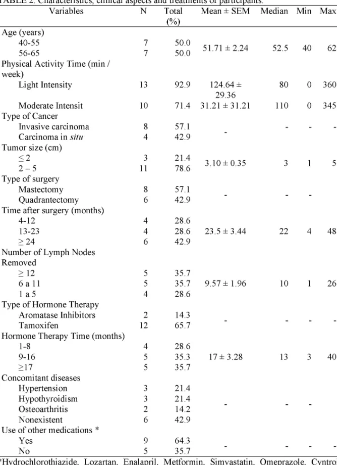
The baseline characteristics of the participants who completed the study are presented in table 2. The mean age was 51.71 ± 2.24 years and the prevalent pattern of physical activity was mild to moderate. Although the predominant type of cancer was invasive carcinoma, there was also a diagnosis of carcinoma in situ. The mean tumor size was 3.10 ± 0.35 cm and the surgical treatment, by mastectomy or quadrantectomy, had been done between 4 months and more than 24 months before the beginning of the intervention described in this study. All participants were lymphadenectomized and were on hormone therapy predominantly with tamoxifen, most of them for more than 9 months. Although the absence of concomitant diseases has been the most prevalent condition, some of the participants had other pathological conditions and made continuous use of other drugs.
[TABLE 2]
Before and after intervention, the consumption of carbohydrates (198.66 ± 15.62 vs. 197.74 ± 12.05; p= 0.96; d= 0.21), lipids (62.65 ± 5.64 vs. 58.93 ± 4.90; p= 0.541; d= 0.18) and proteins (74.54 ± 6.21 vs. 70.07 ± 3.55;p= 0.502; d= 0.23), given in g/day, as well as the energy intake (1646.32 ± 111.28 vs. 1597.21 ± 76.88; p= 0.621; d= 0.16), given in kcal/day, were not statistically different. However, there was an increase in LM and FFM and a decrease in FM and %BF, but without significant changes in BM, BMI, AB and WHR after the intervention reported in this study (Table 3).
[TABLE 3]
the high density lipoprotein cholesterol (HDL-C) and in the basophil counts. The lymphocyte, neutrophil and eosinophil counts did not change significantly with the exercise program. Although serum levels of the analyzed cytokines also have not changed significantly, after the intervention there was a significant increase in the salivary IgA concentration. In addition, we did not obtain plasma IL-6 in the sample. It is important to note that after the intervention there was also a reduction (p=0.021) in the concentration of low-density lipoprotein cholesterol (LDL-C).
[TABLE 4]
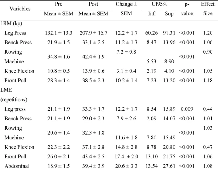
Table 5 shows the variables analyzed to measure the strength and the localized muscular endurance (LME) of the participants. After the intervention, in all exercises, there were significant increases in the load of 1RM (force) and in the number of repetitions completed with 50% of the load of 1RM (LME). The mean percentages of increase in load and number of repetitions in the tests of all the exercises were of 39.16% and 65.66%, respectively. This means that for the exercises performed there were mean increases in load and number of repetitions of
1.09% and 1.82%, respectively, on each training day. [TABLE 5] DISCUSSION
The main objective of this pilot study was to investigate the effects of a nonlinear resistance training (NLRT) protocol on biochemical, hematologic and inflammatory parameters in BC survivors under hormone therapy. Although no serum cytokine showed significant change after 12 weeks of intervention, there was a significant increase in salivary IgA, improvement in body composition, lipid profile, increased basophils, reduced plasma and monocytes and physical fitness of the participants. Other studies that investigated the effects of RT isolated on inflammatory profiles in BC survivors also did not show significant changes in serum inflammatory markers (12, 31, 34). But these studies followed a periodization model of the linear type, with initial intensities of 60% 1RM to 80% 1RM, without control and load readjustments. Therefore, our work was the first to report results after non-linearly periodic RT intervention (11) in this population
also play a determining role in the inflammatory response (27), as is also observed in BC survivors. In the study by Jones et al. (16), IL-6 and C-reactive protein (CRP) were positively associated with BM and BMI. This makes sense, since an increase in body fat may play a determining role in increasing serum levels of proinflammatory cytokines (13, 27).
To date, it has not been proven that increased muscle mass and reduced body fat content could promote an anti-inflammatory environment in BC survivors, but it is known that such anthropometric changes provide protection against the incidence of concomitant pathological processes, as insulin resistance (6, 9). Interestingly, studies that investigated the influence of RT on cancer survivors (12, 31, 34) and did not detect the occurrence of changes in serum cytokine levels, also did not report changes in body composition. But this does not mean that the hypothesis of improvement in the inflammatory profile from reductions in FM and increase in LM in response to RT should not be rejected in this population. Much of the previous studies, as well as the present study, may not have achieved sufficient time for the occurrence of the metabolic adaptations that could influence the inflammatory pathways. In addition, there are still many difficulties in the investigation of serum cytokines, such as laboratory limitations in the quantification process itself, which in fact occurred with IL-6 in the present study and with TNF-a in the study by Hagstrom et al. (2016) (12).
Surprisingly, this was the first study to report that one type of TR increases salivary IgA concentration in patients in BC recovery and under hormone therapy (Table 4). This is very important because a decrease in the level of salivary IgA is associated with an increase in the incidence of diseases of the upper respiratory tract (25), which may mean that salivary IgA may be a useful biological marker for assessing the clinical predisposition also for other diseases (1). Paradoxically, the only other study investigating the influence of exercise on salivary inflammatory response in BC patients observed that yoga practice one week before and four weeks after BC surgery resulted in a significant decrease in salivary IgA concentration (30). It is possible that this contradiction in the results is due to differences in the type, intensity and time of training, as well as differences in the health condition of the participants. Indeed, in the healthy elderly (n=45), the application of 12 weeks of moderate RT, three times a week, was able to significantly increase (36.84%) the salivary IgA concentration (2). In any case, this is a question that should be better investigated, since the tumor burden is directly related to serum IgA levels in cancer patients (1).
subpopulations of NK lymphocytes (12). The increase in basophil count reported in this study is a beneficial change to the population of BC survivors, as these cells play an important role in inflammation and simple allergic responses, although they constitute only 0.5 to 1% of total blood leukocytes (17). The decrease in circulating platelet count may be related to its use in the hemostasis of muscle micro-lesions, caused by TRNL, and also in the angiogenesis process (24). As the TR does not provide alterations in the population of monocytes (4), the decrease in the monocyte count in this study could be due to the increase in the differentiation of monocytes to macrophages rate.
In relation to the lipid profile, the influence of RT on the improvement of the lipid profile in healthy people is well known, as a result of increased mobilization and oxidation of fatty acids (20). In BC survivors, the application of 15 weeks of combined (aerobic and resistance) training was able to reduce blood levels of TGC (5%), t-C (6.8%) and LDL-C (9.7%), as well as increase HDL-C levels (4.5%) (26). The magnitudes of these changes were similar to those described in this study, but the increase in the blood level of HDL-C reported here (17.98%) was substantially higher, and this is very significant, because each increase of 1 mg/dL in HDL C is equivalent to 2-3% reduction in risk of cardiovascular events and decreased chance of recurrence of cancer (21).
The mean increase in muscle strength reported in this study (39.16%) after 12 weeks of intervention was higher compared to the results of other intervention studies with isolated RT after 12 (12, 31) and even after 24 weeks duration (25- 26%) in BC survivors (34). The best efficiency in increasing muscle strength reported in this study may be associated with the periodization model used (11). As the studies that investigated the influence of RT in this population did not evaluate LME (12, 31, 34, 38), establishing comparisons with literature results is not possible, but it is an interesting evaluation method to determine the physical condition of this population, mainly due to fatigue installed after treatment.
The present study has limitations, among which it is worth mentioning the absence of a group whose intervention was a traditional linear physical training protocol, in order to allow an effective comparison between the protocols. But even so, this pilot study also well serves the primary purpose of testing the applicability of NLRT in BC survivors and stimulating the development of new research on the subject.
addition, although there was no improvement in the inflammatory profile of the participants, there were improvements in other health indicators, such as salivary IgA and lipidemia, which
are very relevant for the prevention of concomitant diseases.
CONFLICTS OF INTEREST
M. A. Leite was supported by a scholarship from the “Coordenação de Aperfeiçoamento de Pessoal de Nível Superior” (CAPES). N. Penha-Silva was supported by a scientific productivity grant from “Conselho Nacional de Pesquisa e Desenvolvimento” (CNPq). The authors of the present study have no conflicts of interest to report. There was no participation of professionals or companies that could benefit from the results of the present study. The results of this study are unpublished and were presented clearly, honestly and without improper data manipulation. All authors participated equally in all stages of the study.
REFERENCES
1. Ahmad S, Faruqi NA, Arif SH, Akhtar S. Serum immunoglobulin levels in neoplastic disorder of breast. J Indian M ed Assoc 2002;100(8):495-6.
2. Akimoto T, Kumai Y, Akama T, et al. Effects of 12 months of exercise training on salivary secretory IgA levels in elderly subjects. Br J Sports M ed 2003;37(1):76-9. 3. Alexopoulos CG, Pournaras S, Vaslamatzis M, Avgerinos A, Raptis S. Changes in
serum lipids and lipoproteins in cancer patients during chemotherapy. Cancer Chemother Pharmacol 1992;30(5):412-6.
4. Bobeuf F, Labonté M, Khalil A, Dionne IJ. Effect of Resistance Training on Hematological Blood Markers in Older Men and Women: A Pilot Study. ResearchGate 2009;2009(1687-7063):156820.
5. Brown LE, Weir JP. ASEP Procedures recommendation I: accurate assessment of muscular strength and power. P rof Exerc Physiol 2001;4(11)
6. Caan BJ, Kwan ML, Shu XO, et al. Weight Change and Survival after Breast Cancer in the After Breast Cancer Pooling Project. Cancer Epidemiol Biomark Prev Publ Am Assoc Cancer Res Cosponsored Am Soc Prev Oncol 2012;21(8):1260-71.
7. Coussens LM, Werb Z. Inflammation and cancer. Nature 2002;420(6917):860-7. 8. Dewan MZ, Terunuma H, Takada M, et al. Role of natural killer cells in hormone
9. Dodson S, Baracos VE, Jatoi A, et al. Muscle wasting in cancer cachexia: clinical implications, diagnosis, and emerging treatment strategies. Annu Rev M ed 2011;62:265-79.
10. Fairey AS, Courneya KS, Field CJ, Bell GJ, Jones LW, Mackey JR. Randomized controlled trial of exercise and blood immune function in postmenopausal breast cancer survivors. JApplPhysiolBethesdaM d 1985 2005;98(4):1534-40.
11. Fleck SJ. Non-Linear Periodization for General Fitness & Athletes. J Hum Kinet 2011;29A:41-5.
12. Hagstrom AD, Marshall PWM, Lonsdale C, et al. The effect of resistance training on markers of immune function and inflammation in previously sedentary women recovering from breast cancer: a randomized controlled trial. Breast Cancer Res Treat 2016;155(3):471-82.
13. Heled Y, Dror Y, Moran DS, et al. Physical exercise increases the expression of TNFalpha and GLUT 1 in muscle tissue of diabetes prone Psammomys obesus. Life Sci 2005;77(23):2977-85.
14. Janelsins MC, Davis PG, Wideman L, et al. Effects of Tai Chi Chuan on Insulin and Cytokine Levels in a Randomized Controlled Pilot Study on Breast Cancer Survivors. Clin Breast Cancer 2011;11(3):161-70.
15. Johnson-Kozlow M, Sallis JF, Gilpin EA, Rock CL, Pierce JP. Comparative validation of the IPAQ and the 7-Day PAR among women diagnosed with breast cancer. Int J Behav Nutr Phys Act 2006;3:7.
16. Jones SB, Thomas GA, Hesselsweet SD, Alvarez-Reeves M, Yu H, Irwin ML. Effect of Exercise on Markers of Inflammation in Breast Cancer Survivors: The Yale Exercise and Survivorship Study. Cancer PrevRes PhilaPa 2013;6(2):109-18.
17. Knol EF, Olszewski M. Basophils and mast cells: Underdog in immune regulation? Immunol Lett 2011;138(1):28-31.
18. Lee B-N, Dantzer R, Langley KE, et al. A cytokine-based neuroimmunologic mechanism of cancer-related symptoms. Neuroimmunomodulation 2004;11(5):279-92. 19. Makari-Judson G, Braun B, Jerry DJ, Mertens WC. Weight gain following breast cancer
diagnosis: Implication and proposed mechanisms. World J Clin Oncol 2014;5(3):272- 82.
21. McGrowder D, Riley C, Morrison EYSA, Gordon L. The Role of High-Density Lipoproteins in Reducing the Risk of Vascular Diseases, Neurogenerative Disorders, and Cancer [Internet]. Cholesterol 2011;2011 [cited 2017 May 12 ] Available from: http://www.ncbi.nlm.nih.gov/pmc/articles/PMC3065895/
22. Meneses-Echâvez JF, Correa-Bautista JE, Gonzâlez-Jiménez E, et al. The Effect of Exercise Training on Mediators of Inflammation in Breast Cancer Survivors: A Systematic Review with Meta-analysis. Cancer Epidemiol Biomark Prev Publ Am Assoc Cancer Res Cosponsored Am Soc Prev Oncol 2016;25(7):1009-17.
23. Mills PJ, Ancoli-Israel S, Parker B, et al. Predictors of Inflammation in Response to Anthracycline-Based Chemotherapy for Breast Cancer. Brain Behav Immun 2008;22(1):98-104.
24. Mosca MJ, Rodeo SA. Platelet-rich plasma for muscle injuries: game over or time out? Curr Rev Musculoskelet M ed 2015;8(2):145-53.
25. Neville V, Gleeson M, Folland JP. Salivary IgA as a risk factor for upper respiratory infections in elite professional athletes. M edSci Sports Exerc 2008;40(7):1228-36. 26. Nuri R, Mahmudieh B, Akochakian M, Moghaddasi M, Nuri R. Effect of 15 weeks
Combination exercise training on lipid profile and fatty liver indices in Postmenopausal women with breast cancer. Braz JBiomotricity 2012;6(4):297-303.
27. Ost M, Coleman V, Kasch J, Klaus S. Regulation of myokine expression: Role of exercise and cellular stress. Free Radic Biol M ed 2016;98:78-89.
28. Pedersen BK, Febbraio MA. Muscles, exercise and obesity: skeletal muscle as a secretory organ. Nat Rev Endocrinol 2012;8(8):457-65.
29. Petersen AMW, Pedersen BK. The anti-inflammatory effect of exercise. J Appl Physiol BethesdaM d 1985 2005;98(4):1154-62.
30. Rao RM, Nagendra HR, Raghuram N, et al. Influence of yoga on mood states, distress, quality of life and immune outcomes in early stage breast cancer patients undergoing surgery. Int J Yoga 2008;1(1):11-20.
31. Schmidt ME, Meynkohn A, Habermann N, et al. Resistance Exercise and Inflammation in Breast Cancer Patients Undergoing Adjuvant Radiation Therapy: Mediation Analysis From a Randomized, Controlled Intervention Trial. Int J Radiat Oncol Biol Phys 2016;94(2):329-37.
33. Sewell HF, Halbert CF, Robins RA, Galvin A, Chan S, Blamey RW. Chemotherapy- induced differential changes in lymphocyte subsets and natural-killer-cell function in patients with advanced breast cancer. Int J Cancer 1993;55(5):735-8.
34. Simonavice E, Liu P-Y, Ilich JZ, Kim J-S, Arjmandi B, Panton LB. The effects of a 6- month resistance training and dried plum consumption intervention on strength, body composition, blood markers of bone turnover, and inflammation in breast cancer survivors 1. ApplPhysiolNutr Metab 2014;39(6):730-9.
35. Slater B, Philippi ST, Marchioni DML, Fisberg RM. Validation of Food Frequency Questionnaires - FFQ: methodological considerations. Rev Bras Epidemiol 2003;6(3):200-8.
36. Tsavaris N, Kosmas C, Vadiaka M, Kanelopoulos P, Boulamatsis D. Immune changes in patients with advanced breast cancer undergoing chemotherapy with taxanes. Br J Cancer 2002;87(1):21-7.
37. White JP, Wilson JM, Austin KG, Greer BK, St John N, Panton LB. Effect of carbohydrate-protein supplement timing on acute exercise-induced muscle damage. J Int Soc Sports Nutr 2008;5:5.
38. Winters-Stone KM, Dobek J, Bennett JA, Nail LM, Leo MC, Schwartz A. The effect of resistance training on muscle strength and physical function in older, postmenopausal breast cancer survivors: a randomized controlled trial. J Cancer Surviv Res Pract 2012;6(2):189-99.
39. World Health Organization. International Agency for Research on Cancer. 2013;13. 40. Zanetti HR, da Cruz LG, Lourenço CL, et al. Nonlinear Resistance Training Enhances
Legends of Figures
TABLE 1. Order of exercises, per day, from muscle stimuli.______________________ _______ Stimuli_____________ Monday___________ Wednesday____________ Friday
1. Strength a 2. Hypertrophy b 3. Resistance c
Chest Press Bench Press Knee Flexion Front Pull
Rowing Machine Abdominal
Knee Flexion Front Pull
Rowing Machine Abdominal Leg Press Chest Press
Rowing Machine Abdominal Leg Press Chest Press Knee Flexion Front Pull a Strenght: 3 sets of 4-6 repetitions at 85% 1RM with 2-3 minutes rest between sets.
TABLE 2. Characteristics, clinical aspects and treatments of participants.
Variables N Total Mean ± SEM Median Min Max (%)
Age (years)
40-55 7 50.0 51.71 ± 2.24 52.5 40 62
56-65 7 50.0
Physical Activity Time (min / week)
Light Intensity 13 92.9 124.64 ± 80 0 360
29.36
Moderate Intensit 10 71.4 31.21 ± 31.21 110 0 345 Type of Cancer
Invasive carcinoma 8 57.1 - -
-Carcinoma in situ 4 42.9
Tumor size (cm)
< 2 3 21.4 3.10 ± 0.35 3 1 5
2 - 5 11 78.6
Type of surgery
Mastectomy 8 57.1
Quadrantectomy 6 42.9
Time after surgery (months)
4-12 4 28.6
13-23 4 28.6 23.5 ± 3.44 22 4 48
> 24 6 42.9
Number of Lymph Nodes Removed
> 12 5 35.7
6 a 11 5 35.7 9.57 ± 1.96 10 1 26
1 a 5 4 28.6
Type of Hormone Therapy
Aromatase Inhibitors 2 14.3
Tamoxifen 12 65.7
Hormone Therapy Time (months)
1-8 4 28.6
9-16 5 35.3 17 ± 3.28 13 3 40
>17 5 35.7
Concomitant diseases
Hypertension 3 21.4
Hypothyroidism 3 21.4
Osteoarthritis 2 14.2
Nonexistent 6 42.9
Use of other medications *
Yes 9 64.3
No 5 35.7
TABLE 3. Anthropometric measures of participants before (pre) and after (post) intervention.
Variables
Pre Post
Change ± SEM
CI 95%
Effect Size Mean ±
SEM
Mean ±
SEM Inf Sup
p-value
TABLE 4. Lipid profile, immunological cell counts and inflammatory markers before (pre) and after (post) intervention.
Variables Pre Post Change ± CI95% p- Effect
Mean ± SEM Mean ± SEM SEM Inferior Superior value Size Lipidogram (mg/dL)
Cholesterol 208.8 ± 6.0 198.75 ± 5.9 -6.1 ± 4.7 -15.92 -4.19 0.004 0.45 Triglycerides 163.9 ± 16.8 140.35 ± 13.6 -23.6 ± 6.6 -37.93 -9.29 0.003 0.37 LDL-C 125.6 ± 8.2 112.78 ± 6.3 -11.0 ± 5.2 -22.26 -2.26 0.021 0.47 HDL-C 47.0 ± 2.4 55.50 ± 2.6 8.5 ± 2.0 4.10 12.81 <0.001 0.49 Cell count (103/uL)
Platelets 218.0 ± 14.5 200.8 ± 9.3 -17.2 ± 7.2 -32.68 -1.75 0.032 0.27 Lymphocytes 1.6 ± 0.1 1.7 ± 0.1 0.08 ± 0.11 -0.14 0.31 0.484 0.02 Neutrophils 1.6 ± 0.8 1.7 ± 0.7 0.16 ± 0.23 -0.33 0.66 0.446 0.20 Monocytes 0.18 ± 0.02 0.11 ± 0.02 -0.11 ± 0.04 -0.21 -0.02 0.020 0.62 Eosinophils* 0.07 ± 0.07 0.06 ± 0.12 -0.03 ± 0.02 -0.01 0.06 0.209 0.33 Basophils 0.016 ± 0.002 0.033 ± 0.007 0.02 ± 0.01 0.00 0.03 0.037 0.76 Inflammatory
markers (pg/mL)
IFN-y* 228.1 ± 466.8 177.5 ± 722.7 -44.2 ± 60.3 -174.39 86.06 0.247 0.31 IL-4* 86.2 ± 121.8 77.4 ± 85.2 -40.9 ± 42.9 -133.71 51.77 0.752 0.08 IL-10* 244.2 ± 426.6 173.8 ± 439.2 79.9 ± 115.4 -169.34 329.18 0.937 0.02 IL-17* 11.9 ± 126.2 1.1 ± 57.91 -30.0 ± 20.4 -74.07 14.10 0.123 0.41
TNF-a* 0.3 ± 117.8 7.1 ± 1358.2 225.2 ± -448.83 899.32 0.790 0.07 IL-1Ra* 22.6 ± 293.7 0 ± 0
312.0
-87.7 ± 87.9 -277.54 102.14 0.176 0.36 IgA 152.3 ± 73.0 436.1 ± 175.8 260.0 ± 47.3 85.12 286.37 <0.001 0.75 * Variables that violated normality even with statistical normalization and were expressed as median ± IQR (Q3-Q1) and analyzed by non-parametric inferential statistics.
TABLE 5. Strength and localized muscular endurance of the participants before (pre) and after (post) the intervention.
Variables Pre Post Change ± CI95% p- Effect
Mean ± SEM Mean ± SEM SEM Inf Sup value Size 1RM (kg)
Leg Press 132.1 ± 13.3 207.9 ± 16.7 12.2 ± 1.7 60.26 91.31 <0.001 1.20 Bench Press 21.9 ± 1.5 33.1 ± 2.5 11.2 ± 1.3 8.47 13.96 <0.001 1.06 Rowing
Machine 34.8 ± 1.6 42.4 ± 1.9
7.2 ± 0.8
5.53 8.90 <0.001
0.90
Knee Flexion 10.8 ± 0.5 13.9 ± 0.6 3.1 ± 0.4 2.19 4.10 <0.001 1.05 Front Pull 28.3 ± 1.4 38.5 ± 2.3 10.2 ± 1.4 7.23 13.20 <0.001 1.18 LME
(repetitions)
Leg press 21.1 ± 1.9 33.3 ± 1.7 12.2 ± 1.7 8.54 15.89 0.009 0.44 Bench Press 21.1 ± 1.9 29.0 ± 2.3 7.9 ± 2.6 2.09 14.07 <0.001 1.01 Rowing
Machine 20.6 ± 1.4 32.3 ± 1.8 11.6 ± 1.8 7.80 15.49 <0.001
1.03