Mafalda Maria Robalo de
Azevedo Aleixo Pereira
Licenciada em Bioquímica
Role of sympathetic
innervation in obesity
Dissertação para obtenção do
Grau de Mestre em Bioquímica
Orientador: Ana I. Domingos, Investigadora Principal,
Instituto Gulbenkian de Ciência
Co-orientador: Gonçalo J. L. Bernardes, Investigador
Principal, Instituto de Medicina Molecular
ii
Mafalda Maria Robalo de
Azevedo Aleixo Pereira
Licenciada em Bioquímica
Role of sympathetic
innervation in obesity
Dissertação para obtenção do
Grau de Mestre em Bioquímica
Orientador: Ana I. Domingos, Investigadora Principal,
Instituto Gulbenkian de Ciência
Co-orientador: Gonçalo J. L. Bernardes, Investigador
Principal, Instituto de Medicina Molecular
iii
Role of sympathetic innervation in obesity
Copyright © Mafalda Maria Robalo de Azevedo Aleixo Pereira, Faculdade de Ciências e Tecnologia, Universidade Nova de Lisboa.
iv
Acknowledgements
First of all, I would like to thank to Alekos Athanasiadis for introducing me to Ana Domingos. Then, obviously my acknowledgment goes to Ana Domingos for giving me the opportunity to develop my project in her lab and for teaching me that we are always able to do it on our own. I also thank to Gonçalo Bernardes for believing in this project and for being available every time I asked for help.
From the bottom of my heart, I thank to the “real” people in the lab: Elsa, Nadiya, Roksana, Andreia, Inês and Imogen. You were the ones that were really there, the ones that taught me everything and the ones that helped me every time I needed - sometimes I did not have to ask, you were already there. You girls rock!
Apart from the lab, other people were important during this 14 months journey: people from other labs that were always helpful, Krzysztof (who supported me during countless weekends), people from Unit of Citometry and Imaging (Gaby, Ânia, Pimpão, Cláudia 1, Cláudia 2 and Rui), animal house facility caretakers and technicians, histology technicians, canteen people (mainly Sofia that provided food at strange hours), maintenance team (especially João for being like a privative engineer), Pedro Cal from Gonçalo Bernardes’ Lab and many other people that I am not being able to mention.
Funding agencies were fundamental to allow this work to be performed, namely Instituto Gulbenkian de Ciência, European Molecular Biology Organization and Fundação para a Ciência e Tecnologia.
I could not leave behind Professor Alice that since the beginning accepted to be my “connection” to the Faculty and was always (meaning even in the evening!) available to clarify my doubts and to give me support to keep going.
At last (but for sure no least!), my biggest thank goes to my family, to my boyfriend and to my friends. You all know that I would not survive to this without you!
“E compreendes que é desta fibra que são feitos os vencedores. Que é a persistência, a abnegação, o espírito de sacrifício e um certo autismo que separam os que conseguem dos
v
List of publications
Part of the results presented in this thesis were published in the following reference (DOI 10.1016/j.cell.2015.08.055): Wenwen Zeng*, Roksana M. Pirzgalska*, Mafalda M.A. Pereira, Nadiya Kubasova, Andreia Barateiro, Elsa Seixas, Yi-Hsueh Lu, Albina Kozlova,
Henning Voss, Gabriel G. Martins, Jeffrey M. Friedman and Ana I. Domingos. Sympathetic Neuro-adipose Connections Mediate Leptin-Driven Lipolysis. Cell 163, 84-94 (2015).
vi
Abstract
Obesity is considered a world epidemic, but no efficient therapy is available so far. Besides, the anatomy of the adipose organ was not described in detail. Thus, different mechanisms were proposed to explain obesity condition. One of those is the MONA LISA hypothesis that states that Most Obesities kNown Are Low In Sympathetic Activity. However, this theory is based on measurements of norepinephrine decrease in the heart and there is no direct evidence of the role of sympathetic nervous system (SNS) in obesity.
In this work, PEGyDT-mediated sympathectomy was developed to specifically ablate the SNS, without affecting the central nervous system. This was achieved by PEGylation of diphtheria toxin, a strategy that is approved by Food and Drug Administration and used to modulate pharmacokinetics of several biopharmaceuticals clinically approved.
The results show that sympathectomy leads to irreversible obesity, without affecting food intake. SNS ablation also causes glucose tolerance and thermogenesis impairment. Moreover, it was discovered that the SNS is required for amphetamine effects and this is an additional mechanism of action for this anti-obesity drug, as it was thought that it had an exclusive effect in the brain to suppress appetite. Furthermore, the anatomy of adipose organ was revealed in detail using optical projection tomography coupled to tissue clearing. It was shown that not only the inguinal adipose organ contains nerve bundles and a network of vasculature, but also that in the proximity of these structures there is a lymph node. In the epididymal fat, granular substructures are present but not nerve bundles or lymph node.
In conclusion, a strategy to study the role of the SNS in obesity was developed and causality of MONA LISA hypothesis was demonstrated. Moreover, an additional mechanism of action for amphetamine was shown and neuroanatomy of adipose organs was described for the first time.
Keywords: Obesity, Sympathetic Nervous System, PEGylation, Amphetamine, Anatomy,
vii
Resumo
A obesidade é considerada uma epidemia mundial, mas não se encontra disponível uma terapia eficiente. Além disso, a anatomia do orgão adiposo não foi descrita detalhadamente. Assim, diferentes mecanismos foram propostos para explicar a obesidade. Um deles é a hipótese da MONA LISA que defende que a maioria dos obesos apresentam baixa actividade simpática. Contudo, esta teoria baseia-se em medições da diminuição da norepinefrina no coração, não existindo evidência directa da função do sistema nervoso simpático (SNS) na obesidade.
Neste trabalho, a simpatectomização mediada pela PEGyDT foi desenvolvida para remover especificamente o SNS, não afectando o sistema nervoso central. Isto foi conseguido recorrendo à PEGilação da toxina da difteria, sendo esta uma estratégia aprovada pelo órgão que controla os alimentos e medicamentos e utilizada para modular a farmacocinética de vários fármacos aprovados clinicamente.
Os resultados mostram que a simpatectomização conduz a obesidade irreversível, sem afectar a ingestão de alimentos. A remoção do SNS altera a tolerância à glucose e a termogénese. Foi ainda descoberto que o SNS é necessário para os efeitos da anfetamina, sendo este um mecanismo adicional deste fármaco anti-obesidade uma vez que foi mostrado que este teria um efeito exclusivamente no cérebro, como supressor do apetite. A anatomia do órgão adiposo foi revelada detalhamente usando tomografia de projecção óptica associada à transparentização do tecido. Foi mostrado que o órgão adiposo inguinal não só contém nervos e uma rede de vasculatura, mas também que existe um nódulo linfático na proximidade destas estruturas. Na gordura epididimal, estão presentes sub-estruturas granulares, mas não existem nervos nem nódulos linfáticos.
Concluindo, foi desenvolvida uma estratégia para estudar a função do SNS na obesidade e foi desmonstrada a causalidade da hipótese da MONA LISA. Foi ainda mostrado um mecanismo de acção adicional para a anfetamina e a neuro-anatomia dos órgãos adiposos foi descrita.
Palavras-chave: Obesidade, Sistema Nervoso Simpático, PEGilação, Anfetamina, Anatomia,
viii
Aims
The main aim of this work is to develop a tool that allows the ablation of sympathetic nervous system, without damaging the central nervous system.
After this is accomplished, the goal is to study the effects of sympathectomy on mice, in order to understand the role of sympathetic nervous system in obesity.
ix
List of contents
Acknowledgements ... iv
List of publications ... v
Abstract ... vi
Resumo ... vii
Aims ... viii
List of abbreviations ... xiii
1. Introduction ... 1
1.1. Obesity ... 1
1.2. Anti-obesity therapy ... 1
1.2.1. Fen and AMPH ... 2
1.3. Fat characterization and Optical Projection Tomography (OPT) ... 3
1.4. MONA LISA hypothesis ... 4
1.5. Ablation of central and peripheral nervous systems ... 5
1.6. Protein modifications ... 7
2. Materials and Methods ... 10
2.1. DT modification ... 10
2.2. Sodium dodecyl sulfate polyacrylamide gel electrophoresis (SDS-PAGE) ... 10
2.3. Mass spectrometry (MS) ... 10
2.4. Cell culture of HeLa cells ... 11
2.5. Bone marrow (BM)-derived macrophages from Rosa26Cre-ER(T2); LSL-DTR mice... ... 11
2.6. Mice and housing conditions... 12
2.7. PEGyDT-mediated sympathectomy ... 12
2.8. Functional tests on sympathetic ablated mice ... 13
2.9. Anti-obesity drugs ... 13
2.10. OPT ... 14
2.11. Statistical analysis ... 14
3. Results ... 15
3.1. PEG and Mal bind to DT ... 15
3.2. PEGyDT, but not DT Mal, remains functional after modification ... 16
3.3. PEGyDT does not kill catecholaminergic neurons in the brain, but it kills sympathetic nerves... 18
3.4. Sympathectomy leads to irreversible obesity, without changing food intake ... 20
3.5. Sympathectomy affects glucose tolerance, without affecting blood glucose and insulin... 21
3.6. Sympathectomy affects thermogenesis ... 22
3.7. AMPH prevents obesity, independently of food intake ... 23
3.8. SNS is required for AMPH effects ... 24
x
4. Discussion ... 29
5. Conclusion and Future perspectives ... 33
6. References ... 34
xi
List of Figures
Figure 1.1 – Fen (A) and AMPH (B) chemical structures. ... 3
Figure 1.2 – Schematic representation of MONA LISA hypothesis 41. ... 4
Figure 1.3 – Schematic representation of TH-Cre; LSL-DTR mice 48. ... 6
Figure 1.4 – Representation of DT structure (adapted from 50). ... 6
Figure 1.5 – Strategy to avoid DT in the brain. ... 7
Figure 1.6 – Representation of a general PEGylation reaction (adapted from 54). ... 8
Figure 1.7 – Representation of a general maleimidation reaction (adapted from 68). ... 9
Figure 3.1 – Assessment of DT modification state. ... 15
Figure 3.2 – DT functionality after modification tested on in vitro HeLa cell line culture. ... 16
Figure 3.3 – DT functionality after modification tested in vitro on primary cell culture of macrophages derived from the BM of Rosa26Cre-ER(T2); LSL-DTR mice after tamoxifen was administered in vivo. ... 17
Figure 3.4 – Differences in weight and in food intake between TH-Cre; LSL-DTR mice injected with DT and PEGyDT. ... 18
Figure 3.5 – PEGyDT does not kill catecholaminergic neurons in the brain. ... 19
Figure 3.6 – PEGyDT kills sympathetic nerves. ... 19
Figure 3.7 – Sympathectomy leads to obesity. ... 20
Figure 3.8 – Obesogenic effect of sympathectomy is irreversible and independent of food intake. ... 21
Figure 3.9 – Sympathectomy affects glucose tolerance, without affecting blood glucose and insulin. ... 22
Figure 3.10 – Sympathectomy affects thermogenesis. ... 22
Figure 3.11 – AMPH prevents obesity, independently of food intake. ... 23
Figure 3.12 – SNS is required for AMPH effects. ... 24
Figure 3.13 – Schematic representation of the OPT setup. ... 24
Figure 3.14 – Optical Projection Tomography (OPT) image acquisition and analysis.… 25 Figure 3.15 – OPT of inguinal fat. ... 25
Figure 3.16 – Segmentation of structures within inguinal fat. ... 26
xii
Figure 3.18 – OPT of epididymal fat. ... 26
Figure 3.19 – Segmentation of structures within epididymal fat. ... 27
Figure 3.20 – Granular substructures confined to a region in epididymal fat. ... 27
Figure 3.21 – Graphical abstracts summarizing the work. ... 28
Figure 7.1 – DT concentration test on HeLa cells. ... 39
Figure 7.2 – DT functionality after modification tested on in vitro HeLa cell line culture. ... 39
Figure 7.3 – HeLa cell line culture incubated with BSA. ... 40
Figure 7.4 – DT functionality after modification tested in vitro on primary cell culture of macrophages derived from the BM of Rosa26Cre-ER(T2); LSL-DTR mice after tamoxifen was administered in vivo. ... 40
Figure 7.5 – PBS administration does not affect food intake and body weight. ... 40
Figure 7.6 – DT, but not PEGyDT, administration affects normal movement of mice (*** p < 0.0001, n = 3). ... 40
Figure 7.7 – Food intake during HFD feeding 2 weeks after SNS ablation (n = 5-6). ... 40
Figure 7.8 – Differences in weight and in food intake on a HFD regimen. ... 40
xiii
List of abbreviations
3D Three-dimensional 5-HT 5-hidroxitriptamine β3-Tubβ3-Tubulin
ADHD Attention deficit hyperactivity disorder ADP Adenosine diphosphate
AMPH Amphetamine
ANS Autonomic nervous system BAT Brown adipose tissue BBB Blood-brain barrier BM Bone marrow BMI Body mass index BSA Bovine serum albumin
C Enzymatic or catalytic domain of diphtheria toxin CNS Central nervous system
CT Computed tomography
Dapi 4’,6-diamidino-2-phenylindole
DMEM Dulbecco’s modified Eagle medium
DMSO Dimethyl sulfoxide DNA Deoxyribonucleic acid DT Diphtheria toxin
DTR Diphtheria toxin receptor EF-2 Elongation factor 2
ELISA Enzyme-linked immunosorbent assay
EM Electron microscopy ER Estrogen receptor
Fab Fragment antigen binding
FACS Fluorescence-activated cell sorting FBS Fetal bovine serum
FDA Food and Drug Administration Fen Fenfluramine
GTT Glucose tolerance test HFD High fat diet
iDTR Cre-inducible diphtheria toxin receptor IGC Instituto Gulbenkian de Ciência
IMDMIscove’s modified Dulbecco’s medium
xiv
MALDI-TOF Matrix-assisted laser desorption / ionization – time of flight M-CSF Macrophage colony stimulating factor
MIP Maximal intensity projection
MOG Myelin oligodendrocyte glycoprotein
MONA LISA Most Obesities kNown Are Low In Sympathetic Activity
MRI Magnetic resonance imaging MS Mass spectrometry
MW Molecular weight
NAD Nicotinamide adenine dinucleotide NCDs Non-communicable diseases ND Normal diet
NE Norepinephrine
NHS N-hydroxysuccinimide
OPT Optical projection tomography ORF Open reading frame
PBS Phosphate buffered saline PEG Polyethylene glycol
PEGyDT PEGylated diphtheria toxin
Phen Phentermine
PNAC Protein & Nucleic Acid Chemistry R Receptor-binding domain of diphtheria toxin RT Room temperature
SCID Severe combined immunodeficiency
SDS-PAGE Sodium dodecyl sulfate polyacrylamide gel electrophoresis
SEM Standard error of the mean SNS Sympathetic nervous system
Sulfo-SMCC Sulfosuccinimidyl 4-(N-maleimidomethyl)cyclohexane-1-carboxylate
T Transmembranar or translocation domain ofdiphtheria toxin
TH Tyrosine hydroxylase TNF Tumor necrosis factor
1
1. Introduction
1.1. Obesity
Although only recognized by the American Medical Association in 2013 as a disease, obesity is now considered as the largest and fastest world global epidemic - the “globesity”1,2.
It is a public health challenge that remains unsolved and consequences of this epidemic will lead to serious health disorders, if nothing is done immediately to overcome this problem 3,4.
Each year, overweight or obesity are the cause of 3 million deaths in the world 5. In
2000, 300 million people worldwide were considered obese or overweight and, in 2008, the number was set at 2 billion 1,2,4. Obesity is not restricted to developed countries, being estimated
that 115 million people suffer from obesity-related problems in developing countries 1,2,4,6.
Furthermore, 10 % of children and adolescents are obese and this number is predicted to double in 2025 1.
Overweight and obesity result in impaired quality of life as physical disabilities and psychological problems are caused by these conditions. Besides, they are also considered as risk factors for chronic diseases that are known as non-communicable diseases (NCDs), such as diabetes mellitus, cardiovascular diseases and cancer 1,2,6–8. Overweight and obesity also
lead to adverse metabolic effects on blood pressure, cholesterol, triglycerides and insulin resistance 4,5,9. In addition, obesity causes premature mortality and large healthcare costs 1,2,7,8.
A simple definition for obesity is to consider it as excessive fat accumulation, leading to health risk 9. This is achieved when consumed calories are higher than expended, resulting in
energy imbalance 1,9. More precisely, obesity is measured through body mass index (BMI),
which is calculated dividing the weight (in kilograms) by the square of height (in meters) 1,9,10. If
BMI is 30 kg/m2 or higher, the person is considered obese 1,10,11.
A body weight loss of 5 to 10 % improves insulin resistance and hypertension and this can be achieved with a lifestyle change (reduction in caloric intake and increase in physical activity) 2,8,12. The problem is that compliance to adapt to a new lifestyle is very limited, so
pharmacological approaches are seen as a tool to achieve weight loss 2,6,12. As the awareness
of healthier eating habits and exercise was not effective, drugs regimens are needed to achieve a meaningful and sustainable reduction in body weight 2.
1.2. Anti-obesity therapy
There are now three main approaches to cope with obesity that suffer from a number of limitations:
- Changes in lifestyle (diet and physical activity), which are not effective. Most patients regain weight shortly after therapy end and, in some, regain occurs even during therapy.
- Bariatric surgery (bypass or gastric banding), which is effective for weight loss because it reduces the size of the stomach, thus reducing food intake, but it has drawbacks such as surgical complications, need for reoperation and perioperative mortality. Moreover, it is a very expensive procedure.
- Pharmacotherapy, that is used to promote weight loss and also to increase patients’ compliance to lifestyle changes and will be discussed in detail below 1,2,8,10,13.
Drugs are used to assist and maintain weight loss because a lifestyle modification is not effective. In 2009, 1.5 million obesity-related prescriptions were dispensed 10. Weight loss
pharmacotherapy is prescribed to those with a BMI of 30 kg/m2 or above or to those with a BMI
of 27 kg/m2 or above with an obesity-related condition (for instance, high blood pressure and
diabetes) 14.
An anti-obesity drug is considered efficient when it is effective for 10 % weight reduction and improves overweight-dependent conditions; it is safe for long-term use without losing efficiency; the side effects are tolerable or transitory; it is not addictive; and its mechanism of action is known 1,2,12,15,16. None of the existing drugs fulfill the mentioned characteristics 8. As a
consequence, there is an urgent need for the development of more selective, safer and more efficient drugs for obesity treatment 12.
Until 1940, amphetamine (AMPH) was used as an anti-obesity drug, but after World War II, it became a drug of abuse, leading to the search for safer alternatives in the field of sympathomimetic drugs, which inhibit norepinephrine (NE) re-uptake 17,18. Between 1973 and
2 year later was withdrawn because of cardiac toxicity. Ephedrine was used for asthma treatment, but it was proven to increase thermogenesis and reduce appetite, so it was also used as an anti-obesity drug. In 1994, Jeffrey M. Friedman discovered leptin and it was demonstrated that the lack of this hormone leads to obesity and also that treatment with leptin reverses this situation. After this, more centrally acting drugs were developed but, so far, none of them proved to be efficient 17.
The classification of agents to treat obesity is done according to their mechanisms of action. Hence, these drugs can be divided in three groups: those that reduce food intake; those that alter metabolism and those that increase thermogenesis or energy expenditure 12,19:
Appetite suppressant
Food intake reduction is achieved either by causing a delay in the onset of a meal or by achieving earlier satiety. This group of drugs includes monoamines acting on noradrenergic, serotonin, dopamine, and histamine receptors. Some peptides and neuropeptides also inhibit the appetite such as leptin; neuropeptide-y receptor antagonists; cholecystokinin; ghrelin antagonist; and pancreatic hormones (glucagon and insulin) 19.
The activation of the α1- and β2-adrenoceptors decreases food intake. These receptors
are activated if drugs that release NE or block NE re-uptake are used. The same effect in food intake is detected if 5-hidroxitriptamine (5-HT), most known as serotonin, receptors (5-HT1 and 5-HT2) are activated. This activation is achieved using drugs that block 5-HT re-uptake, such as fluoxetine, sertraline, and fenfluramine (Fen) or providing 5-HT to the receptors 17,19.
Sympathomimetic drugs approved by FDA include: benzphetamine, phentermine (Phen) and diethylpropion (release NE); mazindol and sibutramine (block NE re-uptake); and phenylpropanolamine (activates adrenoceptors) 13,17,19. Sympathomimetic drugs that were not
approved are in the market carrying a warning label (AMPH and methamphetamine) or were never on the market (fenproporex and chlobenzorex) 19.
Alter metabolism (to induce catabolism and inhibit anabolism)The strategies used so far were directed to pre and post-absorptive mechanisms in order to modify fat absorption or metabolism. Pre-absorptive mechanisms aim to influence digestion and absorption of macronutrients, while post-absorptive mechanisms aim to enhance lipolysis, inhibit lipogenesis and affect fat distribution between inguinal and visceral sites. Xenical (orlistat), that inhibits pancreatic lipase and reduces intestinal digestion of fat, is the only drug of this group approved by FDA 1,2,10,12–14,17,19.
Increase thermogenesis
This type of drugs aims to mimic the physiologic effects of exercise, that increases energy expenditure 19. Although this type of drugs were not approved for obesity treatment,
ephedrine was approved to relax bronchial smooth muscle in asthma patients and caffeine inhibits adenosine receptors and phosphodiesterase. Both were proved to increase thermogenesis and were used as anti-obesity treatment 12,17,19.
1.2.1. Fen and AMPH
In the 70s and 80s, Fen (Figure 1.1 A) was widely used as an anti-obesity therapy. It is known to increase serotonin release and inhibit serotonin re-uptake, so it is considered as a nonselective serotonin agonist, acting on 5-HT2A, 5-HT2B, and 5-HT2C receptors 8,13,19. A
therapy using Fen together with the Phen adrenergic drug resulted in a more effective weight loss with fewer side effects when compared to single drug therapies. Despite approval of both drugs by FDA, the combined therapy was not approved but was widely used and became known as the Fen/Phen therapy. The unwanted side effects arose in the form of valvular heart disease and this led to the withdrawal of Fen and dexfenfluramine, in 1997 1,19,20.
AMPH, the contracted form of α-methyl-β-phenethylamine (Figure 1.1 B), was primarily used in the treatment of attention deficit hyperactivity disorder (ADHD) and narcolepsy, and only later it was used as an anti-obesity drug. In the beginning, it was freely available without prescription but then it became a highly restricted controlled drug 17,19,21. Amphetamines were
first used to treat obesity in the 30s and after that many AMPH-like agents were approved 22. Its
mechanism of action consists in stimulating the release of NE and dopamine, acting on the satiety centers of the brain 6. This is a possible mechanism because amphetamines are
3 as dopamine, NE and epinephrine 6,17,19,21–23. It is also established that NE is responsible for the
appetite suppressant effect, while dopamine is responsible for the risk of habituation 17,19. Apart
from the therapeutic efficacy, AMPH has psychiatric-related adverse effects for the reason that this is a sympathomimetic drug, thus stimulating the central nervous system (CNS). Many cases of recreational abuse and addiction were reported, as well as cardiovascular effects such as, tachycardia and increased blood pressure 6,17,19,21–23. In addition, the long term (one year or
more) efficacy is not proven 22,24,25. All these drawbacks limited the therapeutic use and,
nowadays, amphetamines are not clinically approved as a weight loss therapy 6,22,23.
Many sympathomimetic drugs were clinically approved but were withdrawn from the market because of its undesirable side effects (the major concern is the cardiovascular toxicity). Therefore, many anti-obesity drugs are being developed and tested and their approval is being carefully done 1,19,23.
It has been suggested that some patients are insensitive to pharmacological agents, leading to the theory that there are several chemical mechanisms causing obesity 8. In addition
to the absence of efficient treatment, until the discovery of leptin hormone, the adipose tissue was considered only as an energy depot and was not accepted as an endocrine organ 26. As an
endocrine organ, adipose tissue is responsible for the synthesis and secretion of several hormones active in a variety of processes, such as control of nutritional intake, sensitivity to insulin, mediation of inflammatory processes or stimulation of signaling pathways 27,28. The
adipose organ is considered as an inguinal or a visceral organ, based on body location 28–30.
Although it is now considered as an organ, no anatomical characterization was made so far to describe the adipose tissue.
1.3. Fat characterization and Optical Projection Tomography (OPT)
The anatomy of adipose organ has been mostly studied with classical histology methods or electron microscopy (EM) 31. These methods are suitable for histological analysis at a
microscopic spatial scale, but do not give a three-dimensional (3D) perspective of the organization of the organ as a whole. The same applies to optical methods such as confocal or multiphoton microscopy 32,33. At macroscopic spatial scale, methods such as magnetic
resonance imaging (MRI) or computed tomography (CT) allow the measurement of whole body fat distribution 34–36. However, all of these methods lack the spatial resolution that is required for
visualizing structures within an organ. 3D description is important as the health of an organ can be accessed through visualization of its anatomical features.
OPT is a technique with physical principles similar to X-ray CT/gamma radiation, which uses a different wavelength (mainly, in the visible range) 37. A full series of projections of the
whole sample is acquired from multiple angles, typically 800 to 1600 angles, and from this series of projections a stack of axial slices is reconstructed through back-projection reconstruction. The slice reconstruction assumes perfect parallel projection, so scattering of light passing through tissues is minimized by clearing of the specimen 38. Unlike most methods currently available,
OPT coupled to tissue clearing is the only method suitable for imaging whole-mount samples with a spatial scale that lies in the order of centimeters.
The non-efficient anti-obesity therapy associated with lack of knowledge regarding adipose organ anatomy, led scientific community to search for different mechanisms to explain obesity condition. One hypothesis, released in 1991, states that Most Obesities kNow Are Low In Sympathetic Activity (MONA LISA), meaning that obesity is related to a decrease in sympathetic nervous system (SNS) activity 39.
Figure 1.1 – Fen (A) and AMPH (B) chemical structures.
4
1.4. MONA LISA hypothesis
In the process of trying to understand the control of body composition, many theories were developed. Some of them are listed below:
- Glucostatic hypothesis – states the role of blood glucose in the food intake. According to this hypothesis, an increase in blood glucose level leads to a reduction of food intake;
- Lipostatic hypothesis – says that food ingestion is decreased after leptin action on the hypothalamus;
- Gastrointestinal control of food intake hypothesis – defends that gastrointestinal hormones are able to lower food intake;
- Thermoregulatory hypothesis – states that the autonomic nervous system (ANS) is involved in the control of food intake through the production and loss of heat. This implies that body temperature and weight are strictly associated. According to this hypothesis, subjects with a higher body temperature and/or lower sympathetic activity eat a higher quantity of food to reverse this condition 40.
Apart from body composition control, the mechanisms specifically underlying obesity were also a focus of research. The systems involved on this control are: the endocrine system involving adrenal glucocorticosteroids, hyperphagia and SNS 39.
There were reasons to believe that the activation of the SNS would cause an increase in energy expenditure and a decrease in food intake. Therefore, the involvement of the ANS in the regulation of energy balance and body weight has been proposed 39. In addition, it was
proven that there are alterations of the SNS in the pathophysiological mechanisms of obesity. This is how MONA LISA hypothesis, an acronym for Most Obesities kNown Are Low In Sympathetic Activity, arose (Figure 1.2). This hypothesis states that obesity is related with an alteration in the sympathetic activity 39,41.
Ultimately, it is the nutrient intake that influences the body weight stability and the regulatory processes, but food intake and energy expenditure are highly controlled. In addition, metabolic and neuroendocrine mechanisms also play a role in body weight maintenance. It is believed that to maintain energy balance there is a need to post-prandially (following food ingestion) activate the peripheral SNS. This activation depends on the integration of signals related to food intake in the brain. Interestingly, the level of sympathetic activation depends on diet composition, which becomes important in the pathogenesis of obesity and metabolic syndrome 40. As consequence, a fasting state has implications in sympathetic activity, namely:
- The decrease of sympathetic activity could be a mechanism by which body conserves calories. Therefore, its increase could be a useful obesity treatment;
- The “fight or flight” survival response is compromised in subjects with a lower sympathetic activity 42.
The reduction of food intake by post-ingestional activation of sympathetic activity has been proved experimentally. In case of a lesion in the ventromedial hypothalamus (VMH), a reduction of sympathetic activity occurs. Consequently, a reduced sympathetic activity induces a decrease in energy expenditure that leads to a reduction of post-ingestional thermogenesis, which in turn contributes to obesity. In summary, both a reduction of satiety signals and a decrease in post-ingestional energy expenditure can explain an increase of body weight. Therefore, an increase in food intake associated to a reduced activity of the SNS could be the cause of obesity. Experiments that measured the sympathetic activity and food intake simultaneously were also performed to demonstrate the feedback between the SNS and food intake. It was shown that, before food intake ending, sympathetic activity rises, meaning that the sympathetic discharge is a satiety signal 39,40. Experiments also proved that activity of the
Low SNS activity
5 rat SNS is reduced after two days of fasting and that this is reversed when the animals are in the re-fed period. On the other hand, increased sympathetic activity was reported in the heart of mice that were overfeed 39,42,43.
The measurement of the low-frequency of heart rate variability was presented as a non-invasive marker of sympathetic activity, but it was also reported as a poor marker because different situations, such as ischemia and exercise, modify the cardiac sympathetic drive 40,44.
Also, techniques using NE turnover were developed to measure the sympathetic activity. One of those techniques injects [3H]-NE and the rate of disappearance is measured. Another method
to assess NE turnover uses α-methylparatyrosine to inhibit tyrosine hydroxylase (TH), an
enzyme involved in NE biosynthesis. After inhibition of NE biosynthesis, the rate of NE decrease is measured 42. These techniques allow the in vivo quantification of sympathetic activity in an
organ-specific manner 42,43. A direct way to analyze the role of SNS activity in obesity would be
to ablate it, but there is no way to do it without affecting the CNS as well.
1.5. Ablation of central and peripheral nervous systems
In order to study the function of a gene, not only the mouse genome should be altered by conventional transgenic and gene-targeted approaches, but also conditions such as timing, cell-type, and tissue specificity of genes should be controlled. The Cre/loxP system was generated to create tissue-specific knockout mice. Two different genetically engineered mouse lines are needed to have a tissue-specific gene deletion: Cre- and loxP-containing strains. These lines are crossed in order to have the tissue-specific gene knockout in those mice that inherit both the floxed gene and the Cre-expressing transgene. The Cre mice expresses the Cre (“creates”) recombinase under the control of a promoter that is specific for a particular cell or tissue type. The lox-P (“locus of crossover P1”) contains a targeted gene flanked by two loxP sites (“floxed gene”) in a direct orientation. When the Cre recombinase is expressed, the deoxyribonucleic acid (DNA) segment flanked by the loxP sites is excised and consequently inactivated. Contrarily, when the Cre recombinase is not expressed, the gene flanked by loxP sites remains active. Cre recombinase is an enzyme that recognizes specifically loxP sites and catalyzes DNA recombination between these 45.
In the Cre/loxP system, promotor activation of Cre recombinase leads to permanent activation of a reporter gene. If the Cre enzyme is fused to the estrogen receptor (ER), an additional control of reporter activation will be achieved, because this fusion prevents the nuclear localization of Cre-ER proteins. The expression is spatially and temporally controlled by the administration of an estrogen analog (such as tamoxifen) as, after binding to Cre-ER, there is a translocation of the recombinase into the nucleus, where it recombines with the loxP site
46,47.
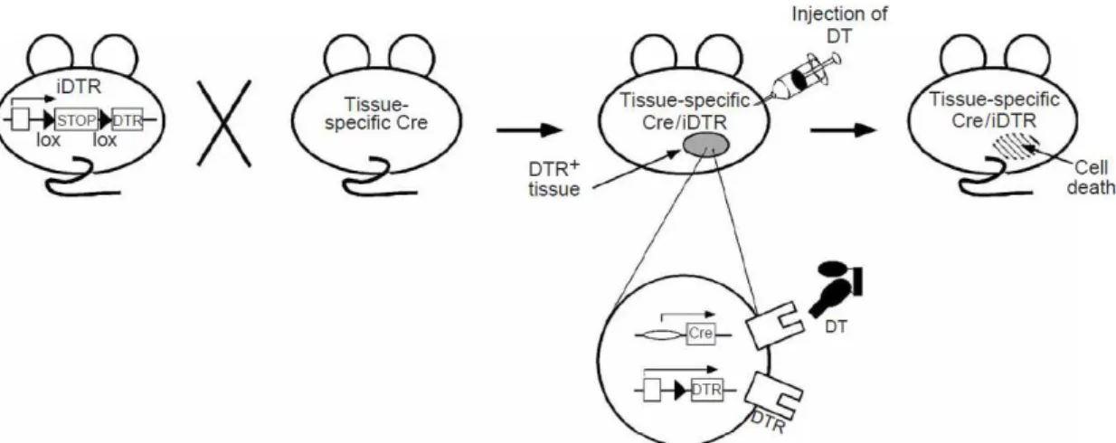
The Cre/loxP recombinase system can be also used for activation of gene expression. Lineage ablation allows the investigation of cell function, so the transgenic expression of diphtheria toxin receptor (DTR) to turn diphtheria toxin (DT)-resistant mouse cells into DT-sensitive cells was developed as a strategy of inducible lineage ablation. In 2005, Buch et al.
developed a Cre-inducible DTR (iDTR) transgenic mice, in which Cre-mediated excision of a STOP cassette turns cells sensitive to DT (Figure 1.3) 48. This mouse allows to study in vivo cell
lineage ablation after application of DT that has an effect only in tissues or cells expressing DTR.
6 Myelin loss after DT injections was observed in a MOGi-Cre/iDTR, which expresses Cre recombinase under the control of the oligodendrocyte-specific promoter of the gene encoding the myelin oligodendrocyte glycoprotein (MOG). This shows that DT crosses the blood-brain barrier (BBB) and promotes cell ablation in the CNS. Therefore, iDTR cell ablation system is a valuable tool to study the function of both peripheral and CNS cells 48.
To achieve the ablation of peripheral and CNS cells, DT has to be administered to mice expressing DTR. DT was purified, crystallized, and characterized in many laboratories and the first publication of its crystal structure occurred in 1992 (Figure 1.4) 49–51. DT was described in
the culture medium of Corynebacterium diphtheriae, being naturally excreted by strains of
diphtheria bacilli and it is toxic for many species such as man, guinea pigs, rabbits, and some birds, but animals like rats and mice are resistant to DT 49,51,52.
DT, a protein of 58,342.0 Da, is a single polypeptide chain, containing 535 aminoacids that, after proteolytic cleavage (with trypsin, pronase or “natural proteases”), can split into two distinct fragments, connected through a disulfide bond. Fragment A, with a molecular mass of 21,167.0 Da, is enzymatically active, while the carboxyl-terminal fragment B (37,195.0 Da) is needed for attachment of toxin to cells and to facilitate the delivery of fragment A to the cytosol
49–53. Fragment A is highly stable with little loss of activity when heated at 100 oC or rapidly
exposed to pH values between 2 and 12; fragment B promptly denatures and precipitates after dissociation from fragment A 49,51,52.
DT has three functional and structural domains implicated in cell intoxication:
- The enzymatic or catalytic (C) domain – corresponding to the first 193 amino acids residues (hydrophobic N-terminal) of fragment A, that is inserted into the plasma membrane. It is responsible for the nicotinamide adenine dinucleotide (NAD)-dependent adenosine diphosphate (ADP)-ribosylation of elongation factor 2 (EF-2), resulting in inhibition of protein synthesis in the cytosol. Therefore, EF-2 was shown to be DT target and substrate;
- The receptor-binding (R) domain - located at the hydrophilic C-terminal (residues 389 to 535), of fragment B and responsible for recognition and interaction with specific surface receptors on sensitive targeted cell membranes. The binding to the receptor allows toxin internalization into acidic intracellular compartments;
- The transmembranar or translocation (T) domain - localized in the fragment B (residues 200 to 379) and responsible for the internalization of domain C of DT to the cytoplasm by membrane penetration 49–53.
C
Fragment A Fragment B
186
S-S201 461S-S471
NH2 T R COOH
Figure 1.3 – Schematic representation of TH-Cre; LSL-DTR mice 48.
7 The DT mechanism of action has been studied extensively 53. The mechanisms of
enzymatic activity, receptor binding, and receptor mediated endocytosis are known in detail, but mechanisms for C domain translocation and release of an active enzyme into the cytosol are not known yet 50–52.
The effect of DT on protein synthesis was first described during respiration studies on HeLa cells by the observation of protein accumulation 49. DT enters cells via receptor-mediated
endocytosis. The R domain binds to specific cell-surface receptors and C domain (fragment A) is internalized into endosomes, leaving fragment B outside of the cell 50–53.
For its biological properties, DT is now being exploited to design new biotechnological tools and therapeutics. Also the engineering of proteins with therapeutic value using the properties of DT is being developed. One of the ideas under development consists in using DT as a way of translocate proteins other than the C domain of DT into cells. DT was the first engineered bacterial toxin to reach the market (FDA-approved therapy for lymphomas) and it is expected that, in the future, useful biotechnological and medical applications of DT will reach the market 50.
A TH-Cre mice have the TH promotor directing the expression of Cre recombinase to catecholaminergic cells, so central and peripheral nervous systems are ablated, when DT is administered to TH-Cre; LSL-DTR mice. Thus, to achieve ablation of SNS without affecting CNS, a strategy to avoid DT in the brain is needed. This strategy involves protein modification in such a way that it does not cross the BBB (Figure 1.5).
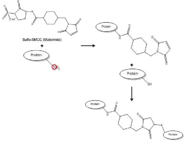
1.6. Protein modifications
The use of proteins as therapeutics agents has been expanded in the last years as a result of discovery of novel proteins, better understanding of their mechanism of action, improvements in expression, and discovery of molecule-altering technologies that have the ability to deliver polypeptides in vivo54. Proteins are now used to diagnostic, monitor and treat
several diseases, but parenteral administration possess disadvantages that limit their usefulness. Some of these limitations are: short circulating half-lives, low solubility, immunogenicity, susceptibility to proteolytic enzymes destruction, and rapid clearance. This implies that several administrations are needed for the drug to be effective, leading to cost increase and higher risk of adverse reactions 54–56.
Chemical reactions are now used to site-selective modify proteins of interest and overcome the aforementioned disadvantages 57. Different strategies have been employed, such
as manipulation of amino acid sequence, conjugation to immunoglobulins and serum proteins, incorporation into drug delivery vehicles and conjugating to natural or synthetic polymers 54,57.
The major concern in these modifications is the possible occurrence of structural modifications and thus different functions 58.
The process of covalent linking one or more polyethylene glycol (PEG) chains to modify a protein, peptide or non-peptide molecule, leaving a N-hydroxysuccinimide (NHS) group, is called PEGylation (Figure 1.6) 56,58–62. In 1977, Davis and Abuchowski performed first
PEGylation reaction in albumin and catalase 55,58–60. Nowadays, it is a well-established, widely
Diphtheria toxin
Diphtheria toxin after modification Lys
8.42 % Lys
5.85 % Lys
7.64 % Lys Catalytic domain
Transmembranar domain
Receptor binding domain
8 employed and fast growing technology that overcomes safety and efficiency requirements of drugs 59. PEGylation is considered a protein modification methodology to improve biomedical
efficacy, bioactivity and physicochemical properties, for instance, of pharmacological agents
54,59,61. It was used in several therapeutic molecules, such as peptides, proteins, antibodies,
antibody fragments, oligonucleotides, small drugs, cofactors, lipids, and saccharides 56,60,63.
PEG is formed by the link of repeating units of ethylene glycol to form polymers with different molecular masses and usually its reactive electrophilic functional group is attached to a specific site of the molecule of interest. In most cases, protein amines (nucleophilic groups) are used as conjugation sites: Ɛ-amino groups of lysines and the N-terminus of polypeptide
molecules (α-amino group). However, the sulfhydryl group of cysteine residues is also widely
used 54–56,58–61,63,64. Lysines are polar and relatively abundant amino acids residues usually
located on the protein surface, which make them prone to chemical reactions with PEG reagents
59.
PEG is a biocompatible polymer that offers several advantages, such as being nontoxic, non-immunogenic, non-antigenic and highly soluble in water. It is a FDA-approved compound that has been used as an excipient in many pharmaceutical and cosmetic formulations 55,56,58– 60,63,65,66. PEG-drug conjugates also present advantages such as a prolonged plasma circulation
in the body, decreased protein immunogenicity, and a decreased degradation by metabolic enzymes 55,56,58–60,62–67. The PEG to protein ratio used in the PEGylation reaction determines the
reaction yield and the number of attached PEGs to a protein 61. Properties of PEG-drug
conjugates depend on: the number of PEG chains attached to the drug, structure and shape of PEG used, the location of the PEG sites on the polypeptide, and the chemistry used to attach PEG 54–56,59,61.
PEG polymer attached to a protein increases its hydrodynamic radius, thus reducing renal filtration. In addition, PEG shields the protein from proteases degradation and immune system recognition. It also increases water solubility, which turns conjugates more polar and renders BBB crossing more difficult 54–56,60,62–65.
PEGylation is one of the most successful strategies to improve the delivery of therapeutic molecules 55,60. The first PEGylated product, PEG-adenosine deaminase
(PEGylated form of adenosine deaminase), was FDA-approved, in 1990, to treat severe combined immunodeficiency (SCID). After that, at least eight PEGylated peptide and protein conjugates have been approved as therapeutic agents 56,58–61,63,65–67. Some examples are
PEG-asparaginase (to treat acute lymphoblastic leukemia), PEGylated fragment antigen-binding (Fab) fragment of tumor necrosis factor (TNF) inhibitor monoclonal antibody (mAb) (to treat Crohn’s disease and rheumatoid arthritis), PEG-granulocyte colony stimulating factor (to stimulate the proliferation, differentiation and survival of neutrophils that are depleted during chemotherapy) 56,58–60,62,65. Apart from approved drugs, more PEGylated products that are now
in clinical trials are expected to be in the market in the next years. The use of successful different PEGylated products is an indication of their usefulness, efficacy and safety 54,55,58–60.
9 Another approach to modify proteins includes taking advantage of the conjugation between serum albumin and α-maleimide (Mal)-functional polymers (Figure 1.7) 68. This
strategy allows the increase in size of DT, in order to minimize clearance. Nevertheless, it was also reported that maleimidation technique leads to heterogeneous mixtures and may result in denaturation 69.
10
2. Materials and Methods
2.1. DT modification
1 mg of DT unnicked, Corynebacterium diphtheriae (Calbiochem, #322326) was
reconstituted in 1 mL of conjugation phosphate buffered saline (PBS) 1X (concentration 16 µM). PBS 1X was prepared by Instituto Gulbenkian de Ciência (IGC) services and is constituted by 137 mM sodium chloride, 2.7 mM potassium chloride, 10 mM sodium phosphate and 2 mM potassium phosphate. Both modifications were performed using 80 equivalents of modifying agent in relation to DT. Hence, 0.001269 mmol of each crosslinker were needed.
PEGylation: MS(PEG)4 methyl-PEG-NHS-Ester (Life Technologies, #22341), after
being fully equilibrated to room temperature (RT), was reconstituted in 1.1 mL of dimethyl sulfoxide (DMSO, Sigma-Aldrich, #D8418), leading to a concentration of 250 mM PEG, according to manufacturer instructions. 250 mM PEG was diluted 1:100 in DMSO and then the appropriate volume was transferred into DT solution. The mixture was incubated for 4 h, at RT, under shaking. Reaction to produce PEGylated DT (PEGyDT) was monitored by electrophoresis of samples collected every hour (see electrophoresis section).
Maleimidation using sulfosuccinimidyl 4-(N-maleimidomethyl)cyclohexane-1-carboxylate (sulfo-SMCC): 1.5 mg of sulfo-SMCC (Life Technologies, #22322) were dissolved
in 272.7 µL of ultra-pure water. The appropriate volume was then dispensed into DT solution. Reaction to generate DT Mal occurred for 4 h, at RT, under shaking. Every hour a sample was taken to be accessed by electrophoresis (see electrophoresis section).
Purification of PEGyDT and DT Mal: In order to remove the excess of cross-linker, gel
filtration chromatography using the gravity protocol of PD MidiTrapG-25 column (GE Healthcare, #28-9180-08) was performed according to manufacturer indications. Briefly, column was prepared and equilibrated with buffer. Sample was applied to the column, the flow-through was discarded and eluate was collected.
In vitro DT Mal and bovine serum albumin (BSA) conjugation: 1 mg/mL of BSA
fraction V (NZYtech, #MB04601) was added to DT Mal. DT Mal BSA was produced by incubation for 30 min, under shaking, at RT.
Concentration determination: The concentrations of PEGyDT, DT Mal and DT Mal
BSA were determined using a NanoDrop ND-2000 UV-Visible spectrophotometer (Fisher Scientific, #11840301), measuring the absorbance at 280 nm of 1 µL-samples and using PBS buffer as blank.
2.2. Sodium dodecyl sulfate polyacrylamide gel electrophoresis (SDS-PAGE)
All the materials needed to perform the experiment described in this section were kindly provided by Alekos Athanasiadis’ lab (special acknowledgment to Krzysztof Kus). 10 µL of each sample were mixed with 1X diluted NuPAGE LDS Sample Buffer 4X (Life Techonologies, #NP0007). The mixtures were boiled at 90 oC, for 3 min. 10 µL of Novex Sharp Unstained
Protein Standard (Life Technologies, #LC5801) and 15 µL of each sample were applied in a gel NuPAGE Novex 12 % Bis-Tris Protein Gels, 1.0 mm, 15 well (Life Technologies, #NP0343BOX). Separation occurred at 200 V, for around 1 h with running buffer 1X diluted NuPAGE MOPS SDS Running Buffer 20X (Life Technologies, #NP0001). The gel was washed with water for 5 min, 3 times and stained for 20 min with SimplyBlue SafeStain (Life Technologies, #LC6060). After staining, gel was washed with water, shaking (2 days).
2.3. Mass spectrometry (MS)
11 prepared according to manufacturer instructions (centrifuged for 4 min, 3500 g, with PBS 1X).
DT, PEGyDT and DT Mal were centrifuged 3500 g until appropriate concentration (10 µM).
2.4. Cell culture of HeLa cells
HeLa cells (a human cell line) were kindly provided by Lars Jansen’s lab (special acknowledgment to João Mata). Cells were maintained in Dulbecco’s modified Eagle medium (DMEM) high glucose with L-glutamine and sodium pyruvate (Biowest, #L0104) supplemented with 10 % fetal bovine serum (FBS) heat inactivated (Biowest, #S181BH) and 1 % penicillin-streptomycin (Biowest, #L0022-100) – DMEM complete medium. Cells were maintained in humidified atmosphere, 5 % CO2 at 37 oC. To maintain cells in culture: first, a wash with PBS
1X was done, then 1 to 3 mL TrypLE Express Enzyme 1X, phenol red (Life Technologies, #12605) were applied and the cells were incubated for 5 min, in humidified atmosphere, 5 % CO2 at 37 oC. To inactivate TrypLE, 5 mL of DMEM complete medium was added and the cells
were centrifuged for 5 min, 300 g, at RT. Finally, cells were resuspended in 5 mL of DMEM
complete medium and plated in a cell culture flask (Corning, #CORN430825). Cells were plated in Costar 12 and 24 Well Clear TC-Treated Multiple Well Plates (Corning, #3513 and #3526) following a procedure similar to the one used to maintain cells, but after centrifugation and resuspension, the cells were counted in a hemocytometer (Neubauer Cell with Double Grid, Fisher Scientific, #10350141) using 1 % Trypan Blue solution (Sigma-Aldrich, #T8154). 105 cells
per well (2 mL/well) and 6x10⁴ cells per well (1 mL/well) were plated in the Costar 12 and 24
plates, respectively.
DT concentration test: Three different concentrations of DT were tested: 1 µg/mL, 3
µg/mL and 5 µg/mL and controls without DT were used. Fluorescence-activated cell sorting (FACS) analysis (for details see FACSCalibur analysis section) was done at 4 h, 6 h, 12 h, 24 h and 48 h. For 1 µg/mL, also 72 h was analyzed.
DT, PEGyDT, DT Mal and DT Mal BSA: All incubations used a concentration of 3
µg/mL (except controls, in which only medium was added). The time points were prepared at 4 h, 6 h, 12 h, 24 h, 48 h and 72 h.
BSA: This assay was done to prove that DT Mal would be the responsible for HeLa cell
death and not BSA. Therefore, 2.25 mg/mL were incubated for 24 h, 48 h and 72 h.
FACSCalibur analysis: Samples were prepared for analysis in a Becton Dickinson
Flow Cytometer FACSCalibur. Data were acquired using CellQuest software (Becton Dickinson) and analyzed using FlowJo. The medium was collected and cells were washed with PBS 1X, 250 to 500 µL of TrypLE were added and incubated in humidified atmosphere, 5 % CO2 at 37 oC. DMEM complete medium (250 to 400 µL) were added and cells were centrifuged for 4 min,
3250 g, at 4 oC. Cells were resuspended in 150 µL FACS buffer (2 % FBS, 0.02 % sodium
azide-1-15N (Sigma-Aldrich, #609374) in PBS 1X) and 25 µL propidium iodide (Sigma-Aldrich,
#P4170) were added. The medium collected before preparation procedures was also analyzed.
2.5. Bone marrow (BM)-derived macrophages from Rosa26Cre-ER(T2); LSL-DTR mice
Tamoxifen (Hefei Joye) was administered in vivo to Rosa26Cre-ER(T2); LSL-DTR mice
and, after 1 month, the BM cells were isolated to be differentiated in vitro into primary
macrophages. The back legs of aforementioned mice were cleaned and the BM cells inside the bone were flushed into a Petri dish (Nunc, #150318) with DMEM high glucose with L-glutamine and sodium pyruvate cell culture medium. After homogenization, cells were centrifuged for 5 min, 300 g, at RT. Cells were resuspended and maintained in Iscove’s modified Dulbecco’s
medium (IMDM) GlutaMAX (Life Technologies, #31980-022) supplemented with 10 % FBS; 1 % penicillin-streptomycin; 0.1 % 2-mercaptoethanol (Life Technologies, #31350-010); 0.5 % sodium pyruvate (Life Technologies, #11360-070) – IMDM complete medium. To stimulate differentiation, 30 % macrophage colony stimulating factor (M-CSF) (kindly provided by Elsa Seixas) was added to the medium. Petri dishes were incubated in humidified atmosphere, 5 % CO2 at 37 oC for 7 days. After differentiation, the macrophages were harvested and centrifuged
12 in a hemocytometer using trypan blue, as previously described. 5x10⁵ cells were plated per well
(1 mL/well) in a Costar 24 Well Clear TC-Treated Multiple Well Plate.
Tamoxifen in vivo: A stock solution of tamoxifen at 20 mg/mL in corn oil
(Sigma-Aldrich, #C8267) was prepared and homogenized by shaking, at 37 oC for 2 to 3 h. To have a
final concentration of 1 mg/mL, a dilution of the stock solution was done in corn oil. An intraperitoneal (i.p.) injection of 50 µL was given, in milky spot, to Rosa26Cre-ER(T2); LSL-DTR pups that were one day older for 5 to 6 consecutive days.
DT and PEGyDT: 3 µg/mL of DT and PEGyDT were prepared in IMDM complete
medium. FACSCalibur analysis was performed at 4 h, 6 h, 12 h and 24 h.
FACSCalibur analysis: Macrophages were harvested and then centrifuged for 4 min,
3250 g, at 4 oC. FACSCalibur analysis was performed as previously described (see Cell culture
of HeLa cells section for details).
2.6. Mice and housing conditions
Mice were housed at controlled temperature and humidity, under a 12 h light/dark cycle. Food and water were supplied ad libitum, except if something different is mentioned. The
animals were euthanized using carbon dioxide. All procedures were approved by IGC ethical committee. All controls were injected with vehicle (PBS 1X). C57BL/6 mice were obtained from the Mice Production Facility at IGC. TH-Cre (#008601) and LSL-DTR (#007900) mice were purchased from Jackson Laboratory. LSL-DsRed mice was imported from Rockefeller University. Rosa26Cre-ER(T2) animals were kindly provided by Miguel Soares’ lab (special acknowledgment to Sílvia Cardoso and Sofia Rebelo).
2.7. PEGyDT-mediated sympathectomy
Cre; LSL-DTR mice were used for this experiment and LSL-DTR, C57BL/6 and TH-Cre mice were used as controls. During the experiment, animals and food were weighted every day. Both DT and PEGyDT were injected i.p.. Injections of 25 ng/g of DT or PEGyDT were administered once a day for 8 consecutive days. TH-Cre; LSL-DTR mice were also injected i.p. with PBS 1X for 8 consecutive days. During the experiment, animals and food were also weighted every day.
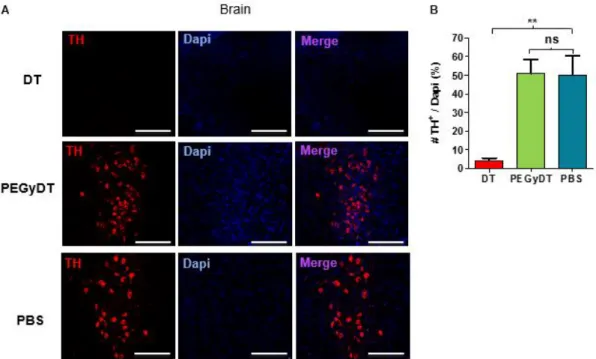
Assessment of SNS ablation by confocal microscopy: Mice were perfused with PBS
1X followed by 4 % formaldehyde (VWR, #9713.9010). The brain was removed and fixed in 4 % formaldehyde for 2 days, at 4 oC. Using a vibratome VT 1000S (Leica), 50 µm thick slices of
ventral tegmental area (VTA) were obtained. These slices were at RT for 1 h, shaking in blocking buffer (1 % BSA, 0.1 % tween 20 (Sigma-Aldrich, #P2287), 0.05 % sodium azide in PBS 1X). The slices were incubated with primary antibody TH chicken (1:1000, Aves Lab, #TYH) o/n, shaking, at 4 oC. After washing, secondary antibody Alexa Fluor 594 Goat Anti-chicken IgY
(H+L) (1:500, Life Technologies, #A-11042) was applied for 1 h, shaking, at RT. After secondary antibody incubation, slices were washed and mounted with Dapi-Fluoromount-G (Southern Biotech, #0100). The microscope slides were dried at RT and then kept at 4 oC.
Fibers isolated from inguinal fat were fixed in 2 % formaldehyde for 2 h, at RT, shaking. After blocking for 1 h, at RT, fibers were incubated with primary antibodies rabbit polyclonal to neuron specific β3-Tubulin (β3-Tub) (1:1000, Abcam, #AB18207) and TH chicken, o/n, shaking,
at 4 oC. After washing, secondary antibodies Alexa Fluor 488 Goat Anti-rabbit IgG (H+L) (1:500,
Life Technologies, #A-11008) and Alexa Fluor 594 Goat Anti-chicken were applied for 1 h, shaking, at RT. After secondary antibodies incubation, fibers were washed and mounted with Fluoromount-G. The microscope slides were dried at RT and then kept at 4 oC.
Confocal images were acquired in Leica TCS SP5 Inverted and Upright Microscopes. Analysis and quantification of acquired images was performed using FIJI 70.
Scoring test: After administration of 25 ng/g of DT or PEGyDT once a day for 8
13
2.8. Functional tests on sympathetic ablated mice
From this point forward, TH-Cre; LSL-DTR strain was used as experimental mice and LSL-DTR strain was used as control mice (unless something else is specified).
Fasting: Food was taken out from the cages and mice were weighted every 12 h during
48 h. After this period, food ad libitum was provided to mice.
High Fat Diet (HFD) studies: Animals were weighted and normal diet (ND) was
replaced by HFD. Mice were weighted every week during 4 weeks and photographs were taken. C57BL/6 animals that were on HFD for 4 weeks were weighted. HFD (ssniff, #E15742-347) contains 60 kJ % fat.
Yo-yo diet (HFD - ND - HFD - ND) and food intake: HFD was replaced by a ND
regimen, then animals were fed with HFD followed by another period of ND. During this period, mice were weighted and food intake was measured.
BMI: Nose-to-anal distance was measured and mice were weighted to calculate the
BMI. This measurement was performed in ND and HFD regimens.
HFD 2 weeks after sympathectomy and food intake: ND was replaced by HFD 2
weeks after sympathectomy was performed. Mice weight and food intake were measured every day.
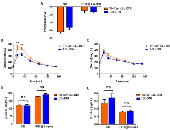
Fasting glucose and Glucose Tolerance Test (GTT): After 12 h fasting during dark
cycle, glucose was measured using Accu-Chek Aviva Glucose (Roche Diagnostics, #05987431023) and the mice were weighted. After this, an i.p. injection of 2 mg/g D-(+)-glucose (Sigma-Aldrich, #G8270) was administered and glucose level was measured at 15 min, 30 min, 60 min, 90 min, 120 min and 150 min post-injection. These procedures were performed in ND and repeated for 5 weeks in HFD regimen.
Fasting insulin: Blood samples were collected using heparin (LEO Pharmaceutical
Products, #0014425-02) after a dark cycle with no access to food. Samples were centrifuged for 15 min, 9400 g, at 4 oC and plasma was collected. Centrifugation was repeated once. Mouse
Ultrasensitive Insulin Enzyme-Linked Immunosorbent Assay (ELISA) (ALPCO #80-INSMSU) was used to measure insulin and the protocol was followed according to manufacturer instructions. These procedures were performed in ND and repeated after 5 weeks in HFD regimen.
Cold exposure: Body temperature was measured using an electronic thermometer ama-digit ad 15 th (Precision, #E910 560) when the animals were housed at RT and animals were weighted. After this, animals were housed at 4 oC with ND food and water ad libitum. Body
temperature was measured and animals were weighted every hour for 8 h.
2.9. Anti-obesity drugs
Fen hydrochloride (Sigma-Aldrich, #F8507) and AMPH hydrochloride (Asiba Pharmatech, #10296-HCl) were used to induce sympathetic activation. Unless stated otherwise, C57BL/6 animals were used and food and water were provided ad libitum. Controls were
administered with vehicle (PBS 1X).
Fen: Mini-osmotic pumps Model 2004 (Alzet, #298) releasing 6 mg/kg/day of Fen
14
AMPH: AMPH hydrochloride was prepared every day. Mice on HFD were injected i.p.
with AMPH hydrochloride 20 mg/kg every day during 7 weeks. Animals that were pair-fed to the ones on AMPH hydrochloride were injected with PBS 1X. Animals and food were weighted every day. After ceasing AMPH administration, mice and food were weighted during 4 weeks.
TH-Cre; LSL-DTR (after sympathetic ablation) and LSL-DTR mice on HFD received an i.p. injection of AMPH hydrochloride 20 mg/kg, every day during 4 weeks. Another group of TH-Cre; LSL-DTR (after sympathetic ablation) and LSL-DTR mice on HFD received an i.p. injection of PBS 1X, every day during 4 weeks. Animals and food were weighted every day.
2.10. OPT
As stated before, this technique was applied to characterize fat and to show its innervation.
Organ dissection and preparation: C57BL/6 mice were perfused with PBS 1X and
after that, inguinal and epididymal fats were carefully removed (with Dumont #5 Forceps) and cleaned. Fats were fixed in 4 % formaldehyde for 3 h, under shaking, at RT. Then, fats were washed and placed in a 1 % agarose, low gelling temperature (Sigma-Aldrich, #A9414) at RT, protected from dust. The agarose volume was approximately 24.5 cm3 (3.5 cm height and 1.5
cm diameter).
Clearing: Fats inside agarose were immersed in 10 % methanol p.a. Normapur (VWR,
#VWRC2084.307) for 2 h, at RT, under shaking. Afterwards, 10 % increasing steps were performed every day until dehydration in 100 % methanol was reached (RT with agitation). The replacement of the methanol with 1:2 mixture of benzyl alcohol (Merck Millipore, #822259) and benzyl benzoate (Merck Millipore, #818701) was performed in 15 % increasing steps every day until clarification was reached. Photographs were taken before and after clearing and measurements of fats were done.
OPT acquisition and analysis: OPT acquisition was performed by Gabriel Martins and
Ânia Gonçalves from Unit of Imaging and Citometry (UIC) of IGC. Briefly, images of the whole fat tissue were acquired using a 1X lens, mounted on an Infinitube tube lens, and projected into a Hamamatsu FlashLT sCMOS camera. A total of 1600 images were acquired for a full rotation (0.25 o steps). The series of projections were then pre-processed using FIJI 70 in order to remove
hot pixels, re-align the axis of rotation in relation to the camera-chip, and finally the back-projection reconstruction was done using the Skyscan’s nrecon software. The stack of slices was further processed with FIJI, to increase contrast and saved to posterior analysis with the software Amira V5.3. Using Amira V5.3, 3D reconstructions and image segmentation were performed to identify and reconstruct individual parts of the fat organs.
2.11. Statistical analysis
15
3. Results
3.1. PEG and Mal bind to DT
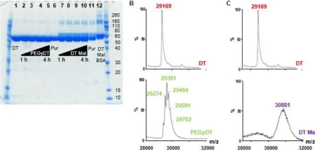
DT modification state was analyzed by SDS-PAGE and by MS. SDS-PAGE shows that after gel filtration chromatography a slight increase in molecular weight (MW) is present both in PEGyDT and in DT Mal. When DT Mal is attached to BSA the shift is even higher(Figure 3.1 A).
By MS technique, it is possible to see that between 1 and 5 PEG molecules were attached to DT and that around 15 Mal molecules were attached to DT (Figure 3.1 B and C). The attachment of 1 molecule of PEG or Mal increases DT MW by 218 g/mol. Thus, the attachment of 5 PEG molecules increases DT MW by 1,090.0 g/mol and the attachment of 15 Mal molecules increases DT MW by 3,270.0 g/mol.
16
3.2. PEGyDT, but not DT Mal, remains functional after modification
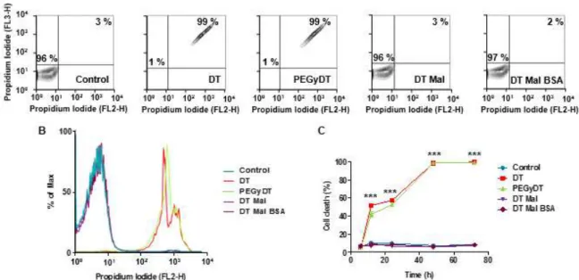
First, a DT concentration test was performed to determine the concentration that should be used to cause cell death in HeLa cells (Figure 7.1 in Appendix). Although 1 µg/mL of DT was enough to cause 100 % cell death, to further test DT and its modified derivatives 3 µg/mL was used.
FACS analysis was performed to access DT and its modified derivatives capacity to cause cell death. After 24 h, DT and PEGyDT already induce around 50 % of cell death, while after 48 h or more, the cell death is almost 100 % (*** p < 0.0001). The same did not happen with DT Mal and DT Mal BSA that did not present a significant difference from control (Figure 3.2).
To demonstrate that BSA was not responsible for the obtained results in cell culture when Mal was tested an experiment was performed (Figure 7.3 in Appendix). The results indicate that BSA does not cause cell death of HeLa cells. As consequence, in case DT Mal would cause cell death, this would be exclusively a consequence of DT maleimidation and not to the presence of BSA.
Also primary cells were used to confirm the results obtained for the cell line (Figure 3.3). The primary cells used were BM-derived macrophages from Rosa26Cre-ER(T2); LSL-DTR mice, to whom tamoxifen was administered. Although the cell death was higher than expected in control, in the presence of DT and PEGyDT it was significantly higher (*** p < 0.0001).
17
Figure 3.3 – DT functionality after modification tested in vitro on primary cell culture of macrophages derived from the BM of Rosa26Cre-ER(T2); LSL-DTR mice after tamoxifen was administered in vivo. A) Schematic representation of the experiment; B) Representative contour plots