Rita João Rosado Serranito Ramos
Degree in Biology
Evaluation of Neuroprotective Effects of
Natural Extracts obtained from Portuguese
Agro-food Residues
Dissertation to obtain a Master Degree in Biotechnology
Supervisor: Ana Matias, Ph.D, IBET/ITQB-UNL
Co- Supervisor: Cláudia Santos, Ph.D, IBET/ITQB-UNL
Júri:
Presidente: Prof. Doutor Pedro Miguel Calado Simões
Arguente: Prof. Doutora Margarida Castro Caldas Braga
Rita João Rosado Serranito Ramos
Degree in Biology
Evaluation of Neuroprotective Effects of
Natural Extracts obtained from Portuguese Agro-food
Residues
Dissertation to obtain a Master Degree in Biotechnology
Supervisor: Ana Matias, Ph.D, IBET/ITQB-UNL
Co- Supervisor: Cláudia Santos, Ph.D, IBET/ITQB-UNL
Júri:
Presidente: Prof. Doutor Pedro Miguel Calado Simões
Arguente: Prof. Doutora Margarida Castro Caldas Braga
iii
Evaluation of neuroprotective effects of natural extracts obtained from Portuguese agro-food residues
Copyright Rita Ramos, FCT/UNL, UNL
v
Acknowledgments
Gostaria de agradecer a todas as pessoas que direta ou indiretamente contribuíram para a realização deste trabalho e sem as quais seria impossível conclui-lo. Em primeiro lugar, gostaria de agradecer à Dra. Ana Matias que foi incansável ao longo de todo este processo, para o qual esteve sempre disponível para compartilhar as suas ideias e conhecimentos, tendo sempre uma palavra de incentivo e de apoio quando as coisas corriam menos bem. E por todas as palavras de motivação e reconhecimento quando tudo corria realmente bem. Obrigada por toda a confiança e apoio Ana. Gostaria de agradecer à Dra. Cláudia Santos por todo o apoio, não menos importante. Por todas as palavras de confiança em situações mais difíceis e por ter sempre uma palavra de apoio e conforto, por todas as ideias e opiniões. Obrigada por tudo o que me ensinou Cláudia. Obrigada a ambas, foi um prazer enorme trabalhar convosco. À Dra. Catarina Duarte por ter acreditado em mim e neste trabalho, por ter sempre uma palavra de motivação e de apoio. Obrigada pelas portas que me abriu. Ao Dr. Ricardo Boavida Ferreira pela oportunidade pela confiança. Agradeço à Dra. Maria do Rosário Bronze, à Joana Antunes e a toda equipa do Laboratório de Química Analítica pelo apoio essencial na identificação de compostos. A todas as pessoas do laboratório de Nutracêuticos e Libertação Controlada e do laboratório de Biologia da Doença e do Stress que tornaram o desenvolvimento deste trabalho em muito mais que um simples trabalho. À Dra. Teresa Serra por estar sempre disponível para ajudar e por tudo o que me ensinou. À Sara Nunes por o tempo despendido, por todas as opiniões sempre importantes e por toda a ajuda. À Joana Poejo por toda a paciência, companhia e ajuda. À Ana Nunes pelo conhecimento que partilhou comigo e por todas as viagens de comboio! Ao Agostinho por toda a ajuda boa disposição. À Lucélia Tavares por todo o conhecimento partilhado. À Dra. Marta Alves pelos conselhos e companhia. À Inês Figueira por tudo o que me ensinou e por toda a cumplicidade, obrigada. À Carolina Jardim e à Diana Macedo pela companhia. À Andreia Gomes, ao Daniel Lopes, ao João Jorge e ao Pedro Ferreira por todas as conversas e pela companhia espetacular que foram em mais uma fase deste processo. Sem vocês não teria sido tão gratificante. Gostaria também de agradecer a todos os meus amigos pelo suporte que são e que sempre foram, pela companhia, por todos os momentos de descontração e acima de tudo, por estarem sempre presentes. Às meninas, Cátia Gomes e Nádia Grilo por todas os momentos, opiniões e apoio. Pela excelente companhia e amizade. Obrigada por terem feito parte desta etapa da minha vida. Ao Hélder, que foi incansável. Pelo apoio incondicional e por toda a paciência, carinho e boa disposição. Obrigada, sem ti não era a mesma coisa! À minha família: Aos meus avós, tios e primas por serem a melhor família do mundo e por todo o orgulho e carinho. Aos meus pais e à minha avó pelo enorme esforço que fizeram para eu conseguir chegar até aqui e por sempre acreditarem em mim. Espero que um dia seja possível retribuir tudo aquilo que já fizeram por mim. À minha irmã que esteve sempre presente e mais uma vez por ter sido imprescindível para concluir mais uma etapa da minha vida!
vii
Abstract
Countries are currently faced with problems derived from changes in lifespan and an increase in lifestyle-related diseases. Neurodegenerative disorders such Parkinson’s (PD) and Alzheimer’s (AD) diseases are an increasing problem in aged societies. Data from World Alzheimer Report 2011 indicate that 36 million people worldwide are living with dementia. Oxidative stress has been associated with the development of AD and PD. Therefore there is interest to search for effective compounds or therapies to combat the oxidative damage in these diseases. Current evidence strongly supports a contribution of phenolic compounds present in fruits and vegetables to the prevention of neurodegenerative diseases such AD and PD.
The industrial processing of a wide variety of fruits results in the accumulation of by-products without commercial value. Opuntia ficus-indica (cactus pear) is consumed fresh and processed
like in juice. Prunnus avium (sweet cherry) is consumed fresh but the organoleptics
characteristics of the fruits leads to the smaller and ragged fruits have no commercial value. Fruit extracts of both species has described to be rich in phenolic compounds and to have high antioxidant activities due to its composition. The aim of this work was assessing the efficacy of
O. ficus-indica and P. avium by-products extracts obtained with conventional solvent extraction
and pressurized liquid extraction in a neurodegeneration cell model. All extracts have protected neuroblastoma cells from H2O2-induced death at low, non-toxic levels, which approach to
physiologically-relevant serum concentration. However, cherry extract has a slighter neuroprotective activity. The protective effect of Opuntia extracts are not conducted by a direct
antioxidant activity since there are not decreases in intracellular ROS levels in cell treated with extracts and challenged with H2O2, while cherry extract neuroprotection seems to be due to a
direct scavenging activity. Extracts from different biological matrixes seems to protect neuronal cells trough different cellular mechanisms.
Keywords: Neuroprotection, Agro-food residues, Opuntia ficus-indica, Prunus avium, Natural
ix
Resumo
Em países desenvolvidos, o envelhecimento da população está a aumentar assim como a incidência de doenças relacionadas com a idade e estilo de vida como as doenças neurodegenerativas. O stress oxidativo tem sido associado ao desenvolvimento de doenças neurodegenerativas como a doença de Alzheimer (AD) e a doença de Parkinson (PD). Assim sendo existe um interesse crescente na busca por novos compostos ou terapias eficazes no combate aos danos provocados pelo stress oxidativo nestas doenças. Evidências apoiam fortemente um contributo de compostos fenólicos na prevenção de doenças neurodegenerativas. O processamento industrial de uma grande variedade de frutos resulta na acumulação de subprodutos sem qualquer valor comercial. Opuntia ficus-indica (figo da índia) é
consumido fresco ou por exemplo processado em sumo. Prunnus avium (cereja) é consumido
preferencialmente fresco, mas o tamanho e as características visuais necessárias leva a que frutos pequenos e de forma irregular não tenham valor comercial. Extratos de ambos os frutos têm sido descritos como ricos em compostos fenólicos e com elevada capacidade antioxidante, devido à sua composição. O objetivo principal desta dissertação é avaliar os efeitos neuroprotectores de extratos naturais obtidos a partir de resíduos agro-industriais destas duas espécies com recurso à extração convencional ou com fluidos pressurizados. A avaliação da capacidade neuroprotectora é realizada recorrendo à linha celular neuronal, proveniente de um neuroblastoma, SK-N-MC, submetida a um stress oxidativo induzido por uma solução de H2O2.
Todos os extratos demonstraram um efeito neuroprotector em concentrações baixas, não tóxicas, fisiologicamente relevantes por se encontrarem perto de concentrações séricas. O efeito protetor dos extratos de Opuntia parece não se dever a uma atividade antioxidante
direta, enquanto a neuroproteção exercida pelo extrato de cereja parece ser devida a uma ação antioxidante direta. Pode-se concluir que diferentes extratos de matrizes biológicas distintas parecem proteger as células neuronais por diferentes mecanismos celulares.
Palavras-chave: Neuroprotecção, Resíduos agroindustriais, Opuntia ficus-indica, Prunus
xi
I
ndex
1. Introduction ... 1
1.1. Neurodegenerative diseases ... 1
1.2. Neurodegeneration and Oxidative Stress ... 1
1.2.1. Neurodegeneration Cell Model ... 2
1.3. Nutraceuticals and Functional Foods ... 3
1.4. Phytochemicals ... 4
1.5. Extraction Technologies ... 9
1.6. Opuntia ficus-indica ... 10
1.7. Prunus avium ... 12
2. Materials and Methods ... 15
2.1. Materials ... 15
2.2. Raw Materials ... 15
2.3. Preparation of Prunus avium and Opuntia ficus-indica Extracts ... 15
2.3.1. Opuntia ficus-indica Extracts ... 15
2.3.2. Prunus avium Extract... 16
2.4. Extracts Characterization ... 17
2.4.1. Total Phenolic Content (TPC) ... 17
2.4.2. Total Flavonoid Content (TFC) ... 18
2.4.3. HPLC – DAD – ED analysis ... 18
2.4.4. TLC analysis ... 19
2.5. Extracts Chemical Antioxidant Activity ... 19
2.5.1. Oxygen Radical Absorbance Capacity (ORAC) ... 19
2.5.2. Hydroxyl Radical Adverting Capacity (HORAC) ... 19
2.6. Cell Based Assays... 20
2.6.1. Cell Culture ... 20
2.6.2. Cytotoxicity Profile determination ... 20
2.6.3. Evaluation of Neuroprotective effect ... 20
2.6.4. Intracellular reactive oxygen species (ROS) production determination ... 21
2.6.5. Glutathione (GSH) and glutathione disulphide (GSSG) quantification ... 21
2.7. Statistical analysis ... 22
3. Results and Discussion ... 23
xii
3.1.1. Chemical Characterization ... 23
3.1.2. Chemical Antioxidant Capacity ... 29
3.1.3. Cytotoxicity Profile Determination ... 30
3.1.4. Evaluation of Neuroprotective Effect ... 31
3.1.5. Intracelular ROS production determination ... 33
3.1.6. Glutathione (GSH) and Glutathione disulphide (GSSG) Quantification ... 34
3.2. Prunus avium ... 35
3.2.1. Chemical Characterization ... 35
3.2.1. Chemical antioxidant activity ... 38
3.2.2. Cytotoxicity profile determination ... 38
3.2.3. Neuroprotective effect evaluation ... 39
3.2.4. Intracellular ROS production determination ... 40
3.2.6. Glutathione (GSH) and Glutathione disulphide (GSSG) Quantification ... 42
4. Final Conclusions ... 43
5. Bibliography ... 45
xiii
Index of Figures
Figure 1.1 Page 2 The approximate balance of antioxidants and reactive species in
vivo.
Figure 1.2 Page 4 The term "nutraceutical" was coined from "nutrition" and
"pharmaceutical" in 1989 by Stephen DeFelice, MD, founder and chairman of the Foundation for Innovation in Medicine (FIM), Cranford, NJ. Adapted from Pandey et al. 2010.
Figure 1.3 Page 6 Classification of dietary phytochemicals (Adapted from Liu, 2004).
Figure 1.4 Page 10 Chemical structure of Amberlite® XAD 16
Figure 1.5 Page 11 Images of Opuntia ficus-indica cladodes and fruits.
Figure 1.6 Page 12 Cherries (Prunus avium) from Saco cultivar collected in “Cova da
Beira” region, Portugal.
Figure 3.1 Page 25 Chromatographic profile of O. ficus-indica from Beja/Serpa fruit
by-products extract (OBS-CSE) obtained using Conventional Solvent Extraction (CSE) at 280, 360, 420, AND 480 nm.
Figure 3.2 Page 26 Chromatographic profile of O. ficus-indica from Beja/Serpa fruit
by-products extracts obtained using Pressurized Liquid Extraction (PLE), at 280 nm. PLE – A, PLE – B, and PLE – C.Piscidic acid (1),
Eucomic acid (2), Ferulic acid (3) and Isorhamnetin (4) were identified.
Figure 3.3 Page 26 Chromatographic profile of O. ficus-indica from Beja/Serpa fruit
by-products extracts obtained using Pressurized Liquid Extraction (PLE), at 360 nm. PLE – A, PLE – B, and PLE – C.
Figure 3.4 Page 27 Chromatographic profile of O. ficus-indica from Beja/Serpa fruit
by-products extracts obtained using Pressurized Liquid Extraction (PLE), at 480 nm. PLE – A, PLE – B, and PLE – C.
Figure 3.5 Page 25 Chromatographic profile of O. ficus-indica fruit by-products extracts
obtained with Pressurized Liquid Extraction (PLE) using ED. PLE – A, PLE – B, and PLE – C. Ferulic acid (1) and Isorhamnetin (2).
xiv
Figure 3.7 Page Chemical antioxidant capacity of Opuntia ficus-indica extracts
measured by ORAC and HORAC assays. (a) ORAC (■) expressed as µmol TE.g-1
dw, and HORAC (■) expressed as µmol CAE.g-1dw.
(b) ORAC (■) expressed as µmol TE.g-1 GAE, and HORAC (■) expressed as µmol CAE.g-1 GAE. All values are expressed as mean
of triplicates ± SD.
Figure 3.8 Page Cytotoxicity profile of Opuntia ficus-indica extracts, assessed by
CellTiter-Blue Reagent® Cell Viability Assay (Promega). SK-N-MC neuroblastoma cells were incubated for 24 h with the extract (0-250 µg GAE mL-1). Cell viability is expressed as percentage of viable cells. Values are expressed as percentage relatively to control (without extract). All values are mean ± SD.
Figure 3.9 Page Neuroprotective effect of Opuntia ficus-indica extracts, assessed by
CellTiter-Blue Reagent Cell Viability Assay (Promega). OBS – CSE (a),PLE – A (b), PLE – B (c), and PLE – C (d). Cell viability is expressed as percentage of viable cells. SK-N-MC cells were pre-incubated with 0.25, 0.5, 1, 2.5, and 5 µg GAE.mL-1 of all extracts for 24 h and then injured by 300 µM H2O2 for 24 h. Statistical differences
between treatments and stressed cells are denoted as *p < 0.05, **p < 0.01, ***p < 0.001 and statistical differences between treatments and not-treated cells are denoted as #p < 0.05, ##p < 0.01, ###p < 0.001. All values are means of three independent experiments ± SD.
Figure 3.10 Page Relative ROS production by SK-N-MC neuroblastoma cells
pre-incubated with O. ficus-indica extracts for 2 (■) and 24 h(■) and
submitted to an oxidative stress (300 µM H2O2 for 30 min)ROS were
detected by fluorimetry using H2DCFDA as probe. Statistical
differences in relation with cells not treated are denoted as *p < 0.05, **p < 0.01, ***p < 0.001. All values are mean ± SD, n=3.
Figure 3.11 Page Quantifications of GSH and ratio GSH/GSSG. After pre-treated for 24
h with O. ficus-indica extracts, SK-N-MC neuroblastoma cells were subjected to 300 µM H2O2 for 24 h. cells were collected and
analyzed for their content in GSH and GSSG by HPLC. The values of GSH are expressed in nmol GSH.mg -1 protein. Statistical differences in relation with cells not treated are denoted as *p < 0.05, **p < 0.01, ***p < 0.001. All values are mean ± SD, n=3.
polyphenol-xv
rich cherry extract (PRCE), expressed as mg GAE.g-1 of extract in a dry weight basis.
Figure 3.13 Page 36 Chromatographic profile of Polyphenol-Rich Cherry Extract (PRCE)
at 280, 360 e 527nm. Chlorogenic Acid (1), Cyanidin-3-O-glucoside
(2), Cyanidin-3-O-rutinuside (3), Peonidin-3-glucoside (4), Rutin (5),
and Quercetin (6) were indentified.
Figure 3.14 Page Chromatographic profile of Polyphenol-Rich Cherry Extract (PRCE)
using ED. Chlorogenic acid (1), glucoside (2), Cyanidin-3-rutinoside (3), Rutin (4), Quercetin (5).
Figure 3.15 Page Antioxidant activity of polyphenol-rich cherry extract (PRCE)
measured by ORAC (■) (μmol TE.g-1 dw extract) and HORAC (■)
(μmol CAE.g-1 dw extract) assays. Statistical difference between
ORAC and HORAC values is denoted with different letters. All values are mean ± SD, n=3.
Figure 3.16 Page Cytotoxicity profile of Prunus avium extract (PRCE), assessed by
CellTiter-Blue Reagent® Cell Viability Assay (Promega). SK-N-MC neuroblastoma cells were incubated for 24 h with the extract (0-250 µg GAE mL-1). Cell viability is expressed as percentage of viable cells. Values are expressed as percentage relatively to control (without extract). All values are mean ± SD.
Figure 3.17 Page Neuroprotective effect of Prunus avium extract (PRCE), assessed by
CellTiter-Blue Reagent® Cell Viability Assay (Promega). Cell viability is expressed as percentage of viable cells. SK-N-MC cells were pre-incubated with 0.25, 0.5, 1, 2.5, and 5 µg GAE.mL-1 of all extracts for
24 h and then injured by 300 µM H2O2 for 24 h. Statistical differences
between treatments and stressed cells are denoted as *p < 0.05, **p < 0.01, ***p < 0.001 and statistical differences between treatments and not-treated cells are denoted as #p < 0.05, ##p < 0.01, ###p < 0.001. All values are means of three independent experiments ± SD.
Figure 3.18 Page Relative ROS production by SK-N-MC neuroblastoma cells
pre-incubated with Prunus avium extract (PRCE) for 2h (■) and 24 h(■)
and submitted to an oxidative stress (300 µM H2O2 for 30 min). ROS
were detected by fluorimetry using H2DCFDA as probe. Statistical
xvi
**p < 0.01, ***p < 0.001. All values are means of three independent experiments ± SD.
Figure 3.19 Page Quantification of GSH and GSH/GSSG ratio. After pre-treated for
24 h with Prunus avium extract PRCE, SK-N-MC neuroblastoma
cells were subjected to 300 µM H2O2 for 24 h. cells were collected
xvii
Index of Tables
Table 1.1 Page 7 Antioxidant effect of polyphenols in in vivo and in vitro models of
neurotoxicity and neurodegeneration.
Table 2.1 Page 11 Molar fraction of Pressurized Liquid Extraction (PLE) solution.
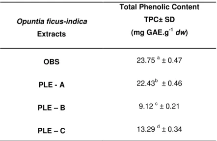
Table 3.1 Page Total phenolic content of Opuntia ficus-indica extracts, assessed by
Folin-Ciocalteau method, expressed as mg GAE.g-1 of extract, in a dry weight basis. Significant statistical differences are denoted with different letters.
Table 3.2 Page Total flavonoid content of Opuntia ficus-indica extracts, assessed by a
xix
List of abbreviations, acronyms and symbols
Abbreviation Full Form
AAPH 2”,2”-Azobis (2-amidinopropane) dihydrochloride
AD Alzheimer’s Disease
ADT Adsorption Technology
ATP Adenosine tri-phosphate
BBB Blood Brain Barrier
CAE Caffeic Acid Equivalents
CCE Crude Cherry Extract
CE Catechin Equivalents
CNS Central Nervous System
CO2 Carbon Dioxide
CSE Conventional Solvent Extraction
CVD Cardiovascular Diseases
DAD Diode Array Detector
DCFH – DA 2”,7”-Dichlorofluorescin Diacetate
ECACC European Collection of Cell Cultures
ED Electrochemical Detector
EGCG Epigallochatequine -3- gallate
EMEM Eagle Minimum Essential Medium
EtOH Ethanol
ETC Electron Transport Chain
EU European Union
FBS Fetal Bovine Serum
FDA Food and Drug Administration
FL Disodium Fluoresceín
GAE Gallic Acid Equivalents
GPx Glutathione Peroxidase
GR Glutathione Reductase
GSH Glutathione
GSSG Glutathione disulphide
H2O Water
H2O2 Hydrogen Peroxide
HORAC Hydroxyl Radical Adverting Capacity
HPLC High Performance Liquid Chromatography
KA kainate
MeOH Methanol
xx
OBS – CSE Opuntia Beja/Serpa – Conventional Solvent Extract
O2- Superoxide anion
OGD Oxygen-Glucose Deprivation
OH. Hydroxyl Radical
ORAC Oxygen Radical Absorbance Capacity
PBS Phosphate Buffer Saline
PD Parkinson’s Disease
PGI Protected Geographical Indication
PLE Pressurized Liquid Extraction
PLE – A Pressurized Liquid Extract – A
PLE – B Pressurized Liquid Extract – B
PLE – C Pressurized Liquid Extract – C
ROS Reactive Oxygen Species
SOD Superoxide Dismutase
SPE Solid Phase Extraction
TE Trolox Equivalents
TFC Total Flavonoid Content
Evaluation of Neuroprotective Effects of Natural Extracts obtained from Portuguese Agro-food Residues
1
1. Introduction
1.1. Neurodegenerative diseases
Countries are currently faced with problems derived from changes in population demography and an increase in lifestyle-related diseases. Neurodegenerative disorders such Parkinson’s (PD) and Alzheimer’s (AD) diseases are an increasing problem in aged societies like the Europe. There is an increased prevalence of both diseases with age (1). Neurodegenerative
diseases are a heterogeneous debilitating group of degenerative disorders characterized by a slow and progressive loss of neuronal cells. Such phenomena lead to gradual and progressive impairments of selective functions of central nervous system (CNS), depending upon the involved type of neuronal cells, and so far are incurable (2). Data from World Alzheimer Report
2011 indicate that 36 million people worldwide are living with dementia, with numbers doubling every 20 years to 66 million by 2030, and 115 million by 2050. Alzheimer’s disease has becoming the most common neurodisorder of today (3, 4). And at financial and social level, AD
represents an impact of €160 billion for the UE27 and €177 billion for whole Europe.
1.2. Neurodegeneration and Oxidative Stress
Neurodegenerative disorders appear to be triggered by multi-factorial events including neuroinflammation, depletion of endogenous antioxidants and increases in oxidative stress.(1,
5, 6). The high metabolic rate and the low concentration of endogenous antioxidants as well as
the proportion of polyunsaturated fatty acids make the brain tissue particularly susceptible to oxidative damage (7). The most effective way to produce adenosine tri-phosphate (ATP) at
cellular level is through oxidative phosphorylation within the mitochondria via the electron transport chain (ETC). The ETC is not completely efficient showing a basal level of electron leak. The reaction of leaked electrons with molecular oxygen produce a short-lived free radicals such superoxide anion (O2-), hydroxyl radical (OH.), and hydrogen peroxide (H2O2) called
reactive oxygen species (ROS)(8, 9). Under normal conditions, mitochondria have an efficient
biochemical defense mechanism to neutralize the effect mediated by these free radicals. Endogenous defenses are composed of glutathione (GSH), glutathione peroxidise (GPx), glutathione reductase (GR), superoxide dismutase (SOD), NADPH, vitamins E and C (10, 11).
Evaluation of Neuroprotective Effects of Natural Extracts obtained from Portuguese Agro-food Residues
2
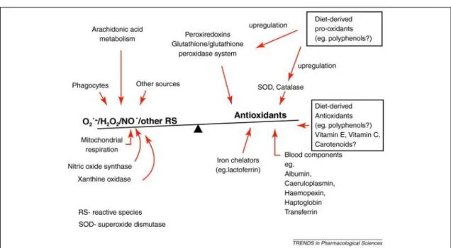
Figure 1.1. The approximate balance of antioxidants and reactive species in vivo.(13)
That is a large body of evidences demonstrating that the accumulation of iron ion species in the brain, the involvement of mitochondria, the decreased levels of endogenous antioxidants, and other ROS-mediated pathways are the major pathological factors that contributes to neurodegenerative diseases like PD and AD (14, 15).
Biological systems have several mechanisms to protect themselves from the ROS consequences. These antioxidant defenses mechanisms include scavenging of ROS species and their precursors, binding catalytic metal ions needed for ROS formation, generating and up-regulating endogenous antioxidant defenses (11). Central nervous system dysfunction has been
observed in all diseases, related to errors in GSH metabolism, suggesting that the brain is particularly susceptible to alterations in GSH homeostasis. The reason for the sensitivity is unknown, although may be consider the possibility of the high susceptibility of the brain to oxidative stress, due to its high oxygen consumption(10).
Glutathione (GSH) plays a central role in the cellular protection mechanism against oxidative and other forms of stress. Glutathione is present in both reduced (GSH) and oxidized (GSSG) state and the redox state of the GSH/GSSG couple can serve as an important indicator of cell redox environment. It has been described that a decrease in neuronal GSH levels leads to mitochondrial dysfunction and apoptosis.
1.2.1. Neurodegeneration Cell Model
Studies to determine biological effect of specific phytochemicals and plant-based foods involve experimentation with in vitro and in vivo systems. In vitro systems present advantages as a first
Evaluation of Neuroprotective Effects of Natural Extracts obtained from Portuguese Agro-food Residues
3
ease of culturing, their relatively low cost, and moderate throughput capabilities (16).
The creation of a cellular model based in the application of oxidative damage in neurons may be helpful to understand the molecular mechanisms underlying in the development of neurodegenerative diseases. Therefore, exposure of cultured neurons to relatively low concentrations of H2O2 induces changes in cell metabolism and lead to a moderate neuronal
death, reproducing what may occur during neurodegeneration process (17).
In the Disease and Stress Biology (DSB) lab, SK-N-MC cell line is used as neuronal cell model and is a continuous cell line, obtained from human metastic neuroblastoma tissue (17-19).
1.3. Nutraceuticals and Functional Foods
It is well known that consumption of plant-based foods such fruits, vegetables, grains and cereals have key role in health promotion and disease prevention (20). There has been an
increased concern for consuming health-promoting food products and two new concepts appear in the market: Nutraceuticals and Functional Foods. The short-term goal of nutraceuticals and dietary supplements is to improve the quality of life and enhance health status while its long-term goal is to increase lifespan while maintaining health (20).
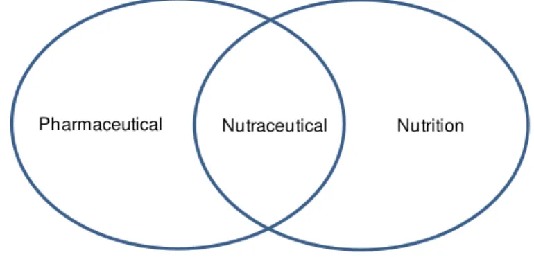
The term nutraceutical was coined from nutrition and pharmaceutical (Fig. 1.2) in 1989 by Stephen DeFelice, founder and chairman of foundation for innovation in medicine, an American organization which encourages medical health (21). Nowadays the term Nutraceutical is defined
as “a product isolated or purified from the food, generally sold in medicinal form not associated with food and demonstrated to have a physiological benefit .It also provides benefit against
chronic disease”(21). Such products may range from isolated nutrients, dietary supplements to
genetically engineered designer foods and herbal products and can be found in presentations similar to drug such pills, extracts or tablets (22).
Figure 1.2. The term "nutraceutical" was coined from "nutrition" and "pharmaceutical" in 1989 by Stephen
DeFelice, MD, founder and chairman of the Foundation for Innovation in Medicine (FIM), Cranford, NJ. Adapted from Pandey et al. 2010 .
Functional foods represents a type of food that when consumed regularly exert a specific health-beneficial effect (i. e., a healthier status or a lower risk of disease) beyond their nutritional properties, and this effect must be scientifically proven (22, 23). From consumers point of view,
Evaluation of Neuroprotective Effects of Natural Extracts obtained from Portuguese Agro-food Residues
4
functional foods and nutraceuticals may offer many benefits such as an increasing of health value of our diet, help people to live longer, help to avoid particular medicinal conditions, be perceived to be more "natural” than traditional medicine and less likely to produce unpleasant side-effects and present food for population with special needs. Some of the most common bioactive ingredients found in nutraceutical and functional food market are bioactive non-nutrient plant compounds. These compounds called phytochemicals, have raised interest in human nutrition because of their potential effects as antioxidants, antiestrogenics, anti-inflammatory, immunomodulatory and anticarcinogenics (22, 23). Nutraceuticals are able to
exert neuroprotection via a wide range of proposed mechanisms, such as scavenging of free radicals and ROS, metal chelation, modulation of cell-signalling pathways, and inhibition of inflammation (24). Most of phytochemical compounds with biological activities present in
nutraceuticals and functional foods are isolated from natural sources using several extraction techniques.
1.4. Phytochemicals
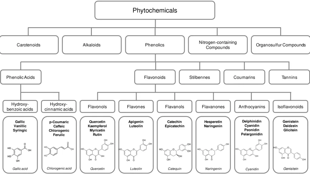
The composition of foods cannot be reduced to the macronutrients and essential micronutrients. Foods also contain a large number of other compounds that, although not essential, also have influence in human health. Several hundreds of phytochemicals such as polyphenols, carotenoids, glucosinolate, saponins or alkaloids have been identified in foods of plant origin. Some of these compounds may contribute to explain the beneficial health effects of consuming fruits and vegetables (25).
Polyphenols constitute a large group of phytochemicals with more than 8000 indentified compounds (26) and generally, involved in defence against ultraviolet radiation or aggression by
phatogens (27). Polyphenols may be classified in several ways such as a classification based
on structure and function. Simple phenols are substances containing only one aromatic ring and having at least one phenolic hydroxyl group and have the possibility of have other substituent. Phenols and polyphenols may occur as unconjugated aglycones or, as conjugates, frequently with sugar or organic acids. Flavonoids is the most extensively studied polyphenols, all characterized by a C6-C3-C6 structure, subdivides by the nature of C3 element into anthocyanins,
flavanols, flavanones, flavones, flavonols, isoflavones as shown in figure 1.3 (28).
Epidemiological studies suggest that high dietary intake of polyphenols is associated with decreased risk of a range of diseases including cardiovascular disease (CVD), specific forms of cancer and neurodegenerative diseases such AD (29) and PD (30-31). A regular dietary intake
of flavonoid-rich foods and/or beverages has been associated with 50% reduction in the risk of dementia, a preservation of cognitive performance with ageing, a delay in the onset of Alzheimer’s disease and a reduction in the risk of developing Parkinson’s disease (1). Many
studies have reported the bioavailability of polyphenolic compounds in the systemic circulation
(32-34). However, less is known about their degree of brain bioavailability, flavanones such as
Evaluation of Neuroprotective Effects of Natural Extracts obtained from Portuguese Agro-food Residues
5
have also been identified in the cortex and cerebellum of rat and pig following feeding with blueberries.
Together, these results suggest that polyphenols are able to transverse the BBB, in different degrees and depending on their structure. Thus, such compounds are likely to be candidates for direct neuroprotective and neuromodulatory actions (1). The contribution of phenolic
compounds to the protection against degenerative diseases as well as their effects on health has been attributed to their antioxidant activities (36, 37), but also to their cellular modulatory
Evaluation of Neuroprotective Effects of Natural Extracts obtained from Portuguese Agro-food Residues
6
Figure 1.3.Classification of dietary phitochemicals (Adapted from Liu, 2004) . (41)
Flavanones
Hydroxy-benzoic acids Flavonols Flavones Anthocyanins Isoflavonoids
Hydroxy-cinnamic acids Flavanols
Phytochemicals
Carotenoids Alkaloids Phenolics Nitrogen-containingCompounds Organosulfur Compounds
Phenolic Acids Flavonoids Stilbennes Coumarins Tannins
Hesperetin Naringenin
Naringenin Gallic
Vanillic Syringic
Gallic acid
Delphinidin Cyanidin Peonidin Pelargonidin
Cyanidin
Genistein Daidzein Glicitein
Genistein Apigenin
Luteolin
Luteolin Quercetin
Kaempferol Myricetin
Rutin
Quercetin p-Coumaric
Caffeic Chlorogenic
Ferulic
Chlorogenic acid
Catechin Epicatechin
Evaluation of Neuroprotective Effects of Natural Extracts obtained from Portuguese Agro-food Residues
7
Table 1.1. Polyphenols effects in in vivo and in vitro models of neurotoxicity and neurodegeneration (Adapted from Ebrahimi et al. (2012). (14)
Substance Cell line/ Animal model Effect
Aloe-emodin N-methyl-d-aspartate (NMDA)-induced toxicity in
retinal ganglion cells (RGCs) Elevates levels of RNA and protein expression of superoxide dismutase (SOD); Attenuates NMDA-induced apoptosis of RGCs (42)
Curcumin N27 dopaminergic neurons Protects against mitochondrial complex I inhibition (leading to mitochondrial
dysfunction) and NS (43)
Curcumin Homocysteine-induced neurotoxicity in rats Reduces Malondialdehyde (MDA)a and Superoxide anion levels; Reduces lipid
peroxidation; Improves learning and memory in rats (44)
Curcumin N27 dopaminergic neuronal cell line Increases glutathioneb (GSH) levels (45)
Curcumin 3-Nitropropionic acid (3-NP)-induced
neurotoxicity in rats Improves the 3-NP-induced motor and cognitive impairment; Attenuates 3-NP-induced OS (including lipid peroxidation, reduced GSH and nitrite activity);
Restores the decreased succinate dehydrogenasec activity (46)
Epigallocatechingallat
(EGCG) Glucose oxidase-induced neurotoxicity in H 19-7 (a rat neuronal cell line) Enhances cellular resistance to glucose oxidase-mediated oxidative damage; Elevates heme oxygenase-1d (HO-1) mRNA and protein expression; Activates
transcription factor Nrf2e (47)
EGCG Glutamate-induced toxicity in HT22 mouse
hippocampus neuronal cells, Kainic acid-induced neurotoxicity in Rats
Reduces glutamate-induced oxidative cytotoxicity; Inactivates the NF-_B signaling pathway; Reduces ROS accumulation and NF-_B transcriptional activity
(48)
EGCG Transient global cerebral ischemia
C57BL/6 in mice Reduces the development of delayed neuronal death after transient global cerebral ischemia in mouse brain (49)
EGCG Age-associated oxidative damage in rat brain Amplifies the activities of enzymic antioxidants like SOD, catalase, glutathione peroxidase, glutathione reductase and glucose-6-phosphate dehydrogenase; Improves the activity of non-enzymic antioxidants like tocopherol, ascorbic acid
and glutathione;Ameliorates the MDA and protein carbonyl levels (50)
EGCG Progressive neurotoxic model of long-term
serum deprivation in human SH-SY5Y neuroblastoma cells
Decreases protein levels and mRNA expression of the beta subunit of the enzyme prolyl 4-hydroxylase; Decreases protein levels of two molecular chaperones that are associated with HIF regulation, the immunoglobulin-heavy-chain binding
protein and the heat shock protein 90 beta (51)
EGCG SOD1-G93A transgenic mice (a model of ALS) Maintains the normal expression of p85a PI3-K, pAkt, and pGSK-3 (molecular signals of survival); Reduces activation of NF-kB and the cleaved form of caspase-3; Reduces microglial activation; Prolongs the life span; Delays the onset
of symptoms (52)
Mangiferin Glutamate-induced neurotoxicity in rat cerebral
cortex neurons Prevents neuronal death, oxidative stress and mitochondrial depolarization (53)
Mangiferin 1-Methyl-4-phenyl pyridinium (MPP(+))-induced
Evaluation of Neuroprotective Effects of Natural Extracts obtained from Portuguese Agro-food Residues
8
line N2A Quenches reactive oxygen intermediates
Mangiferin Morin Glutamate-induced neurotoxicity in rat primary
culture of neurons Reduces ROS formation; Activates enzymatic antioxidant system; Restores mitochondrial membrane potential (55)
Polyphenol-rich
Hedeoma multiflorum
extract
Biochemical assay on rat brain Homogenates Inhibits lipid peroxidation; Scavenges 2,2_-diphenyl-1-picrylhydrazyl (DPPH) radicals
(56)
Polyphenol-rich osmanthus fragrans extract
Glutamate, arachidonic acid, and 6
hydroxydopamine-induced neurotoxicity in rat primary cortical neurons
Scavenges DPPH and hydroxyl anions Inhibits lipid peroxidation
(57)
Red wine polyphenol
compound Rat model of ischemic cerebral stroke Prevent the burst of excitatory amino acids in response to ischemia; Reduce brain infarct volumes; Enhance the residual cerebral blood flow during occlusion and reperfusion; Modulate expression of proteins involved in the maintenance of
neuronal caliber and axon formation (58)
Resveratrol Lipopolysaccaride (LPS)-induced dopaminergic
neurodegeneration in rat Reduces NADPHactivation; Attenuates the activation of MAPK and NF-_B signaling pathways; g oxidase-mediated generation of ROS; Inhibits microglia
Implies neuroprotection against LPS-induced dopaminergic neurodegeneration (59)
Resveratrol Glutamate-induced toxicity in mice primary culture of neurons; Optimized ischemic-reperfusion stroke model in mice
Induces heme oxygenase 1d (HO-1) in dose- and time-dependent manner; Protects mouse neurons, subjected to an optimized ischemic-reperfusion stroke
model; Protects neurons against ecitotoxicity (60)
Resveratrol 1-methyl-4-phenyl-1,2,3,6 tetrahydropyridine
(MPTP)- induced Parkinson in mice Protects from MPTP-induced motor coordination impairment, hydroxyl radical overloading, and neuronal loss; Scavenges free radicals (61)
Resveratrol MPP(+)-induced neurotoxicity in dopaminergic
neurons of midbrain slice culture Prevents accumulation of ROS, depletion of cellular glutathione, and cellular oxidative damage induced by MPP(+); Activates sirtuin family of NAD-dependent
histone deacetylases (62)
Resveratrol A-beta induced toxicity in neurons from a mouse model (Tg2576 line) and mouse neuroblastoma (N2a) cells
Maintains normal expression of peroxiredoxins and mitochondrial structural genes; Maintains normal mitochondrial function
(63)
Tea polyphenols NMDA-induced neurotoxicity in mice Attenuates the increased production of synaptosomal ROS; Reduces the
Evaluation of Neuroprotective Effects of Natural Extracts obtained from Portuguese Agro-food Residues
9
1.5. Extraction Technologies
Amongst fruits, vegetables and herbs, agricultural and industrial by-products are attractive sources for extraction of compounds with nutraceutical interest such pigments or natural antioxidants. Special attention has been focused on the extraction from inexpensive or residual sources from agricultural industries. Some studies have already been done on by-products, which could be potential sources of antioxidants (65). Once the industrial processing of a wide
variety of fruits results in the accumulation of large quantities of residues, the recovery of these food industry by-products could reduce waste disposal problems and serve as a potential new source of phytochemicals that can be used in nutraceutical and functional food market creating value-added applications. In order to obtain such value compounds, extraction techniques have been investigated. Conventional Solvent Extraction (CSE) is a traditional extraction method characterized by a solid-liquid principle. Solid-liquid extraction can be defined as a mass transport phenomenon in which solids contained in a solid matrix migrate into a solvent brought into contact with the matrix. The single-stage system represents the complete operation of contacting the solid matrix with the fresh solvent. However, these techniques have, generally, negative environmental impact and some drawbacks such as long extraction times, large amounts of initial matrix and organic solvents and lower extraction yields (65).
An attractive and alternative methodology to conventional extraction, to produce extracts for pharmaceutical or nutraceutical application is Pressurized Liquid Extraction (PLE). The technique involves the utilization of water and/or organic solvents at considerable elevated temperatures (313 – 473 K) and pressures (3.3 – 20.3 MPa). Liquid carbon dioxide is found to be a good extraction solvent in food industry due to its physicochemical properties, low toxicity and price. Since carbon dioxide is non-polar specie it is not suitable solvent to extract polar polyphenols. The addition of solvents like ethanol increases the solvating power of carbon dioxide and the extraction yield of phenolic compounds. Ethanol is a permitted co-solvent in food industry.The advantages of PLE technique are usually related with the usage of carbon dioxide (66) that has been described as a “green solvent” and gives to the method improved
characteristics in terms of mass transfer and of solvation properties(66, 67). Extraction time can
be decreased using high temperature and high pressure: there is enhanced diffusivity of the solvent and, at the same time, there is the possibility of working under an inert atmosphere and with protection from light (68).
Evaluation of Neuroprotective Effects of Natural Extracts obtained from Portuguese Agro-food Residues
10
The adsorption capacity of an adsorbent for a solute may vary with processing conditions such as temperature and pH value of solution. The elution step is usually performed with alcohols, in particular ethanol as it is acceptable for food and pharmaceutical applications.
The adsorption processes can be performed in a batch model (70) or in continuous, where the
adsorbent is packet into a column (71). The use of resins for food production proposes is
regulated by the US Food and Drug Administration (FDA) and the Council of Europe.
Figure 1.4. Chemical structure of Amberlite® XAD 16.
The selection of adsorbents is critical for the development of an adsorption process. Adsorbent characteristics, such as particle size, surface, area and porosity should be taken in consideration in each case study. For the purification of polyphenols various adsorbents have been used (72) One of the most commonly applied is a polymeric resin Amberlite® XAD16
(Figure 1.7) (73).
1.6. Opuntia ficus-indica
Opuntia ficus-indica (cactus pear) is the larger member of Cactaceae family that mainly grows in
arid and semi-arid regions and is largely distributed in Mexico, much Latin America, South Africa and Mediterranean area(74). Opuntia fruits are oval and elongated berries of 67 to 216 g weigh.
Cactus pear fruits have a thick pericarp (peel) and a juicy pulp with many seeds. The pulp is the edible part of the fruit and is mainly composed of water (84-90%) and reducing sugars (10-15%). Fruit seeds represent about 10-15% of the edible pulp and are usually discarded as waste after pulp extraction (75). The high season for harvesting Opuntia cactus fruits is between
the end of July and November in the Mediterranean regions (76). Fruit and stems of Opuntia
species have been used in folk medicine for burns, wounds, edema, bronchial asthma, hypertension, indigestion, and type II diabetes (77). They nutritional properties have long been
known and it is also used in traditional medicine. In the industrialized countries of Mediterranean area, cladodes are not consumed but the fruits are largely used as nutritional sources (76). The
plant is used mainly for fruit production and the fruit are consumed as fresh fruit or juice. Although in some countries it is used as a vegetable for fodder (78, 79).
C
C
C
C
C
C
C
H
H
H
H
H
H
H
H
H
H
H
Evaluation of Neuroprotective Effects of Natural Extracts obtained from Portuguese Agro-food Residues
11
Figure 1.5. Images of Opuntia ficus-indica cladodes and fruits.
Cactus pear was largely ignored by the scientific community until the beginning of the 1980’s when several studies were published on their biological functions. In recent years, investigations on the chemical and nutritional value of Opuntia spp. have attracted attention in the food,
nutritional and pharmacological sciences (80, 81) . Based on various studies, fruit pulp is
considered a good source of minerals, especially calcium, potassium and magnesium and phosphorous(74). The concentration in bioactive compounds is dependent on the cultivar,
cultivation site, maturation stage and environmental conditions.
Fruit is commonly consumed fresh but there is increased interest in the transformation into different products such as juice, jam and jelly among others. After being consumed or transformed, fruit peels and seeds are discarded and taking in account they composition in phytochemicals make them very attractive as sources for extraction. After extraction these by-products can be used as addictive in food preparation or in the pharmaceutical and nutraceutical market (82). The nutraceutical benefits of Opuntia spp. fruits are believed to be
related to their content in ascorbic acid and phenolic compounds, including flavonoids, and a mixture of yellow betaxanthin and red betacyanin pigments. Betaxanthins and betacyanins belong to betalains group that are water soluble compounds described as excellent radical scavengers.(79)
In vitro studies have demonstrated that a variety of compounds present in Opuntia sp. fruits are
able to exert neuroprotective activity. The methanolic extract of O. Ficus-indica produced
dose-dependent neuroprotective effects on hydroxyl- and superoxide radical-mediated neuronal damage to mouse primary cultures (83) and the pre-treatment of cultured neurons and glia cells
Evaluation of Neuroprotective Effects of Natural Extracts obtained from Portuguese Agro-food Residues
12
and oxygen-glucose deprivation (OGD)-induced neurotoxicity dose-dependent.(84) Kim and
collaborators (2006) also have demonstrated that when gerbils receive different doses of the Opuntia extract, the neuronal damage in the hippocampus was reduced. Moreover Opuntia
polysaccharides as described to exert a neuroprotective activity against a H2O2-induced
oxidative damage in PC12 cells by maintain cell viability, reduce apoptosis and decrease intracellular ROS levels.(77)
1.7. Prunus avium
Prunus avium (sweet cherries) are very attractive fruits for consumers due to their taste, colour,
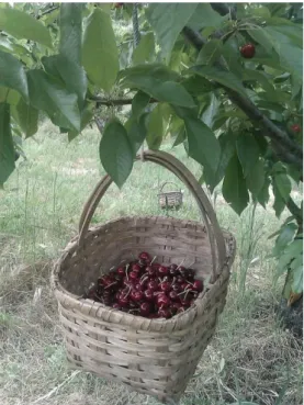
sweetness and wealth of nutrients being easily adapted to a regular diet. Portugal produces more than 15 000 tons of cherries every year. It is in the northeast of the country, in the region of Beira Interior, where the fruit is more cultivated and the cherries has protected geographical indication (PGI) registration according to the European Union (EU) regulations.
Figure 1.6. Cherries (Prunus avium) from Sacocultivar collected in “Cova da Beira” region, Portugal.
Saco cultivar is an old traditional cherry. It is a very promising functional fruit due to its powerful
antioxidant attributed to the phenolic composition (85). Serra et al. evaluated nine “Cova da
Beira” cherry varieties in terms of polyphenolic content and bioactivity (antioxidant properties) and it was shown that Saco cherry extract has a strong antioxidant activity. However there is
little information about cherries biological activities. However, due to its small size and weight the acceptance of Saco cherry by consumers could be compromised since larger fruits are
preferred.
It has been described that sweet cherries cultivars such as Saco contain various anthocyanins
Evaluation of Neuroprotective Effects of Natural Extracts obtained from Portuguese Agro-food Residues
13
collaborators described that sweet cherries contain cyaniding -3- glucoside and cyaniding-3-rutinuside being that 77.3 to 86.6 % of the total anthocyanins content. Among hydroxycynamic acid derivates, cherries had neochlorogenic acid and p-coumaroylquinic acid as the
predominant compounds and the glycosides of peonidin and pelargonidin as minor compounds
(86). Cyaniding -3- glucoside is described as able to transpose the BBB. (87)
Nevertheless, there is little information available on the possible health benefits of phenolic compounds present in fresh cherries on animal cells exposed to cell-damaging oxidative stress.
Taking this in account the major aim of this work is to obtain and characterize natural extracts obtained from Opuntia ficus-indica and Prunus avium, residues using different extraction
technologies, evaluate their neuroprotective effect in a H2O2-induced neurodegeneration cellular
Evaluation of Neuroprotective Effects of Natural Extracts obtained from Portuguese Agro-food Residues
Evaluation of Neuroprotective Effects of Natural Extracts obtained from Portuguese Agro-food Residues
15
2. Materials and Methods
2.1. Materials
For phytochemical characterization: Amberlite® XAD 16 and sodium carbonate (Na2CO3) were
purchased from Sigma- Aldrich (St Quentin Fallavier, France), Folin Ciocalteau reagent was acquired from Panreac (Barcelona, Spain) and gallic acid from Fluka (Germany). Sodium nitrite
(NaNO2) was purchased from Riedel-de-Haën (Seelze, Germany), aluminium chloride (AlCl3)
and sodium hydroxide (NaOH) was obtained from Sigma-Aldrich, in Germany. (+)-Catechin hydrate was purchased from Sigma (Japan).
For antioxidant activity assays chemicals used were: 2‟,2‟-Azobis (2- amidinopropane) dihydrochloride (AAPH), 6-hydroxy-2,5,7,8- tetramethylchroman-2-carboxylic acid (Trolox), caffeic acid (C9H8O4), cobalt floride tetrahydrate (CoF2), hydrogen peroxide (H2O2) and picolinic
acid (C6H5NO2) were purchased from Sigma-Aldrich (St Quentin Fallavier, France). Disodium
fluorescein (FL) was from TCI Europe (Antwerp, Belgium). Sodium chloride (NaCl), potassium chloride (KCl) and monopotassium phosphate (KH2PO4) were from Sigma-Aldrich (St Quentin
Fallavier, France) and sodium phosphate dibasic dehydrate (Na2HPO4⋅2H2O) from Riedel-de
Haën (Seelze, Germany) were used for phosphate buffer solution preparation (PBS).
Cell based assays were performed using: 2’,7’-dichlorofluorescein diacetate (DCFH-DA), hydrogen peroxide (H2O2), sodium dodecyl sulphate (SDS), GSH and GSSG standards,
orthophthalaldehyde (OPA), Eagle Minimum Essential Medium (EMEM), glutamine, non-essential amino acids, sodium pyruvate, were purchased from Sigma-Aldrich (St. Quentin Fallavier, France), Disodium fluorescein was obtained from TCI Europe (Antwerp, Belgium), CellTiter – Blue Reagent® Cell Viability Assay was obtained from Promega (San Luis Obispo, CA, USA). Fetal bovine serum (FBS), Trypsin-EDTA and penicillin-streptomycin (PenStrep) were obtained from Invitrogen (Gibco, Invitrogen Corporation, Paisley, UK). Phenolic standards used were: chlorogenic acid from Sigma-Aldrich (St. Louis, MO, USA), and cyanidin-3-glucoside, cyanidin-3-rutinoside were all from Extrasynthèse (Genay, France). Ultra-pure water
(18.2 MΩ.cm) was obtained from a Millipore Direct Q3 UV system (Millipore, USA).
2.2. Raw Materials
Cactus pear (Opuntia ficus-indica) fruits were collected at Beja/Serpa, Portugal, in September
2011. After remove the spikes, fruits were processed using a kitchen robot (UFESA, LC5005, China) and the peels and seeds were collected. The waste (peels and seeds) was kept at -20oC.
Sweet cherries (Prunus avium), from Saco cultivar, grown in Cova da Beira region, Portugal,
were harvested by hand between May and June of 2011. The whole fruits were processed with seeds and stalks, milled and frieze dried. Milled cherries were kept at – 20 ºC.
2.3. Preparation of Prunus avium and Opuntia ficus-indica Extracts
Evaluation of Neuroprotective Effects of Natural Extracts obtained from Portuguese Agro-food Residues
16
2.3.1.1. Conventional Solvent Extraction
Opuntia ficus-indica fruits were processed into juice and the peels and seeds were collected
and submited to a conventional solid-liquid extraction with a biocompatible mixture: ethanol (EtOH): water (H2O) (1:1 v/v) solution (1:20, w/v). The extraction were processed in the dark for
4 h under constant agitation (200 rpm), at room temperature (IKA® dual-speed mixer RW 20.n, Aldrich, St. Luis, USA) and protected from light. The homogenates were centrifuged at 9000 rpm for 10 min. The supernatants were filtered under vacuum and the solvent was evaporated in a rotary evaporator (Buchi, Switzerland) at 40 ºC. The extract was freeze dried at and kept at – 20 ºC until analyses.
2.3.1.2. Pressurized Liquid Extraction
Extracts obtained during pressurized liquid extraction from Opuntia ficus-indica fruits residues
were kindly provided by Dr. Hermínio de Sousa and his laboratory team group (Laboratory of Polymer Processing and Supercritical Technology, Universidade de Coimbra, Portugal). Three different extracts were obtained at 313 K and 200 MPa using three different EtOH/CO2/H2O
mixtures (table 2.1). The extract was designated as PLE-A for the 0.4/0.3/0.3 mixture, PLE-B and PLE-C for 0.5/0.4/0.1 and 0.5/0.1/0.4 mixtures, respectively.
Table 2.1. Molar fraction of Pressurized Liquid Extraction (PLE) solution.
2.3.2. Prunus avium Extract
2.3.2.1. Conventional Solvent Extractio (CSE)
After dehydratation by freeze dryer, the cherries were homogenized in a grinder (UFESA, LC5005, China). Powdered cherries were extracted in the dark with EtOH:H2O (1:1, v/v) solution
(1:20, w/v), for 2 h at room temperature, under constant agitation (200 rpm) (IKA® dual-speed mixer RW 20.n, Aldrich, St. Luis, USA). The homogenates were filtered under vacuum and the solvent was evaporated in a rotary evaporator (Buchi, Switzerland) at 40 oC. The remaining
extracts were diluted in bi-distilled water. The extract was kept at – 20 º C.
Extracts
Molar Fractions
EtOH CO2 H2O
PLE – A 0.4 0.3 0.3
PLE – B 0.5 0.4 0.1
Evaluation of Neuroprotective Effects of Natural Extracts obtained from Portuguese Agro-food Residues
17
2.3.2.2. Adsorption Technology (AD)
To concentrate in anthocyanins of Prunus avium extract obtained on 2.3.2.1. section, food
grade macroporous resin Amberlite® XAD-16 was used as adsorbent. This resin is allowed for food applications by the U.S. Food and Drug Administration Code of Federal Regulation Title 21
(88).
The preconditioning of the adsorbent was done by an extensive wash with abundant distilled water to remove salts and impurities, and was then dried at 70 oC for 24 h. The dried resin was immersed in EtOH (96%) for 12 h. EtOH was then replaced by bi-distilled water through washing.
The production of cherry polyphenols rich extract was performed in batch mode. Briefly, aqueous crude cherry extract (CCE) obtained previously (section 2.3.2.1) were maintained in contact with the resin in a ratio of 80 mg GAE.g-1 of resin, protected from light and submitted to 200 rpm of agitation, during 4 h. After 4 h of contact, supernatant was removed and resin washed three times with bi-distilled water. Anthocyanins were then eluted two times with EtOH (96 %). The ethanolic fraction were concentrated by evaporation, freeze dried and kept at – 20 ºC.
2.4. Extracts Characterization
The extracts were analyzed for their chemical composition and antioxidant capacity against peroxyl and hydroxyl radicals. Composition in polyphenols was determined using Folin-Ciocalteau colorimetric method and flavonoids content was performed by the AlCl3
complexation method (89) modified by Tavares et al, 2010. Antioxidant activity was assessed
using two different in vitro chemical assays, ORAC and HORAC adapted by Feliciano et al,
2009 and Serra et al, 2011, respectively. Extracts phenolic profile were obtained by High
Performance Liquid Chromatography with Diode Array Detector and Electrochemical Detector (HPLC – DAD – ED).
2.4.1. Total Phenolic Content (TPC)
Total phenolic content present in extracts was determined according to the Folin- Ciocalteau method. To perform the colorimetric method, 20 µL of the appropriate dilutions of extracts were added to 1580 µL of distilled water. Than 100 µL of the Folin Ciocalteau reagent was added. The reaction was neutralized with 300 µL of sodium carbonate. The absorbance, at 765 nm of each sample was measured after 40 min at 40 ºC of incubation, in a Genesys10uv spectrometer (Thermo Spectronic, New York, USA). The results were expressed as means of independent triplicates as mg of Gallic Acid Equivalents (GAE) per gram of dry weight extract (mg GAE.g-1
Evaluation of Neuroprotective Effects of Natural Extracts obtained from Portuguese Agro-food Residues
18
2.4.2. Total Flavonoid Content (TFC)
The total flavonoid content was determined using a spectrophotometric method adapted to 96-well microplate by Tavares et al, 2010. Briefly, 25 µL of each sample, 125 µL of distilled water
and 7.5 µL of NaNO2 5% (v/v) was added to each well of a 96-well plate. The plate was
incubated for 6 min at room temperature. Than 15 µL of a 10% AlCl3 (v/v) solution was added.
After 5 min of incubation, 100 µL of NaOH 1 M was added and the solution of each well mixed. The absorbance was measured at 510 nm in a microplate reader (BioTekTM Power Wave XS).
(+)-Catechin hydrate, minimum 98% (w/w) was used as standard, and the results are expressed in means of triplicates as mg catechin equivalents (CE) per mg of dry weight extract (mg CE.mg-1dw).
2.4.3. HPLC – DAD – ED analysis
HPLC analysis of Opuntia ficus-indica and Prunus avium fruit residues extracts were performed
by Analytical Group of IBET (Oeiras, Portugal) coordinated by Dr. Rosário Bronze. Briefly the analysis were performed on a Waters® Alliance 2695 (Waters) equipped with a quaternary pump, solvent degasser, auto sampler and column oven, coupled to a Photodiode Array Detector Waters 996 PDA (Waters). A pre-column (RP-18, 5 μm, Lichrocart) and reverse phase column (RP-18, 2.5 µm, from Manu-cart) with oven at 35 ºC were used for separation. The gradient mobile phase consisted of 0.1 % phosphoric acid p.a. in ultra pure water (A): 0.1 % phosphoric acid p.a. in LC-MS grade acetonitrile (B) and ultra pure water at a flow rate of 0.30 mL.min-1. The injection volume was 5 μL.Photodiode Array Detector was used to scan wavelength absorption from 210 to 600 nm.
Evaluation of Neuroprotective Effects of Natural Extracts obtained from Portuguese Agro-food Residues
19
2.4.4. TLC analysis
Presence of terpenoids compounds was assessed in the three PLE extracts by thin layer chromatography (TLC) using Lupeol as standard. 10 µL of extracts and standard were carefully placed in a Plate Silica gel 60 F254. The mobile phase was an acetate:methanol:water (77:15:8)
solution. In order to reveal the terpenoid compounds present Liebermann – Burchard reagent were prepared adding carefully 5 mL of acetic anhydride and 5 mL concentrated sulphuric acid to 50 mL absolute ethanol, while cooling on ice. The TLC plate is sprayed with freshly prepared solution, heated for 10 min at 100 ºC and inspected in UV-365nm.
2.5. Extracts Chemical Antioxidant Activity
2.5.1. Oxygen Radical Absorbance Capacity (ORAC)
The oxygen radical absorbance capacity was assessed by ORAC modified by Ou. et al.
2001(90). The oxidation of disodium flourescein (FL) is catalyzed by peroxyl radicals generated
by 2, 2’-azobis(2-amidopropane)dihydrochloride (AAPH) at 37 oC. ORAC assay measure the ability of the antioxidant compounds present in the sample to inhibit that oxidation.
The protective effect of an antioxidant specie present in the sample is obtained by the difference between the area under the fluorescence decay curve (AUC) of the sample and the blank, in which no antioxidant is present. Briefly, sodium fluorescein (150 μL; 2,0x10⁻⁸ M in PBS 75 mM
pH 7,4) and samples or standards (25 μL) were added to a 96-well microplate and incubated
during 10 min at 37 ºC. AAPH (25 μL,1.28x10-2 prepared in PBS 75mM pH 7,4) were added to
each well. FL800 (Bio-Tek Instruments) microplate fluorescence reader was used (λex: 485 nm, λem: 530 nm). The blank contained PBS (pH 7.4, 75 mM) instead of sample and 5 to 50 µM of Trolox was used to the calibration curve. The results were expresses as means of independent triplicades as µmol of Trolox Equivalents (TE) per gram of dry weight extract (µmol
TE.g-1dw).
2.5.2.
Hydroxyl Radical Adverting Capacity (HORAC)HORAC assay was based on a method reported by Ou et al 2002 (91) modified for the FL800
microplate fluorescence reader FL800 (Bio- Tek Instruments). HORAC assay evaluates the hydroxyl radical prevention capacity by monitoring the fluorescence decay of fluoresceín (FL), which is used as a probe. The hydroxyl radical was generated by a Co(II)-mediated Fenton like reaction and the fluorescence decay curve of FL was used to quantify the HORAC value. Briefly, 10
L of each sample were added to 180
L of FL (4 x 10⁻³
M) and then was added 10
L of a 0.55 M H2O2 solution. The reaction starts when it is added 10
L of CoF2 to each well ofa 96-well microplate at 37 ºC. The FL800 (Bio-Tek Instruments) microplate fluorescence reader
Evaluation of Neuroprotective Effects of Natural Extracts obtained from Portuguese Agro-food Residues
20
expressed as means of independent triplicates as µmol of Caffeic Acid Equivalents (CAE) per gram of dry weight extract (µmol CAE.g-1dw).
2.6. Cell Based Assays
2.6.1. Cell Culture
Human Neuroepithelioma from Supra-Orbital Metastasis SK-N-MC cells, obtained from the European Collection of Cell Cultures (ECACC), were routinely grown in EMEM (Eagle Minimum Essential Medium, Sigma) supplemented with 2mM L-Glutamine (Sigma), 10% (v/v) heat-inactivated fetal bovine serum (FBS) (Sigma), 1% (v/v) non-essential amino acids
(NEM)(Sigma), 1 mM sodium pyruvate and 50 U.mL penicillin and 50
g.mL streptomycin. According to ECACC instructions, stock cells were maintained in 25 cm2 T-flask, incubated at 37 ºC and 5% CO2. Cells are split to sub-confluence of 70 – 80 % confluence using 0.05 % (v/v)trypsin-EDTA (Gibco).
2.6.2. Cytotoxicity Profile determination
Extracts toxicity assays were performed using CellTiter-Blue® Reagent. Briefly, SK-N-MC cells were seeded at a density of 2.5x10⁴ cells/well in 96 well plate. After 24 hours, SK-N-M cells were pre-incubated with the extracts in a range 0-250 µg GAE.mL-1 medium, for 24 h. A control was performed with cells incubated only with cell culture medium.
After 24 of incubation with the extracts, 20 µL of CellTiter-Blue® Reagent was added to each well for 3 h. Resazurin present in CellTiter-Blue® Reagent is a dark blue dye that has little intrinsic fluorescence, is reduced by metabolically active cells into a fluorescent product, resorfurin. Therefore, fluorescent signal is proportional to the number of viable cells. The product was quantified by measurement of the fluorescence (λex: 560 nm, λem: 590 nm) on microplate fluorescence reader FL800 (Bio-Tek Instruments).
2.6.3. Evaluation of Neuroprotective effect
To evaluate the neuroprotective effect of each extract, a neurodegeneration cell model was used. The model describes the treatment of SK-N-MC neuroblastoma cells with H2O2 to induce
50% of cell death. Cells were seeded at 2.5 x 10⁴ cells per well. After 24 hours of growth the medium was removed and the cells were washed with PBS. Cells were pre-incubated with non-toxic concentrations of each extract and after 24 hours, cells were washed again with PBS and medium was replaced by medium containing 0,5% (v/v) FBS and 300 µM H2O2 for 24h. Cell