Isolation and characterization of a
reserve protein from the seeds of
Opuntia ficus-indica
(Cactaceae)
Departamento de Bioquímica e Biologia Molecular, Universidade Federal do Ceará, Fortaleza, CE, Brasil
A.F. Uchoa, P.A.S. Souza, R.M.L. Zarate, E. Gomes-Filho and F.A.P. Campos
Abstract
We describe here the isolation and characterization of a major albumin from the seeds of Opuntia ficus-indica (Cactaceae). This protein has a molecular mass of 6.5 kDa and was isolated by a combination of gel filtration chromatography and reverse-phase HPLC. The amino acid composition of this protein was determined and it was shown to have similarities with the amino acid composition of several proteins from the 2S albumin storage protein family. The N-terminal amino acid sequence of this protein is Asp-Pro-Tyr-Trp-Glu-Gln-Arg.
Correspondence
F.A.P. Campos
Departamento de Bioquímica e Biologia Molecular, UFCE Caixa Postal 1065 60001-970 Fortaleza, CE Brasil
Fax: 55 (085) 288-9829 E-mail: bioplant@ufc.br.
Research supported by CNPq (No. 520332/93-8).
Received September 29, 1997 Accepted March 18, 1998
Key words
•Cactaceae
•Prickly-pear
•Opuntia ficus-indica
•Seed proteins
•2S albumins
Introduction
Over the years the prickly-pear (Opuntia ficus-indica) has shown its usefulness as a forage crop for small farmers in the Brazilian northeast. This is especially true in the years of drought, when on many occasions its fleshy phylloclads are the sole source of water and nutrients for farm animals (1,2). The new biotechnological tools are opening new opportunities for manipulating impor-tant agronomic traits of different crops. In our laboratory we are interested in exploring the possibility of using these tools to im-prove the nutritional quality of the prickly-pear for farm animals. Specifically, we want to introduce in it a transgene coding for a protein of good nutritional quality and to have this transgene expressed at high levels in the phylloclads. In order to achieve this we are developing protocols for plant regen-eration and transformation and we are study-ing the pattern of protein deposition in seeds and phylloclads. We present here data on the
isolation and characterization of the major salt-soluble protein from the seeds and dem-onstrate that this protein belongs to the 2S albumin storage protein family. To our knowl-edge, this is the first report on the character-ization of a seed reserve protein from a Cactaceae plant.
Material and Methods
Plant material
Seeds of O. ficus-indica cv. Gigante were obtained from mature fresh fruits collected in May 1996 on a farm in Madalena, Ceará, Brazil.
Protein extraction and purification
Salt-soluble proteins were extracted from seed flour in 0.1 M Tris/HCl and 0.5 M NaCl, pH 8.0, for 2 h. After centrifugation (10000 g, 20 min, 4oC), ammonium sulfate
concentra-tion of 90% and the precipitated proteins were recovered by centrifugation. After di-alysis against distilled water at 4oC and
cen-trifugation, the albumin and globulin tions were obtained. The albumins were frac-tionated on a Sephacryl S-100-HR column (80 x 2.6 cm) equilibrated and eluted with 50 mM ammonium bicarbonate at a flow rate of 30 ml/h and 4.3-ml fractions were collected. This column was previously calibrated using the MW-GF-70 kit for molecular mass 6.5 to 66 kDa from Sigma Chemical CO. (St. Louis, MO). The effluents containing the 6.5-kDa protein as demonstrated by SDS-PAGE were pooled, freeze-dried and fractionated by molecular exclusion HPLC on a Superdex Peptide HR 10/30 column from Pharmacia Biotech (Uppsala, Sweden), developed with an HPLC system from Waters Corporation (Millford, MA). The column was equilibrated and eluted with 50 mM ammonium bicar-bonate, at a flow rate of 0.5 ml/min. A single protein peak was obtained which was further fractionated by reverse phase-HPLC (RP-HPLC) on a 3.9 x 300-mm µ-Bondapak C-18 column (Waters) linked to an HPLC system from Waters Corporation. The column was equilibrated with 0.1% trifluoracetic acid and proteins were eluted with a linear gradi-ent of 0-60% acetonitrile in 0.1% trifluora-cetic acid, run in 60 min. The effluents were monitored at 280 nm in all chromatographic steps. Protein concentration was determined
by the protein-dye binding method of Bradford (3).
Amino acid analysis
Protein samples were resuspended in 500 µl of constant-boiling 6 N HCl containing 0.1% phenol. Ten percent of each sample was brought up to a final volume of 150 µl of constant-boiling HCl (6 N) plus 0.1% phe-nol. Hydrolysis was carried out in evacu-ated, sealed, thick-walled borosilicate glass tubes for 24 h at 120oC. Cooled tubes were
opened and samples were dried under vacuum and stored at -20oC until derivatization.
Samples were derivatized with phenylisothio-cynate (PITC) (Pierce, Millford, IL) by the method of Bidlingmeyer et al. (4). The pro-tein hydrolysates were redried twice with ethanol:water: triethylamine (2:2:1, v/v), and derivatized with ethanol:water: triethylamine: PITC (7:2:2:1, v/v). Samples were analyzed by RP-HPLC on a Waters system equipped with a Waters 712 WISP and a Waters sys-tem interface module linking the HPLC to a Baseline 810 workstation. A Pico-Tag col-umn for amino acid hydrolysate analysis (Waters) maintained at 46oC by a Waters
temperature-control module was used in ac-cordance with the manufacturer’s recom-mended procedure. Hydrolysate amino acid standards (Sigma) were used for peak identi-fication and subsequent calculations. Lysozyme was also analyzed and the results were compared with published values to ensure the accuracy of the technique. Three independent determinations were performed for each sample. Selected replicates were analyzed in duplicate to ensure repeatability.
SDS-PAGE
Gel electrophoresis was performed using the tricine-SDS polyacrylamide gel electro-phoresis method for the separation of low molecular mass proteins described by Schagger and Jagow (5). The proteins
con-A280 2.0
1.5
1.0
0.5
0
Molecular mass (kDa)
60 30 12 5 2
Vo
0 100 200 300 400 500
Volume (ml) Figure 1 - Chromatography of
tained in the MW-SDS-17S kit (Sigma) for molecular mass 3.46 to 16.95 kDa were used as molecular mass markers.
N-Terminal analysis
The homogeneity of the purified protein and the N-terminal sequence were deter-mined by the DABITC/PITC double-cou-pling method (6) following the protocol de-scribed by Campos and Richardson (7).
Results and Discussion
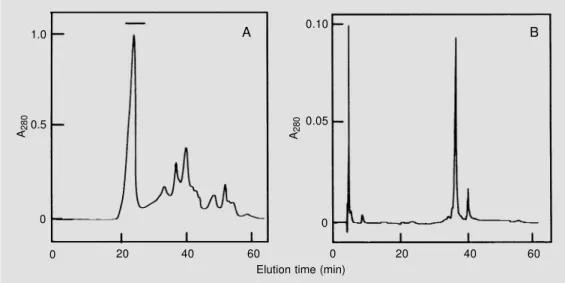
Chromatography of the albumin fraction on a Sephacryl S-100-HR column yielded a major broad protein peak eluting in the mo-lecular mass range of 20 to 5 kDa (Figure 1). The protein profile of every third tube was analyzed by SDS-PAGE (data not shown) and those tubes containing the 6.5-kDa pro-tein at high concentrations were pooled, freeze-dried and applied to a Superdex Pep-tide column equilibrated and eluted with 50 mM ammonium bicarbonate. This yielded a major peak containing the 6.5-kDa protein eluted from 21 to 26 min (see horizontal bar) and several other peaks containing small peptides and other unidentified compounds (Figure 2A). Analysis by SDS-PAGE of the peak containing the 6.5-kDa protein
indi-cated the presence of minor contaminants (data not shown). Final purification was achieved by RP-HPLC (Figure 2B), which yielded a major 6.5-kDa protein band eluted at an acetonitrile concentration of 35.5% and a minor peak eluting at an acetonitrile
A280 1.0
0.5
0
20 40 60
0
0.10
0.05
0
20 40 60
0 A280
Elution time (min)
A B
Figure 2 - A, Chromatography of a fraction enriched with the 6.5-kDa protein on a Superdex Pep-tide HR 10/30 column. Proteins (1.0 mg in running buffer) were eluted with 50 mM ammonium bicarbonate at a flow rate of 0.5 ml/min and samples of 1.0 ml were collected. The fractions in-dicated by the horizontal bar were pooled and subjected to the next purification step. B, RP-HPLC of the fraction enriched with the 6.5-kDa protein on a µ-Bondapak C-18 column. Proteins (200 µg) were eluted with a lin-ear gradient of 0-60% acetoni-trile in 0.1% trifluoracetic acid run for 60 min.
kDa 1 2
16.95
14.44
10.60
8.16
6.21
3.46
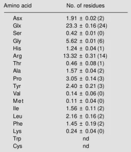
Table 1 - Amino acid composition of the 6.5-kDa protein isolated from the seeds of the prickly-pear, as determined by Pico Tag technology.
Composition is reported as number of residues per mol of protein based on a molecular mass of 6500. The values for cystein and tryptophan were not determined (nd). Number within parentheses represent the assigned integral values.
Amino acid No. of residues
Asx 1.91 ± 0.02 (2)
Glx 23.3 ± 0.16 (24)
Ser 0.42 ± 0.01 (0)
Gly 5.62 ± 0.01 (6)
His 1.24 ± 0.04 (1)
Arg 13.32 ± 0.31 (14)
Thr 0.46 ± 0.08 (1)
Ala 1.57 ± 0.04 (2)
Pro 3.05 ± 0.14 (3)
Tyr 2.40 ± 0.21 (3)
Val 0.14 ± 0.06 (0)
M e t 0.11 ± 0.04 (0)
Ile 1.56 ± 0.11 (2)
Leu 2.16 ± 0.16 (2)
Phe 1.45 ± 0.19 (2)
Lys 0.24 ± 0.04 (0)
Trp nd
Cys nd
concentration of 39.3%. The purity of the 6.5-kDa protein was demonstrated by SDS-PAGE (Figure 3) and by N-terminal analysis that yielded only aspartic acid as the N-terminal amino acid. This protein corresponds to 18% of the total salt soluble protein of the seed.
The protein obtained by the RP-HPLC step was used for amino acid analysis and protein sequencing. The amino acid compo-sition of the protein (Table 1) showed that it is devoid of Ser, Val, Met and Lys and has a high percentage of Arg (23%) and of Glx (40%). The behavior of the protein in non-denaturing gels and in ion exchange media indicates that this protein is very basic (data not shown) and therefore most of the amino
acids identified as Glx are probably Gln. Seed storage proteins are often rich in the amide amino acids glutamine and asparagine, but may also be rich in arginine (8). All three of these amino acids have a high nitrogen-to-carbon ratio, making them particularly suited for nitrogen storage (9). An amino acid com-position search on the ExPASy world wide web server (http://www.expasy.ch/ch2d/ aacompi.html) showed that the amino acid composition of the 6.5-kDa protein has a high degree of similarity with the 2S seed storage protein from Helianthus annus and with the 2S albumin from the seeds of Rici-nus communis. We obtained the N-terminal sequence of the protein by the DABITC/ PITC double-coupling method and found the sequence Asp-Pro-Tyr-Trp-Glu-Gln-Arg and found no matching sequences in the above cited proteins.
Only recently have the 2S albumins be-come recognized as a major group of seed storage proteins. They are known to occur in species as diverse as oil-seed rape (Cruci-ferae), sunflower (Compositae), Brazil nut (Lecythidaceae) and lupin (Leguminosae) (10) and were initially defined as a group on the basis of their sedimentation coefficients (S20w) of 2 (11). The data shown here
References
1. Braga R (1976). Plantas do Nordeste, Especialmente do Ceará. Coleção Mossoroense. Vol. XLII. Escola Superior de Agricultura de Mossoró, Rio Grande do Norte, Brazil.
2. Vietmeyer N (1986). Lesser-known plants of potential use in agriculture and forestry. Science, 232: 1379-1384.
3. Bradford M (1976). A rapid and sensitive method for the quantitation of microgram quantities of proteins utilizing the
prin-ciple of protein-dye binding. Analytical
Biochemistry, 72: 248-254.
4. Bidlingmeyer BA, Cohen SA & Tarvin TL (1984). Rapid analysis of amino acids
us-ing pre-column derivatization. Journal of
Chromatography, 33: 93-104.
5. Schagger H & Jagow G (1987). Tricine-sodium dodecyl sulfate-polyacrylamide gel electrophoresis for the separation of proteins in the range of 1 to 100 kDa. Analytical Biochemistry, 166: 368-379. 6. Chang JY, Brauer D & Wittmann-Liebold
B (1978). Micro-sequence analysis of pep-tides and proteins using 4-NN-dimethyla-minoazobenzene 4'-isothiacyanate/phen-ylisothiocyanate double coupling method. FEBS Letters, 93: 155-161.
7. Campos FAP & Richardson M (1984). The complete amino acid sequence of the α-amylase inhibitor I-2 from seeds of ragi
(Indian finger millet, Eleusine coracana
Gaertn.). FEBS Letters, 167: 221-225.
8. Shotwell MA & Larkins BA (1989). The
biochemistry and molecular biology of seed storage proteins. In: Marcus A (Edi-tor), The Biochemistry of Plants. Vol. 15. Academic Press, New York, 297-345. 9. King JE & Gifford DJ (1997). Amino acid
utilization in seeds of loblolly pine during germination and early seedling growth. Plant Physiology, 113: 1125-1135. 10. Shewry PR, Napier JA & Tatham AS
(1995). Seed storage proteins: structures and biosynthesis. Plant Cell, 7: 945-956. 11. Youle RJ & Huang AHC (1981).