Photos in Pediatrics
Pediatric-onset necrobiosis lipoidica
Q1 JoãoNascimento1andSusanaMachado2Departments of1Pediatrics, and2Dermatology, Centro Hos
Q2 pitalar Porto, Portugal
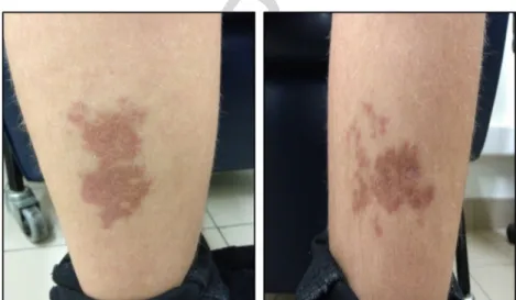
A 14-year-old boy with a 9 year history of type 1 diabetes mellitus (DM; HbA1c, 8.0–11.5%) presented with symmetric pre-tibial red-brown plaques with well-defined borders (Fig.
F1 1)
that had lasted for 6 months. He denied itch, pain or a previous traumatic event, and noted that the skin lesions had started as small papules (<0.5 cm).
Skin biopsy was performed and histopathology indicated extensive necrotic areas of collagen degeneration in the dermis with palisaded
granulomas composed of histiocytes, lymphocytes and fibroblasts (Fig.
F2 2). Thickening of the blood vessel walls was also present. These findings were consistent with necrobiosis lipoidica (NL), a disorder of collagen degeneration strongly associated with DM but also described in non-diabetic patients.1 Pediatric onset is rare.1,2
The etiology of NL remains uncertain, but it seems to involve metabolic and inflammatory changes such as an increase in tumor necrosis factor-α skin level, deposition of glycoproteins in blood vessel walls, antibody-mediated vasculitis and increased collagen cross-linking.1,3
Lesions are usually asymptomatic and the main complaint is the unpleasant cosmetic appearance.
The patient was treated with 0.1% topical tacrolimus with partial regression of the erythema and size of the lesions in 6 months. Treatment for NL is not very effective, partially because the exact etiology remains unknown.1,4Glycemic control is imptant, and topical corticosteroids should be used with caution in or-der to avoid cumulative atrophic effect.
Fig. 1 Pre-tibial red-brown plaque (60 × 25 mm) with indurated borders and atrophic centers.
Fig. 2 Areas of collagen degeneration with palisaded granulomas involving the subcutaneous tissue and dermis.
Correspondence: João Nascimento, MD, Department of Pediatrics, Centro Hospitalar Porto, Rua da Igreja n° 93, Lamaçães Braga 4710-085, Portugal. Email: nascimentojoao10744@gmail.com
Received 15 February 2015; revised 15 August 2015; accepted 29 September 2015.
© 2015 Japan Pediatric Society
Pediatrics International (2015) ••, ••–•• doi: 10.1111/ped.12872 Jour
na l C od e A rti cl e ID Di sp at ch : 23.12.15 C E : P E D 1 2 8 7 2 No . o f P ag es : 2 ME : 1 2 3 4 5 6 7 8 9 10 11 12 13 14 15 16 17 18 19 20 21 22 23 24 25 26 27 28 29 30 31 32 33 34 35 36 37 38 39 40 41 42 43 44 45 46 47 48 49 50 51 52 53 54 55 56 57 58 59 60 61 62 63 64 65 66 67 68 69 70 71 72 73 74 75 76 77 78 79 80 81 82 83 84 85 86 87 88 89 90 91 92 93 94 95 96 97 98 99 100 101 102 103 104 105 106
The main complication of the disease is ulceration that could occur after trauma.
Follow up is advised given that squamous cell carcinoma has been reported in some patients.5
References
1 Reid SD, Ladizinski B, Lee K et al. Update on necrobiosis lipoidica: A review of etiology, diagnosis and treatment options. J. Am. Acad. Dermatol. 2013; 69: 783–91.
2 Erfurt-Berge C, Seitz AT, Rehse C et al. Update on clinical and lab-oratory features in necrobiosis lipoidica: A retrospective multicentre study of 52 patients. Eur. J. Dermatol. 2012; 22: 770–75. 3 Mistry N, Chih-Ho Hong H, Crawford RI. Pretibial angioplasia: A
novel entity encompassing the clinical features of necrobiosis lipoidica and the histopathology of venous insufficiency. J. Cutan. Med. Surg. 2011; 15: 15–20.
4 Clayton TH, Harrison PV. Successful treatment of chronic ulcerated necrobiosis lipoidica with 0.1% tacrolimus ointment. Br. J. Dermatol. 2005; 152: 581–2.
5 Lim C, Tschuchnigg M, Lim J. Squamous cell carcinoma arising in an area of long-standing necrobiosis lipoidica. J. Cutan. Pathol. 2006; 33: 581–3.
2 J Nascimento and S Machado
© 2015 Japan Pediatric Society
54 55 56 57 58 59 60 61 62 63 64 65 66 67 68 69 70 71 72 73 74 75 76 77 78 79 80 81 82 83 84 85 86 87 88 89 90 91 92 93 94 95 96 97 98 99 100 101 102 103 104 105 106 1 2 3 4 5 6 7 8 9 10 11 12 13 14 15 16 17 18 19 20 21 22 23 24 25 26 27 28 29 30 31 32 33 34 35 36 37 38 39 40 41 42 43 44 45 46 47 48 49 50 51 52 53
Author Query Form
Journal: Pediatrics International
Article: ped_12872
Dear Author,
During the copyediting of your paper, the following queries arose. Please respond to these by annotating your proofs with the nec-essary changes/additions.
• If you intend to annotate your proof electronically, please refer to the E-annotation guidelines.
• If you intend to annotate your proof by means of hard-copy mark-up, please use the standard proofing marks. If manually writing corrections on your proof and returning it by fax, do not write too close to the edge of the paper. Please remember that illegible mark-ups may delay publication.
Whether you opt for hard-copy or electronic annotation of your proofs, we recommend that you provide additional clarification of answers to queries by entering your answers on the query sheet, in addition to the text mark-up.
Query No. Query Remark
Q1 AUTHOR: Please confirm that given names (red) and surnames/family names (green) have been identified correctly.
USING e-ANNOTATION TOOLS FOR ELECTRONIC PROOF CORRECTION
Required software to e-Annotate PDFs: Adobe Acrobat Professional or Adobe Reader (version 7.0 or
above). (Note that this document uses screenshots from Adobe Reader X)
The latest version of Acrobat Reader can be downloaded for free at:
http://get.adobe.com/uk/reader/
Once you have Acrobat Reader open on your computer, click on the Comment tab at the right of the toolbar:
1. Replace (Ins) Tool – for replacing text.
Strikes a line through text and opens up a text box where replacement text can be entered.
How to use it
Highlight a word or sentence.
Click on the Replace (Ins) icon in the Annotations section.
Type the replacement text into the blue box that appears.
This will open up a panel down the right side of the document. The majority of tools you will use for annotating your proof will be in the Annotations section, pictured opposite. We’ve picked out some of these tools below:
2. Strikethrough (Del) Tool – for deleting text.
Strikes a red line through text that is to be deleted.
How to use it
Highlight a word or sentence.
Click on the Strikethrough (Del) icon in the Annotations section.
3. Add note to text Tool – for highlighting a section to be changed to bold or italic.
Highlights text in yellow and opens up a text box where comments can be entered.
How to use it
Highlight the relevant section of text. Click on the Add note to text icon in the
Annotations section.
Type instruction on what should be changed regarding the text into the yellow box that appears.
4. Add sticky note Tool – for making notes at specific points in the text.
Marks a point in the proof where a comment needs to be highlighted.
How to use it
Click on the Add sticky note icon in the Annotations section.
Click at the point in the proof where the comment should be inserted.
Type the comment into the yellow box that appears.
USING e-ANNOTATION TOOLS FOR ELECTRONIC PROOF CORRECTION
For further information on how to annotate proofs, click on the Help menu to reveal a list of further options: 5. Attach File Tool – for inserting large amounts of
text or replacement figures.
Inserts an icon linking to the attached file in the appropriate pace in the text.
How to use it
Click on the Attach File icon in the Annotations section.
Click on the proof to where you’d like the attached file to be linked.
Select the file to be attached from your computer or network.
Select the colour and type of icon that will appear in the proof. Click OK.
6. Add stamp Tool – for approving a proof if no corrections are required.
Inserts a selected stamp onto an appropriate place in the proof.
How to use it
Click on the Add stamp icon in the Annotations section.
Select the stamp you want to use. (The Approved
stamp is usually available directly in the menu that appears).
Click on the proof where you’d like the stamp to appear. (Where a proof is to be approved as it is, this would normally be on the first page).
7. Drawing Markups Tools – for drawing shapes, lines and freeform annotations on proofs and commenting on these marks.
Allows shapes, lines and freeform annotations to be drawn on proofs and for comment to be made on these marks..
How to use it
Click on one of the shapes in the Drawing Markups section.
Click on the proof at the relevant point and draw the selected shape with the cursor. To add a comment to the drawn shape,
move the cursor over the shape until an arrowhead appears.
Double click on the shape and type any text in the red box that appears.