Vo l. 8(17), p p . 1789-1792, 23 April, 2014 DOI: 10.5897/ AJMR2014.6687
Artic le Numb e r: 0265F2A44171 ISSN 1996-0808
Co p yrig ht © 2014
Autho r(s) re ta in the c o p yrig ht o f this a rtic le http :/ / www.a c a d e mic jo urna ls.o rg / AJMR
African Journal of Microbiology Research
Full Length Research Paper
Recognition of leptospirosis in dengue-suspected
cases during dengue outbreak in Ceará State, Brazil
Augusto César Aragão OLIVEIRA
1, Raissa Matos FONTES
1, Claudênia Costa PRACIANO
1,
Fernanda Montenegro de Carvalho ARAÚJO
2, Luciano Pamplona de Góes CAVALCANTI
1,3,
Jeová Keny Baima COLARES
1,4, Margarida Maria de Lima POMPEU
1and
Danielle Malta LIMA
1,4*
1
Pathology Department, Federal University of Ceará, Fortaleza, CE, Brazil.
2
Laboratory of Public Health of the Ceará State (LACEN), Brazil.
3
Community Health Department, Federal University of Ceará, Fortaleza, CE, Brazil.
4
University of Fortaleza (UNIFOR), Fortaleza, Ceará State, Brazil.
Received 4 February, 2014; Accepted 14 April, 2014
Dengue fever frequently presents with non specific symptoms, making it difficult to be distinguished from other acute febrile illnesses, including leptospirosis. Given this, serum samples from 82 patients with clinical features of dengue-like illness were evaluated for dengue infection by IgM-enzyme-linked immunosorbent assay (ELISA) and reverse transcription-polymerase chain reaction (RT-PCR). Negative samples for dengue were tested for leptospirosis by IgM-ELISA and PCR. Dengue infection was detected in 35 patients. Six patients were positive to leptospirosis. This result shows that leptospirosis is underestimated in Ceará State, especially during epidemics of dengue with many reports, leading to an apparent low incidence of leptospirosis.
Key words: Dengue, leptospirosis, differential diagnosis, underrecognition, misdiagnosis.
INTRODUCTION
Dengue fever (DF) is an infectious disease caused by the dengue virus (DENV), of the family Flaviviridae and genus Flavivirus. Four serotypes can cause DF: DENV-1, DENV-2, DENV-3 and DENV-4 (Henchal and Putnak, 1990). DF is the most important arboviral disease in the world, and it causes more human illness and death than any of the known arthropod-borne viral infections. More
than 2.5 billion people, in at least 100 countries, are at risk of dengue infections accounting for nearly 100 million cases/year globally (World Health Organization, 2012). Large epidemics have occurred frequently in Brazil, particularly in the northeastern region, including Ceará State, where DF has been reported since 1986. Major outbreaks have occurred in 1994, 2008, 2011 and 2012
*Corresponding author. E-mail: [email protected]. Tel: (+55-85) 3477-3611.
1790 Afr. J. Microbiol. Res.
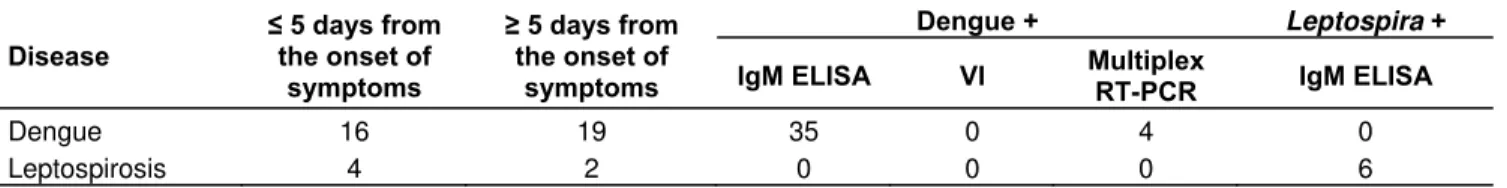
Table 1. Clinical and laboratorial features of patients tested for dengue and leptospirosis.
Disease
≤ 5 days from the onset of
symptoms
≥ 5 days from the onset of
symptoms
Dengue + Leptospira +
IgM ELISA VI Multiplex RT-PCR IgM ELISA
Dengue 16 19 35 0 4 0
Leptospirosis 4 2 0 0 0 6
Dengue +: patients tested positive for dengue; Leptospira +: patients tested positive for leptospirosis; ELISA: enzyme-linked immunosorbent assay; RT-PCR: reverse transcriptase-polymerase chain reaction; IgM: immununoglobulin M; VI: virus isolation.
(Ceará, 2014).
Inendemicareas,clinicalmanifestationsofDFareoften nonspecificandmaybeindistinguishablefromotherfebrile syndromessuchasinfluenza,oropouchefever,hantavirus infectionandleptospirosis(Flanneryetal.,2001; Levettet al.,2000;Manocketal.,2009).
Leptospirosis is a bacterial disease caused by
Leptospira, and its infection may produce a wide clinical spectrum ranging from asymptomatic infections or undifferentiated febrile syndrome (UFS) to multiple organ failure or death (Libraty et al., 2007). Most leptospiral infections are asymptomatic or result in a mild disease, which can lead to a misdiagnosis of DF instead of leptospirosis in regions where both diseases are endemic (Daher et al., 2010).
DF and leptospirosis share many clinical similarities as well as geographic distribution and potentially fatal complications but need different treatments (Conroy et al., 2014). Up to now, there is no specific treatment for DF, but appropriate intensive supportive therapy may reduce mortality to less than 1% in some severe cases. Therefore, accurate and timely diagnosis is very important for patient management in dengue infection (Mehta et al., 2013). The treatment of leptospirosis requires specific antibiotic therapy, such that the earlier the diagnosis is made, the better the potential outcome of treatment, which can be seriously damaged by a misdiagnosis (Ellis et al., 2008).
This study aimed to evaluate the occurrence of leptospirosis in dengue-suspected cases during the 2008 dengue outbreak in Ceará State, Brazil. The study was approved by the Ethics Committee of the Hospital São José de Doenças Infecciosas (no. 031/2009).
MATERIALS AND METHODS
Serum samples collected from 82 patients with a clinical history of acute fever consistent with dengue infection were provided by Laboratory of Public Health of Ceará State and analyzed retrospectively. As previously published by Lima et al. (2011), the samples were assayed for DENV by enzyme-linked immunosorbent assay (ELISA) (Diagnostics PanBio®, Brisbane, Australia), multiplex
reverse-transcription polymerase chain reaction (RT-PCR) (Lanciotti et al., 1992), and virus isolation (VI) using C6/36 cell monolayer cultures with the detection of infection by indirect
immunofluorescence assay. In this work, dengue-negative samples were tested for qualitative detection of IgM antibodies to Leptospira
in the serum using a Leptospira IgM ELISA commercial kit (Diagnostics PanBio®, Brisbane, Australia) and by PCR using LP1 and LP2 primers (Kee et al., 1994) and Ludwig commercial kit (Ludwig Biotechnology Ltd., Rio Grande do Sul, Brazil). All the PCR and RT-PCR fragments obtained were separated in 2% agarose gel, stained with Gel RedTM (Biotium Inc., Hayward, USA), and observed under ultraviolet light.
RESULTS AND DISCUSSION
Of the 82 patients, 35 (42.6%) were seropositive for dengue, of which all were ELISA-reactive and VI-negative (Table 1). Four patients (4.8%) were positive by RT-PCR and ELISA, of which 2 (2.4%) were infected with DENV-2 and 2 (2.4%) with DENV-3.
Serological tests for leptospirosis were performed in only 42 of 47 dengue-negative patients due to insufficient sample volume. Among the 42 patients analyzed, 6 (14.3%) were positive for leptospirosis by ELISA and PCR showing negative results. Thirty-six (85.7%) tested negative for both dengue and leptospirosis (Table 1).
The presence of leptospirosis in dengue-suspected cases in this study, and the rate of leptospirosis-infected patients (14.3%), corroborates several previous studies, which have demonstrated the occurrence of misdia-gnoses between these diseases due to the similarity of the initial clinical features and the increase in the number of cases during rainy seasons, and supports further documented ranges of between 14 to 24% as reported previously in studies from different locations (Sanders et al., 1999; Libraty et al., 2007; Souza et al., 2007). The cases of leptospirosis in this study were detected in two male patients (33.3%) and in four female patients (66.7%). The most common symptoms identified were fever, headache, arthralgia, myalgia, retro-orbital pain, and asthenia; nausea/vomiting were observed less frequently. In some cases, diarrhea and hemorrhagic manifestations, which are not specific characteristic of leptospirosis presentation, were present. One patient died possibly due to improper treatment or undetermined diagnosis.
substantially for dengue and leptospirosis. Early diagnosis also allows the establishment of surveillance and control measures. Effective treatment for leptospirosis must be performed early, because delays or failure in the establishment of proper therapeutic can result in patient death (Daher et al., 2010).
One study limitation was the fact that paired samples were not used and thus, the microagglutination test, considered the gold standard for the diagnosis of leptospirosis, was not performed. Therefore, it is impossible to state categorically that a positive IgM for
Leptospira necessarily implies an active infection, because anti-Leptospira IgM antibodies may remain detectable for several months after initial exposure. These detectable antibodies prevent differentiation between recent infections or a false-positive result (Cumberland et al., 2001). Besides, all samples were negative in PCR, probably due to freeze-thaw cycles of the samples, which may have affected the leptospiral genome detection. On the other hand, the adequate sensitivity of the IgM-ELISA used in this study to detect leptospirosis (PanBio®) decreases the likelihood of false-negative results (Libraty et al., 2007).
Forty patients (48.7%) had an undetermined diagnosis. Other UFS-causing pathogens, not studied here, may have affected these patients. In fact, it is known that patients with DF or leptospirosis can be misdiagnosed with other diseases, such as hantavirus infection, influenza and rubella; all of these could be responsible for the cases of nonspecific diagnosis of UFS (Libraty et al., 2007; Suharti et al., 2009).
This report is a strong indication that misdiagnosis is possible during dengue epidemics due to the lack of laboratory resources and similar non-specific symptoms, causing a false increase in the number of cases of DF and, consequently, an underreporting of other potentially fatal etiologies, such as leptospirosis.
The present study has shown that some cases of leptospirosis were not recognized in the 2008 dengue outbreak in Ceará. Therefore, it is reasonable to assert that leptospirosis can easily be confused with dengue, especially during outbreaks. Thus, differentiation and prompt diagnosis of leptospirosis during an outbreak is essential to enable the establishment of an effective antibiotic therapy and to avoid potentially fatal disease exacerbations due to improper treatment. Furthermore, it reinforces the need for implementation and expansion of surveillance investigations of febrile syndromes to improve the knowledge of the prevalence of leptospirosis, dengue, and other diseases, as they influence the population of Ceará State, Brazil.
Conflict of Interests
The author(s) have not declared any conflict of interests.
Oliveira et al. 1791
ACKNOWLEDGEMENTS
The authors thank Laboratory of Public Health of Ceará State, Laboratory of Parasitology, Department of Pathology and Legal Medicine of Federal University of Ceará, Center for Epidemiological Surveillance of the Secretary of Health of the Ceará State.
This work was financially supported by Conselho Nacional de Desenvolvimento Científico e Tecnológico (CNPq) and Coordenação de Aperfeiçoamento de Pessoal de Nível Superior (CAPES) Brazil. Danielle Malta Lima, was supported by Programa de Apoio a Projetos Institucionais com a Participação de Recém Doutores -PRODOC (CAPES).
REFERENCES
Ceará. State Government of Ceará (2014). Fortaleza (CE): Center for Epidemiological Surveillance, [cited 2014 april 06].
http://www.saude.ce.gov.br/index.php/boletins
Cumberland P, Everard CO, Wheeler JG, Levett PN (2001). Persistence of antileptospiral IgM, IgG and agglutinating antibodies in patients presenting with acute febrile illness in Barbados 1979-1989. Eur. J. Epidemiol. 17:601-608.
Conroy AL, Gélvez M, Hawkes M, Rajwans N, Liles WC, Villar-Centeno LA, Kain KC (2014). Host biomarkers distinguish dengue from leptospirosis in Colombia: a case-control study. BMC Infect. Dis. 14: 35.
Daher EF, Lima RS, Silva-Júnior GB, Silva EC, Karbage NN, Kataoka RS, Carvalho Júnior PC, Magalhães MM, Mota RM, Libório AB (2010). Clinical presentation of leptospirosis: a retrospective study of 201 patients in a metropolitan city of Brazil. Braz. J. Infect. Dis. 14(1):3-10.
Ellis T, Imrie A, Katz AR, Effler PV (2008). Underrecognition of leptospirosis during a dengue fever outbreak in Hawaii, 2001-2002. Vector Borne Zoonotic Dis. 8:541-547.
Flannery B, Pereira MM, Velloso LF, Carvalho CC, Codes LG, Orrico GS, Dourado CM, Riley LW, Reis MG, Ko AI (2001). Referral pattern of leptospirosis cases during a large urban epidemic of dengue. Am. J. Trop. Med. Hyg. 65:657-663.
Henchal EA, Putnak R (1990). The dengue viruses. Clin. Microbiol. Rev. 3:376-396.
Kee SH, Kim IS, Choi MS, Chang WH (1994). Detection of leptospiral DNA by PCR. J. Clin. Microbiol. 32(4): 1035-1039.
Lanciotti RS, Calisher CH, Gubler DJ, Chang GJ, Vorndam AV (1992). Rapid detection and typing of dengue viruses from clinical samples by using reverse transcriptase polymerase chain reaction. J Clin. Microbiol. 30(3):545-551.
Levett PN, Branch SL, Edwards CN (2000). Detection of dengue infection in patients investigated for leptospirosis in Barbados. Am. J. Trop. Med. Hyg. 62:112-114.
Libraty DH, Myint KS, Murray CK, Gibbons RV, Mammen MP, Endy TP, Li W, Vaughn DW, Nisalak A, Kalayanarooj S, Hospenthal DR, Green S, Rothman AL, Ennis FA (2007). A comparative study of leptospirosis and dengue in Thai children.PLoS Negl. Trop. Dis. 1(3): e111.
Lima DM, Sabino-Santos JG, Oliveira AC, Fontes RM, Colares JK, Araújo FM, Cavalcanti LP, da Fonseca BA, Figueiredo LT, Pompeu MM (2011). Hantavirus infection in suspected dengue cases from State of Ceará, Brazil. Rev. Soc. Bras. Med. Trop. 44(6):795-796.
1792 Afr. J. Microbiol. Res.
Manock SR, Jacobsen KH, de Bravo NB, Russell KL, Negrete M, Olson JG, Sanchez JL, Blair PJ, Smalligan RD, Quist BK, Espín JF, Espinoza WR, MacCormick F, Fleming LC, Kochel T (2009). Etiology of acute undifferentiated febrile illness in the Amazon basin of Ecuador. Am. J. Trop. Med. Hyg. 81(1):146-51.
Sanders EJ, Rigau-Pérez JG, Smits HL, Deseda CC, Vorndam VA, Aye T, Spiegel RA, Weyant RS, Bragg SL (1999). Increase of leptospirosis in dengue-negative after a hurricane in Puerto Rico in 1966. Am. J. Trop. Med. Hyg. 61:399-404.
Souza AI, Nogueira JM, Pereira MM (2007). Anti-Leptospira antibodies in patients in the State of Mato Grosso do Sul with clinical suspicion of dengue or viral hepatitis. Rev. Soc. Bras. Med. Trop. 40:431-435.
Suharti C, Van Gorp EC, Dolmans WM, Groen J, Hadisaputro S, Djokomoeljanto RJ, Osterhaus DME, Van der Meer JWM (2009). Hanta Virus infection during dengue virus infection outbreak in Indonesia. Acta Med. Indones. 41:75-80.