ABSTRACT
Objective
This study analyzed the influence of the irrigating solutions ultrasonic activation on the obturator cement penetration into lateral root canals.
Methods
Fifty maxillary molars (palatine root) were randomly assigned to 5 experimental groups (Group 1 (EDTA 17% + manual agitation for 5 minutes), Group 2 (EDTA 17% + ultrasonic activation for 15 seconds), Group 3 (NaOCl 1% + Ultrasonic activation for 15 seconds), Group 4 (EDTA 17% + ultrasonic activation for 15 seconds and NaOCl 1% + ultrasonic activation for 15 seconds) and Group 5 (negative control). The lateral root canals were made in the apical and middle third. The obturation occurred in a single session. The radiographic and microscopic analyzes were performed to evaluate the sealant cement penetration degree. Data were analyzed by the Mann-Whitney and Wilcoxon tests.
Results
Apical and middle third radiographic analysis showed that Groups 2 and 4 presented better penetration of the sealant cement. In the microscopic analysis, Group 4 presented superior results in relation to the other groups in both thirds. In the apical third, radiographic and microscopic analyzes showed significant differences in the comparisons between Group 4 and Groups 5 (p = 0.019) and 3 (p = 0.023) and between Group 5 and Groups 2 (p = 0.012), 3 (P = 0.038) and 4 (p = 0.019), respectively.
Conclusion
It was concluded that the ultrasonic activation of the NaOCl 1% + EDTA 17% irrigation solution provides greater penetration of the endodontic cement in lateral root canals.
Indexing terms: Dental cements. Root canal irrigants. Root canal therapy. Smear layer. Ultrasonics.
RESUMO
Objetivo
Este trabalho analisou a influência da ativação ultrassônica de soluções irrigadoras na penetração do cimento obturador em canais laterais.
Métodos
Cinquenta molares superiores (raiz palatina) foram distribuídos aleatoriamente em 5 grupos experimentais (n=10): Grupo 1 (EDTA 17% + agitação manual por 5 minutos), Grupo 2 (EDTA 17% + ativação ultrassônica por 15 segundos), Grupo 3 (NaOCl 1% + ativação ultrassônica por 15 segundos), Grupo 4 (EDTA 17% + ativação ultrassônica por 15 segundos e NaOCl 1% + ativação ultrassônica por 15 segundos) e Grupo 5 (controle negativo). Os canais laterais foram confeccionados nos terços médio e apical. A obturação ocorreu em sessão única e foram realizadas análises radiográficas e microscópicas para avaliar o grau de penetração do cimento obturador. Os dados foram analisados pelos testes de Mann-Whitney e Wilcoxon.
Influence of the ultrasonic activation of irrigating solutions on sealer
penetration into lateral root canals
influência da ativação ultrassônica de soluções irrigadoras na penetração de cimento obturador em
canais laterais
Thalles Henrique Macedo BARBOSA1
ORCID iD 0000-0002-0763-4378 Markelane Santana SILVA2
ORCID iD 0000-0002-2933-1431 Daylana Pacheco da SILVA2
ORCID iD 0000-0002-1690-6451 Antônio Carlos Mendes de MOURA3
ORCID iD 0000-0002-3576-5092 Maria Ângela Arêa Leão FERRAZ1
ORCID iD 0000-0001-5660-0222 Carlos Alberto Monteiro FALCÃO1
ORCID iD 0000-0001-7787-0280
1 Universidade Estadual do Piauí, Curso de Odontologia. Av. Nossa Sra. de Fatima, s.n., Fátima, 64202-020, Parnaíba, PI, Brasil. Correspondência
para / Correspondence to: THM BARBOSA. E-mail: < [email protected]
2 Universidade Federal do Piauí, Faculdade de Odontologia. Teresina, PI, Brasil.
3 Grupo Pesquisa Piauí Ltda., Setor de Criação, Coordenação Científica. Teresina, PI< Brasil.
Como citar este artigo / How to cite this article
Barbosa THM, Silva MS, Silva DP, Moura ACM, Ferraz MAAL, Falcão CAM. Influence of the ultrasonic activation of irrigating solutions on sealer
temperature until the experiment time. In order to facilitate the root canals instrumentation, the dental crowns were sectioned with double-faced diamond disc (KG Sorensen®,
Cotia, SP, Brazil) mounted in low rotation (INTRAmatic 181, Kavo, Joinville, SC, Brazil). After sectioning, the teeth were marked on the root face at 3mm and 6mm from the apex for perforation of the lateral canals, corresponding to the apical and middle third respectively.
The canals were made perpendicular to the long axis of the teeth using a 10-gauge Kerr (Dentsply®,
Petrópolis, RJ, Brazil) rotating instrument. In order to be able to adapt and fix it to a mandrel for low rotation, the cable was eliminated and at the other extremity, a bevel cut to form a chamfer. Its length was adjusted with cyanoacrylate, forming a trephine.
The canals were submitted to the biomechanical preparation with a Reciproc instrument R50 (VDW, Germany), driven by VDW Silver (VDW, Germany) with 1% sodium hypochlorite solution (Biodinâmica®, Ibiporã, PR,
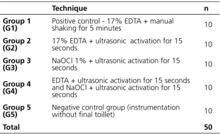
Brazil). After instrumentation and drying, the teeth were randomly distributed into 5 groups (n = 10). The irrigation solution used in the final toillet was activated with a manual instrument (G1) and with the ultrasonic insert E1 - Irrisonic (Helse ®, Santa Rosa de Viterbo, SP, Brazil), according to table 1.
Table 1. Distribution of the groups according to the final toilet. Parnaiba (PI), 2016.
Technique n
Group 1
(G1) Positive control - 17% EDTA + manual shaking for 5 minutes 10
Group 2
(G2) 17% EDTA + ultrasonic activation for 15 seconds 10
Group 3
(G3) NaOCl 1% + ultrasonic activation for 15 seconds 10
Group 4 (G4)
EDTA + ultrasonic activation for 15 seconds and NaOCl + ultrasonic activation for 15
seconds 10
Group 5
(G5) Negative control group (instrumentation without final toillet) 10
Total 50
INTRODUCTION
The smear layer produced during instrumentation modifies the dentin permeability and interferes with the seal of the obturator material [1,2]. The use of auxiliary chemical substances in the final toillet allows the increase of permeability and facilitates the intratubular endodontic cements penetration, which promotes a micromechanical retention of the filling to the dentinal wall [3].
Compounds used in irrigation have been chemically modified and mechanical devices seek to improve the penetration and effectiveness of irrigation [4]. Sequential use of organic solvent (sodium hypochlorite - NaOCl) followed by inorganic solvent (ethylenediaminetetraacetic acid - EDTA) leads to cleaner canals [4,5].
Ultrasound is used as an auxiliary tool to clean the root canals through the activation of irrigation solutions to maximize its effect [6]. Passive Ultrasonic Irrigation (PUI) is based on the use of an ultrasound-activated instrument in the root canal filled with irrigant allowing the solution transport to the apical region. Thus, less debris and smear layer are observed in this region [7,8].
The aim of this work was to evaluate the influence of the ultrasonic activation of the irrigating solutions EDTA and NaOCl in the penetration of the obturator cement in lateral channels.
METHODS
This study was approved by the Research Ethics Committee of the Universidade Estadual do Piauí - UESPI, with protocol number 768.450. We selected 50 healthy extracted upper molars with completely formed roots (selected palatine root). For the sample selection, the teeth were examined with a magnifying lens (8X) that allowed observing the absence of cracks, imperfections or apical resorptions.
The specimens were autoclaved (Cristófoli, São Paulo, SP, Brazil) and stored in saline solution at room
Resultados
A análise radiográfica do terço apical e médio mostrou que os Grupos 2 e 4 apresentaram melhor penetração do cimento obturador. Já na análise microscópica, o Grupo 4 apresentou resultados superiores em relação aos demais grupos em ambos os terços. No terço apical, as análises radiográfica e microscópica exibiram diferenças significativas nas comparações entre o Grupo 4 com os Grupos 5 (p = 0,019) e 3 (p = 0,023) e entre o Grupo 5 com os Grupos 2 (p = 0,012), 3 (p = 0,038) e 4 (p = 0,019), respectivamente.
Conclusão
Conclui-se que a ativação ultrassônica da solução irrigadora NaOCl 1% + EDTA 17% proporciona maior penetração do cimento endodôntico em canais laterais.
After the final toillet and the conduits drying, the canals were sealed by the single cone technique with Sealer 26 (Dentsply®, Petrópolis, RJ, Brazil) with Lentullo
nº 4 drill bit (Dentsply®, Petrópolis, RJ, Brazil) and
guta-percha of the Reciproc R50 System. The teeth were sealed with photopolymerizable composite resin (Dentsply®,
Petrópolis, RJ, Brazil). After the obturation, the roots were kept 48 hours in a humid environment at 37ºC to wait for polymerization of the cement and then to be radiographed.
After 72 hours, the teeth were sectioned with diamond disk (KG Sorensen®, Cotia, SP, Brazil) in the two
transversal planes (middle and apical third), 1 mm from of the artificial channels. A wear of 1 mm thickness with a sanding disk (KG Sorensen®, Cotia, SP, Brazil), medium
grain size, was performed to visualize the degree of sealing cement penetration, analyzed by Operative Microscope (DF Vasconcellos®, Valença, Spain) with a 25-fold increase.
The results were grouped into three scores (Table 2).
Table 2. Distribution of results by degree of the sealant cement penetration. Parnaiba (PI), 2016.
Escore Penetration degree
0 Lack of lateral canal filling
1 Penetration of the cement in the proximal third of the artificial canal
2 Penetration of cement in the middle third of the artificial canal 3 Penetration of the cement to the final third (totally or partially) of the artificial canal
Descriptive analysis was performed, to evaluate the scores, and statistical analysis of the data. The Mann-Whitney test was used to examine the differences between the two groups regarding the degree of lateral canal filling in two-thirds from the radiographic and microscopic analyzes. The Wilcoxon test was used to examine the differences between the degree of lateral canal filling in two thirds within the same group in both analyzes. A p-value <0.05 was used as a criteria for the statistically significant differences. Because it was a small sample (n <30) the exact level of unilateral significance was used for the p value.
RESULTS
According to the radiographic analysis, the groups
activated in which EDTA (Group 2) and EDTA + NaOCl (Group 4) showed a higher number of teeth with a score of 3, both in the apical and middle third. However, group 4 had no specimen with a score of 0, standing out from the others. Group 3 presented a result equal to the negative control and lower than the other groups.
According to the microscopic analysis, group 4 had a higher number of teeth with a score of 3 in both thirds, as well as no specimen with a score of 0, standing out in comparison to the other groups.
According to the Mann-Whitney test, for radiographic and microscopic filling data in the middle third, none of the comparisons between groups showed a statistically significant difference. Data from the radiographic filling analysis in the apical third showed significant differences in the comparisons between group 4 with groups 5 (p=0.019) and 3 (p=0.023). In the data from the microscopic analysis of filling in the apical third, all the groups that used ultrasonic activation presented better results than the negative control. Group 5 showed a significant difference in comparisons with groups 2 (p=0.012), 3 (p=0.038) and 4 (p=0.019) (Table 3).
Table 3. Apex filling up to 3 mm comparison from the radiographic analysis between Groups 5-4 and 3-4 and microscopic analysis between Groups 5-2, 5-3 and 5-4 by the Mann-Whitney Test (U). Parnaiba (PI), 2016.
Radiographic analysis
U Z(U) p (unilateral) p (bilateral)
Groups 5 – 4 22,50 2,079 0,019 * 0,038
Groups 3 – 4 23,500 2,003 0,023 * 0,045
Microscopic analysis
U Z(U) p (unilateral) p (bilateral)
Groups 5 – 2 20,00 2,268 0,012 * 0,023
Groups 5 – 3 26,500 1,776 0,038 * 0,076
Groups 5 – 4 22,500 2,079 0,019 * 0,059
Note: * Statistically significant difference.
Table 4. Filling of the middle and apical thirds comparison from the Group 1 radiographic analysis and the microscopic analysis of groups 2 and 5 by the Wilcoxon Test. Parnaiba (PI), 2016.
Radiographic analysis
Number of
pairs Z p (unilateral) p (bilateral)
Group 1 6 1,887 0,030 *
-Microscopic analysis
Number of
pairs Z p (unilateral) p (bilateral)
Group 2 4 1,826 0,034 * 0,068
Group 5 6 2,201 0,014 * 0,028
Note: * Statistically significant difference
DISCUSSION
In this study, lateral canals were made with trephine obtained with K #10 file. Canals obtained from instruments with this diameter can be detected when sealed. Approximately 43% of lateral canals have approximate diameters of the K #10 [9].
When analyzing the filling of lateral canals, there was a trend towards greater canals filling in the middle third, results that may be related to the fact that, in the cervical and middle thirds, irrigation is facilitated by the diameter of the modeled canal. Fontana et al.3 (2009)
observed the tendency of cements to penetrate more in to the cervical-located dentinal tubules, which presented a higher intratubular penetration index despite of the cements evaluated. The histochemical method used by Bonetti et al. [10] allowed the quantification of the dentin permeability promoted by irrigating solutions in which the cervical third presented the highest penetration of the dye.
The efficacy of PUI as an auxiliary method in the final toillet was evidenced with more artificial channels filled in score 3. Ultrasound was used to aid the stirring of the 1% NaOCl solution alone and alternated with 17% EDTA, the last one achieved better results. Rödig et al.8
(2010) also used PUI in the final irrigation with NaOCl and all the groups that had ultrasonic activation presented significant differences in relation to the negative control. Marques et al. [11] evaluated the removal of the smear layer in the apical third of teeth irrigated with EDTA at 17% with and without agitation, observing that both techniques were effective without significant difference. However, in the study by Hertel et al. [12] it has been shown that sufficient mechanical debridement combined
with NaOCl and PUI results in high success rates compared to EDTA and PUI.
Gründling et al. [13], when evaluating the effect of NaOCl PUI and EDTA on root canals of bovine teeth, concluded that it may help to clean the root canal, however, the main role in the elimination of bacteria is performed by the irrigant. In the study by Koçak et al. [14] PUI significantly increased the efficacy of smear layer removal irrespective of the irrigating solution. All irrigation regimens evaluated were significantly more effective in the coronal and middle thirds compared to the apical third.
Group 4 (EDTA and NaOCl + ultrasonic activation) presented the best results with significant differences in comparison to the control group. However, Carvalho et al. [15] evaluated the effect of 17% EDTA and 2.25% peracetic acid and 10% citric acid on the push-out bond strength of endodontic cements and the authors concluded that the use of different chelating agents did not influence the cements. Already Prado et al. [16], when evaluating the effect on the roughness of the dentin surface of EDTA, NaOCl and chlorhexidine (CHX) solutions in the final toillet, verified that the final irrigation with NaOCl after the use of EDTA leads to increased surface roughness, although, the use of EDTA alone or associated with CHX did not significantly alter the roughness values of this surface.
Bonetti et al. [10] showed that EDTA as a final irrigator associated with different concentrations of NaOCl did not promote changes in dentin permeability in the root canal, the success of endodontic treatment depends mainly on the mechanical preparation. Guerreiro-Tanomaru et al. [17] evaluated the efficacy of PUI in the elimination of Enterococcus faecalis from root canals and concluded that both PUI and conventional irrigation with NaOCl 1% contribute to disinfection but are unable to eliminate E. faecalis.
CONCLUSION
It was concluded that the ultrasonic activation of the NaOCl 1% + EDTA 17% irrigation solution provided greater penetration of the endodontic cement in lateral channels.
Collaborators
Helped and participated in the accom-plishment of all the experimental stages of the work. Also revising it critically for important intellectual contente. Participated in the review of the written part of the article. DP SILVA, was responsible for the bibliographical research and writing of the article. co-interpretation of data and contributions to conception and desing this paper. Revising it critically for important intellectual content. ACM MOURA, acquisition of data, co-interpretation of data and statistical treatment. MÂAL FERRAZ, is a endodontics teacher. She was coauthor
and adviser of the students in the development and preparation of the Course Completion Work. Participated in the orientation, execution of the experiment and the writing of the article. CAM FALCÃO, is a endodontics teacher. He was coauthor and adviser of the students in the development and preparation of the Course Completion Work. Also revising it critically for important intellectual contente. Revising it critically for important intellectual content. Resising and with final approval of the version to be published.
REFERENCES
1. Rathakrishnan M, Sukumaran VG, Subbiya A. To Evaluate the Efficacy of an Innovative Irrigant on Smear Layer Removal - SEM Analysis. J Clin Diagn Res. 2016; 10(4): 104-106. doi: 10.7860/ JCDR/2016/17200.7685
2. Souza RA, Silva SJA. Interferência da camada residual no selamento apical. Rev Bras Odontol. 2001; 58(1):16-19.
3. Fontana CE, Davini F, Takahashi C, Silveira CFM, Cunha RS, Bueno CES, et al. Avaliação em microscopia eletrônica de varredura da penetração intratubular de diferentes cimentos endodônticos. Rev Assoc Paul Cir Dent. 2009; 63(5):396-400. 4. Haapasalo M, Shen Y, Qian W, Gao Y. Irrigation in endodontics.
Dent Clin North Am. 2010; 54(2):291-312. doi: 10.1016/j. cden.2009.12.001
5. Zhang K, Kim YK, Cadenaro M, Bryan TE, Sidow SJ, Loushine RJ, et al. Effects of different exposure times and concentrations of sodium hypochlorite / ethylenediaminetetraacetic acid on the structural integrity of mineralized dentin. J Endod. 2010;36(1):105-9. doi: 10.1016/j.joen.2009.10.020
6. Susin L, Liu Y, Yoon JC, Parente JM, Loushine RJ, Ricucci D, Bryan T, Weller RN, Pashley DH, Tay FR. Canal and isthmus debridement efficacies of two irrigant agitation techniques in a closed system. Int Endod J. 2010; 43(12):1077-90. doi: 10.1111/j.1365-2591.2010.01778.x
7. Cohenca N, Silva LA, Silva RA, Nelson-Filho P, Heilborn C, Watanabe E, et al. Microbiological evaluation of different irrigation protocols on root canal disinfection in teeth with apical periodontitis: an in vivo study. Braz Dent J. 2013; 24:467-473. doi: 10.1590/0103-6440201302179
8. Rödig T, Sedghi M, Konietschke F, Lange K, Ziebolz D, Hulsmann M. Efficacy of syringe irrigation, RinsEndo and passive ultrasonic irrigation in removing debris from irregularities in root canals with different apical sizes. Int Endod J. 2010;43:581-589. doi: 10.1111/j.1365-2591.2010.01721.x
9. Goldberg F, Artaza LP, De Silvio AC. Effectiveness of different obturation techniques in the filling of simulated lateral canals. J Endod. 2001;27(5):362-364.
10. Bonetti MM, Moura CCG, Oliveira MAVC, Biffi JCG. Histochemical evaluation of dentin permeability after using EDTA as auxiliar irrigation of the root canal. Biosci J. 2013;29(1): 231-236.
11. Marques AAF, Garcia LFR, Frota MF, Simões RA, Consani S. Avaliação ultraestrutural da remoção da smear layer em canais radiculares utilizando EDTA 17% com ou sem agitação. Rev Clín Pesq Odontol. 2008; 4(2):71-75
12. Hertel M, Sommer K, Kostka E, Imiolczyk SM, Ballout H, Preissner S. Outcomes of endodontic therapy comparing conventional sodium hypochlorite irrigation with passive ultrasonic irrigation using sodium hypochlorite and ethylenediaminetetraacetate. A retrospective analysis. Open Dent J. 2016;12(10):375-81. doi: 10.2174/1874210616021001375
13. Gründling GL, Zechin JG, Jardim WM, de Oliveira SD, de Figueiredo JA. Effect of ultrasonics on Enterococcus faecalis biofilm in a bovine tooth model. J Endod. 2011; 37:1128-1133. doi: 10.1016/j.joen.2011.05.006
14. Koçak S, Bağcı N, Çiçek E, Türker SA, Can Sağlam B, Koçak MM. Influence of passive ultrasonic irrigation on the efficiency of various irrigation solutions in removing smear layer: a scanning electron microscope study. Microsc Res Tech. 2017 Jan 23. doi: 10.1002/jemt.22829
15. Carvalho NK, Prado MC, Senna PM, Neves AA, Souza EM, Fidel SR, et al. Do smear-layer removal agents affect the push-out bond strength of calcium-silicate based endodontic sealers? Int Endod J. 2016. doi: 10.1111/iej.12662
16. Prado M, Assis DF, Simão RA. Efeito de diferentes soluções utilizadas como irrigante final na superfície dentinária: análise de rugosidade. Rev Odontol UNESP. 2014;43(1):36-40. doi: 10.1590/S1807-25772014000100006
17. Guerreiro-Tanomaru JM, Chávez-Andrade GM, de Faria-Júnior NB, Watanabe E, Tanomaru-Filho M. Effect of passive ultrasonic irrigation on enterococcus faecalis from root canals: an ex vivo study. Braz Dent J. 2015;26(4):342-6. doi: 10.1590/0103-6440201300022