HLA IN BRAZILIAN ASHKENAZIC JEWS WITH CHRONIC DERMATOPHYTOSIS CAUSED BY
TRICHOPHYTON RUBRUM
Aya Sadahiro1*; José Roberto Feresin Moraes2; Maria Elisa Hue Moraes2; Matilde Romero2; Nancy Alves de Lima Gouvea3; Celso José Gouvea3; Mauricio Morishi Ogusku4;
Iphis Campbell5; Clarisse Zaitz6
1Departamento de Parasitologia, Universidade Federal do Amazonas, Manaus, AM, Brasil; 2Laboratório de Imunogenética do
Instituto Nacional do Câncer, Rio de Janeiro, RJ, Brasil; 3Laboratório de Imunogenética, São Paulo, Brasil; 4Instituto Nacional
de Pesquisas da Amazônia, Manaus, AM, Brasil; 5Divisão de Dermatologia, Universidade de Brasília, Brasília, DF, Brasil; 6Departamento de Clínica Médica, Faculdade de Ciências Médicas da Santa Casa de São Paulo, São Paulo, SP, Brasil
Submitted: October 10, 2001; Returned to authors: May 20, 2003; Approved: May 20, 2004.
ABSTRACT
The frequency of HLA (Human Leucocyte Antigens) was analyzed in 25 non-consanguineous Brazilian Ashkenazic Jews, resident in the city of São Paulo, Brazil, suffering from chronic dermatophytosis caused by T. rubrum, and in 25 non-infected individuals belonging to the same ethnic group. Statistically significant values (p<0.05) were observed for HLA-B14 associated with resistance to chronic dermatophytosis and HLA-DQB1*06 (p=0.05) possibly related to susceptibility. These findings suggest that genes on the chromosome 6, in the region of the major histocompatibility complex, may influence the development of chronic dermatophytosis.
Key words: chronic dermatophytosis, Trichophyton rubrum, (HLA) human leucocyte antigens
INTRODUCTION
Dermatophytosis is a superficial skin infection and appendage caused by fungi of the genera Trichophyton, Microsporum and Epidermophyton.
The results of dermatophytosis prevalence studies in the population of different countries are of the most heterogeneous possible. However, in the majority of recent reports, Trichophyton rubrum and Trichophyton mentagrophytes are the more frequent etiologic agents (3,4,10,17,24,26).
According to the latest studies carried out in Brazil, the most isolated species in chronic infections of tineaungueum and pedis has been the T. rubrum (6,9,16,23). Although this disease is not serious in terms of mortality or physical sequelae, it has significant clinical consequences, mainly regarding aesthetic and chronic aspects of the infection, besides therapeutics difficulties (17). A considerable number of patients
fail to respond satisfactorily to instituted medical treatment, thus presenting remissions and relapses (15).
Several factors may be involved in the progression of the chronic form of the disease, from keratinase and mannans production by fungi, to the existence of alterations in the immunological response of the host.
Studies carried out with the purpose of verifying the susceptibility or resistance to chronic infection by T. rubrum are still very controversial. In humans, a relationship between the histocompatibility antigens, HLA (Human Leucocyte Antigens) and the incidence of the disease has been reported. HLA is a fundamental component of the immune system, playing an important role in the antigens presentation process to the lymphocytes T, resulting or not in an effective immunological response. HLA can be divided into: a) HLA-class I (A, B and C), virtually expressed in all cells with nucleus and involved in peptide presentation to lymphocytes TCD8+; b) HLA-class II
(DR, DQ and DP), expressed in antigens presenting cells (APC) and taking part in peptides presentation to the lymphocytes TCD4+; and c) HLA-class III, that codify important molecules
for the immune system, as some components of the Complement system and cytokine (21).
In the first study of HLA system and chronic dermatophytosis caused by T. rubrum, no association between this infection and histocompatibility antigens (22) was found. But later, high frequency of HLA-A26 and HLA-A33 was observed in patients with chronic dermatophytosis of the foot (1). Moreover, in individuals with onychomycosis, the higher frequency was DR52, and in the control group it was HLA-DR53, suggesting an important role of these antigens in the modulation of the immune response in chronic dermatophytosis for T. rubrum (27).
In this context, the present study analyzed the frequency of HLA in a group of Ashkenazic Jews with chronic dermatophytosis of the foot and nails by T. rubrum, directing the study towards a possible association of the disease to histocompatibility antigens.
MATERIALS AND METHODS
Patients
Fifty non consanguineous Brazilian Ashkenazic Jews, living in the city of São Paulo – SP, Brazil were selected, of which 25 were bearers of chronic dermatophytosis in the sole or nails, caused by T. rubrum, and 25 were healthy individuals (control group). The afflicted and the control groups were typed for HLA I class (A, B and C) and class II (DRB1* and DQB1*). This study was carried out on Brazilian Ashkenazic Jews of Central European ancestry, who were considered a homogeneous group (2,5) and presented a well-known HLA’s profile, important for the preliminary identification of some HLA associate to the disease.
Mycological Procedures
Feet skin scales and nail scrapings were examined directly after preparation with 10% potassium hydroxide (KOH) and dimethyl sulfoxide (DMSO) treatment for visualization of hyphal elements by optical microscopy. For culture, skin scales and nail scrapings were inoculated in a Sabouraud agar medium, supplemented with cycloheximide and chloramphenicol. The isolated fungi were identified by: a) slide culture (microslide) by the Riddell method (19), for observation of the structures characteristic of the fungus; b) Urease test by the Philpot method (18), which was negative; c) nutritional tests for dermatophytes, using 7 medium types (agar-trichophyton 1-7), which was positive for the base medium, containing only casaminic acid, free vitamins and medium containing tiamin; and d) pigmentation test in potato agar, T. rubrum produces a red-purple pigment (13).
HLA class I (A, B and C) Typing
Antigens were determined by a standard microlympho-cytotoxicity test. Well-characterized antisera obtained commercially were used for HLA typing (Biotest Diagnostics Corporation, U.S.A. and One Lambda, U.S.A.). In brief, 10 mL of whole blood was collected with heparin. The mononuclear cells of the peripheric blood (PBMC) were obtained by density gradient in ficoll-hypaque. The cells were adjusted to 2x10⁶ cells/
mL concentration and 1uL of this suspension was added in a Terasaki microplate of 96 wells, containing antibodies for different HLA types. After a 30 minute incubation, rabbit complement was added and incubated for an additional 60 minutes. Afterwards, eosin was added, and finally the reaction was stopped with buffered formaldehyde. After letting it stand for an hour, the reaction was visualized by phase contrast optical microscope. The reactions in the wells with 75% of non-viable cells were considered positive.
HLA class II, DRB1 and DQB1 Typing
The HLA class II typing was performed by DNA-based techniques (high resolution) of specific allele oligonucleotide hybridization using the Inno-LiPA kit (Innogenetics N. V., Belgium). In brief, the DNA was extracted from 5 mL of whole blood using the “Salting out” technique. The DNA was amplified by PCR with biotin-labeled primers for DRB1 and DQB1. The amplified products were hybridized with probes which were immobilized in parallel lines on a nitrocellulose strip, specifically for identification of the different alleles. After hybridization, streptavidin alkaline phosphatase conjugate was added followed by cromogenic substratum. The interpretation was performed by analyzing the hybridization profile with each probe, indicating the HLA genes detected.
Each individual expresses two antigens for locus, in other words, the “n” used for the calculations of frequency and percentage of these antigens was 50, indicating the number of alleles observed.
Statistical Analysis
The frequency of antigens and HLA genes was calculated using standard methods and analysis of association by a qui-square test with Yates correction, establishing a significance level in p<0.05.
RESULTS
Table 2. Frequency of HLA-B patients with chronic dermatophytosis and controls in Brazilian Ashkenazic Jews.
HLA-B Patient Control p
n % n %
B7 1 2.0 1 2.0 NS
B8 4 8.0 2 4.0 NS
B13 2 4.0 2 4.0 NS
B14 2 4.0 10 20.0 p<0.05
B18 0 0.0 3 6.0 NS
B27 1 2.0 2 4.0 NS
B35 15 30.0 9 18.0 NS
B37 1 2.0 1 2.0 NS
B38 6 12.0 7 14.0 NS
B41 1 2.0 2 4.0 NS
B44 2 4.0 2 4.0 NS
B50 0 0.0 2 4.0 NS
B51 2 4.0 0 0.0 NS
B52 3 6.0 0 0.0 NS
B35 0 0.0 1 2.0 NS
B57 1 2.0 1 2.0 NS
B60 1 2.0 2 4.0 NS
B61 0 0.0 1 2.0 NS
Blank 8 16.0 2 4.0 NS
Total 50 100.0 50 100.0
NS = Not Significant.
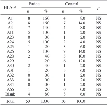
Table 1. Frequency of HLA-A patients with chronic dermatophytosis and controls in Brazilian Ashkenazic Jews.
HLA-A Patient Control p
n % n %
A1 8 16.0 4 8.0 NS
A2 8 16.0 7 14.0 NS
A3 7 14.0 4 8.0 NS
A11 5 10.0 1 2.0 NS
A23 0 0.0 1 2.0 NS
A24 5 10.0 2 4.0 NS
A25 1 2.0 3 6.0 NS
A26 5 10.0 7 14.0 NS
A28 2 4.0 5 10.0 NS
A29 1 2.0 6 12.0 NS
A30 2 4.0 1 2.0 NS
A31 1 2.0 3 6.0 NS
A32 0 0.0 1 2.0 NS
A33 0 0.0 1 2.0 NS
A34 0 0.0 1 2.0 NS
A66 1 2.0 0 0.0 NS
Blank 4 8.0 3 6.0 NS
Total 50 100.0 50 100.0
NS = Not Significant.
Table 3. Frequency of HLA-C patients with chronic dermatophytosis and controls in Brazilian Ashkenazic Jews.
HLA-C Patient Control p
n % n %
Cw1 0 0.0 2 4.0 NS
Cw2 1 2.0 1 2.0 NS
Cw3 0 0.0 2 4.0 NS
Cw4 13 26.0 9 18.0 NS
Cw5 0 0.0 1 2.0 NS
Cw6 4 8.0 8 16.0 NS
Cw7 9 18.0 10 20.0 NS
Blank 23 46.0 17 34.0 NS
Total 50 100.0 50 100.0
NS = Not Significant.
Analyzing the frequency of the antigens HLA-class II, no significant value for HLA-DR was observed (Table 4). However, when the percentages of HLA-DQB1*0602, 0603, 0607, 0609 and 06011 in the patients group were totaled, values proximate to the significant (p=0.05) were detected, possibly related to susceptibility (Table 5).
In the typings by serology (HLA class I), relatively high percentages of “blanks” were detected, mainly in the locus C, with 46% in the patients and 34% in the controls (Table 3). These blanks may represent a homozygosis or commercial available antisera that were not able to identify the expressed antigen. Blanks went less frequent in typing by molecular biology (HLA class II), and in these cases are probably only reflecting a homozygosis.
DISCUSSION
Chronic infections caused by dermatophytes usually involve anthropophilic fungi, such as Trichophyton rubrum. This fungus is capable of invading the corneous stratum and nails. The infections are typically asymptomatic, demonstrating the successful adaptation of this fungus to the human organism (7).
The cellular immunity has an important role for dermatophyte eradication in the skin. Individuals, who have this immunity impaired, frequently develop widespread and recurring fungi infections (8,20).
Immunogenetic mechanisms also seem to execute an important role in pathogenesis of the mycosis. Studies about associations of HLA and mycosis have been carried out, and some HLA class II alleles, DRB1*11 and DQB1*03 were significant in North American Caucasian patients with mycosis fungoides (12). Similar results were observed in another study with Ashkenazic Jews in which allele specifics of HLA-DRB1*1104 and DQB1*0301 were associated to susceptibility (11). Although association with HLA-class I have not been observed in these more recent studies with mycosis fungoides, in our investigation, the HLA-B14 presented a significant value for resistance to chronic dermatophytosis, in other words, the individuals who express this antigen may be more resistant to the disease. In this case, it is possible that expression of HLA-B14 together with peptides of T. rubrum can modulate positively the performance of the immune cells of the host against the fungus.
Regarding HLA class II, in our investigation no allele associate to disease was found. However, gathering all the different subtypes of DQB1*06 (DQB1*0602, 0603, 0607, 0609 and 06011), values close to the significant (p=0.05) for susceptibility was observed. Probably one or more common regions of the molecule of HLA-DQB1*06, but not the molecule in its totality, can bind itself to antigens of T. rubrum, modulating negatively the immune response to fungus by T cell, in other words, by presenting fungal peptides that do not induce a protective cellular response (Th1), limiting the action on the fungus, and consequently allowing it to remain in the human organism.
Table 4. Frequency of HLA-DRB1 patients with chronic dermatophytosis and controls in Brazilian Ashkenazic Jews.
HLA-DRB1 Patient Control p
n % n %
DRB1*0101 1 2.0 2 4.0 NS
DRB1*0102 3 6.0 5 10.0 NS
DRB1*0301 4 8.0 3 6.0 NS
DRB1*03011 1 2.0 0 0.0 NS
DRB1*0401 1 2.0 0 0.0 NS
DRB1*0402 5 10.0 7 14.0 NS
DRB1*0403 3 6.0 0 0.0 NS
DRB1*0404 0 0.0 1 2.0 NS
DRB1*0408 1 2.0 0 0.0 NS
DRB1*0701 5 10.0 12 24.0 NS
DRB1*08031 1 2.0 0 0.0 NS
DRB1*1001 0 0.0 2 4.0 NS
DRB1*1101 7 14.0 5 10.0 NS
DRB1*1104 3 6.0 2 4.0 NS
DRB1*1201 1 2.0 1 2.0 NS
DRB1*1301 4 8.0 1 2.0 NS
DRB1*1302 1 2.0 1 2.0 NS
DRB1*1303 2 4.0 0 0.0 NS
DRB1*1305 1 2.0 1 2.0 NS
DRB1*1306 1 2.0 0 0.0 NS
DRB1*1408 0 0.0 1 2.0 NS
DRB1*1501 1 2.0 1 2.0 NS
DRB1*1502 4 8.0 0 0.0 NS
Blank 0 0.0 5 10.0 NS
Total 50 100.0 50 100.0
NS = Not Significant.
Table 5. Frequency of HLA-DQB1* patients with chronic dermatophytosis and controls in Brazilian Ashkenazic Jews.
HLA-DQB1 Patient Control p
n % n %
DQB1*0201 9 18.0 13 26.0 NS
DQB1*0301 14 28.0 9 18.0 NS
DQB1*0302 6 12.0 7 14.0 NS
DQB1*03032 1 2.0 1 2.0 NS
DQB1*0304 1 2.0 0 0.0 NS
DQB1*0305 1 2.0 0 0.0 NS
DQB1*0501 5 10.0 8 16.0 NS
DQB1*0502 1 2.0 0 0.0 NS
DQB1*05031 0 0.0 1 2.0 NS
DQB1*0602 1 2.0 1 2.0 NS
DQB1*0603 6 12.0 2 4.0 NS
DQB1*0607 1 2.0 0 0.0 NS
DQB1*0609 1 2.0 1 2.0 NS
DQB1*06011 3 6.0 0 0.0 NS
Blank 0 0.0 7 14.0 NS
Total 50 100.0 50 100.0
NS = Not Significant.
Our results suggest that in this population, the system HLA certainly plays an important role in the immunological response of cells T for fungi antigens, with the HLA-B14 controlling the resistance for chronic dermatophytosis by T. rubrum and HLA-DQB1*06, possibly for susceptibility.
ACKNOWLEDGMENTS
We are grateful for the cooperation of Megumi Sadahiro (Fundação “Alfredo da Mata”, Manaus-AM), Laboratório de Micologia da Faculdade de Medicina da Santa Casa de São Paulo, Laboratório de Micologia Médica do Instituto de Medicina Tropical de São Paulo and Laboratório de Imunogenética da Fundação Pró-Sangue Hemocentro de São Paulo and George Nakamura for revision in English.
RESUMO
Antígenos Leucocitários Humanos (HLA) em Judeus Ashkenazitas Brasileiros portadores de
dermatofitose crônica causada por
Trichophyton rubrum
A freqüência dos HLA foi analisada em 25 Judeus Ashkenazitas, não consangüíneos, residentes em São Paulo, Brasil, com dermatofitose crônica causada por T. rubrum e em 25 indivíduos sadios, pertencentes ao mesmo grupo étnico dos pacientes. Observou-se valor estatisticamente significante (p<0,05) para HLA-B14 associado a resistência à dermatofitose crônica enquanto HLA-DQB1*06 (p=0,05) possivelmente relacionado a susceptibilidade. Estes achados indicam que o desenvolvimento da dermatofitose crônica pode ser influenciado por genes localizados no cromossomo 6, na região do complexo principal de histocompatibilidade.
Palavras-chave: dermatofitose crônica, Trichophyton rubrum, HLA (Antígeno Leucocitário Humano)
REFERENCES
1. Ahmed, A.R.; Schreiber, P.; Aiello, J.; Tiwari, J.L.; Terasaki, P.I. A preliminary report on the role of some immunologic factors in persistence of chronic tinea pedis. Clin. Exp. Derm., 10: 45-50, 1985. 2. Amar, A.; Kwon, O.J.; Motro, U.; Uit, C.S.; Bonne-Tamir, B.; Gabison, R.; Brautbar, C. Molecular analysis of HLA class II polymorphisms among different ethnic groups in Israel. Hum. Immunol., 60: 723-730, 1999.
3. Anane, S.; Aoun, K.; Zallagua, N.; Bouratbine, A. Onychomycosis in the Tunis area. Epidemiological and mycological data. Ann. Dermatol. Venerol., 128: 733-736, 2001.
4. Aste, N.; Pau, M.; Aste, N.; Biggio, P. Tinea pedis observed in Cagliari, Italy, between 1996 and 2000. Mycosis, 46: 38-41, 2003. 5. Bonne-Tamir, B.; Bodmer, J.G.; Bodmer, W.F.; Pickbourne, P.; Brautbar,
C.; Gazit, E.; Nevo, S.; Zamir, R. HLA polymorphism in Israel: an overall comparative analysis. Tissue Antigens, 11: 235-250, 1978.
6. Costa, M.; Passos, X.S.; Souza, L.K.H.; Miranda, A.T.B.; Lemos, J.A.; Oliveira-Junior, J.G.; Silva, M.R.R. Epidemiologia e etiologia das dermatofitoses em Goiânia, GO, Brasil. Rev. Soc. Bras. Med. Trop., 35: 19-22, 2002.
7. Dahl, M.V.; Grando, S.A. Chronic dermatophytosis: What is special about Trichophyton rubrum? Adv. Derm., 9: 97-109, 1994. 8. Faergemann, J.; Gisslen, H.; Dahlberg, E.; Westin, J.; Roupe, G.
Trichophyton rubrum abscesses in immunocompromised patients. A case report. Acta Derm. Venereol., 69: 244-247, 1989.
9. Fernandes, N.C.; Akiti, T.; Barreiros, M.G. Dermatophytoses in children: study of 137 cases. Rev. Inst. Med. trop. S. Paulo, 43: 83-85, 2001.
10. Gupta, A.K.; Chang, P.; Del Rosso, J.Q.; Adam, P.; Hofstader, S.L. Onychomycosis in children; prevalence and management. Pediatr. Dermatol., 15: 464-471, 1998.
11. Hodak, E.; Lapidoth, M.; Kohn, Y.; David, M.; Brautbar, C.; Kfir, B.; Narinski, R.; Safirman, C.; Maron, L.; Klein, T. Mycosis fungoides: HLA class II associations among Ashkenazi an non-Ashkenazi Jewish patients. Br. J. Dermatol., 145: 974-980, 2001.
12. Jackow, C.M.; McHam, J.B.; Friss, A.; Alvear, J.; Reveille, J.R.; Duvic, M. HLA-DR5 and DQB1*03 class II alleles are associated with cutaneous T-cell lymphoma. J. Invest. Dermatol., 107: 373-376, 1996. 13. Lacaz, C.S.; Porto, E.; Martins, J.E.C. Micologia Médica, fungos, Actinomicetos e Algas de interesse médico. Editora Sarvier, São Paulo, 1991, 695p.
14. McGregor, J.M.; Hamilton, A.J.; Hay, R.J. Posible mechanisms of immune modulation in chronic dermatophytoses: an in vitro study. Br. J. Dermatol., 127: 233-238, 1992.
15. Meehan, K.J.; Miller, C. The clinical challenge of onychomycosis. JAAPA. 14: 43-46, 2001.
16. Mezzari, A. Frequency of dermatophytes in the metropolitan area of Porto Alegre, RS, Brazil, Rev. Inst. Med. Trop. S. Paulo., 40: 71-76, 1998.
17. Perea, S.; Ramos, M.J.; Garau, M.; Gonzales, A.; Noriega, A.R.; Palacio, A. Prevalence and risk factors of tinea unguium and tinea pedis in the general population in Spain. J. Clin. Microbiol., 38: 3226-3230, 2000. 18. Philpot, C.M. The differentiation of Trichophyton mentagrophytes from Trichophyton rubrum by a simple urease test. Sabouraudia, 5: 189-193, 1967.
19. Riddell, R.W. Permanent stained mycological preparations obtained by slide culture. Mycologia, 42: 265-270, 1950.
20. Staughton, T. Skin manifestations in AIDS patients. Br. J. Clin. Pract., 71(Supp.): 109-113, 1990.
21. Stites, D.P.; Terr, A.J.; Parslow, T.G. Imunologia Médica, Editora Guanabara Koogan, Rio de Janeiro, 2000, 689p.
22. Svejgaard, E.; Jakobsen, B.; Svejgaard, A. HLA studies in chronic dermatophytosis caused by Trichophyton rubrum. Acta Dermatovener. 63: 254-255, 1983.
23. Vilani-Moreno, F.R.; Arruda, M.S.P.; Claro, S.G.; Marcos, E.V.C.; Ura, S. Dermatophytosis: Association between ABO Blood groups and reactivity to the trichophytin. Rev. Inst. Med. Trop. S. Paulo, 41: 285-289, 1999.
24. Weitzman, I.; Chin, N.X.; Kunjukunju, N.; Della-Latta, P.A Survey of dermatophytes isolated from human patients in the United States from 1993 to 1995. J. Am. Acad. Dermatol., 39: 255-261, 1998. 25. Woolfolk, J.A.; Sung, S.S.J.; Benjamin, D.C.; Lee, J.K.; Platts-Mills,
T.A.E. Distinct human T cell repertoires mediate immediate and delayed-type hypersensitivity to the Trichophyton antigen, Tri r 2. J. Immunol., 165: 4379-4387, 2000.
26. Zaias, N.; Glick, B.; Rebell, G. Diagnosing and treating onychomycosis. J. Fam. Pract., 42: 513-518, 1996.