405
Brazilian Journal of Microbiology (2012): 405-417ISSN 1517-8382
CLONING OF A NOVEL XYLANASE GENE FROM A NEWLY ISOLATED FUSARIUM SP. Q7-31 AND ITS EXPRESSION IN ESCHERICHIA COLI
Zhan-Ling Xie 1,2,*, Hai-Yan Gao 2,Qian Zhang 2,Huan Wang 2, Ying Liu 1
1
Gansu Agricultural University, Lanzhou 730000, China; 2Qinghai University, Xining 810016, China.
Submitted: March 31, 2011; Returned to authors for corrections: May 06, 2011; Approved: August 30, 2011.
ABSTRACT
A strain ofQ7-31 was isolated from Qinghai-Tibet Plateau and was identified as Fusarium sp.based on its
morphological characteristics and ITS rDNA gene sequence analysis. It has the highest capacity of
degrading cell wall activity compared with other 11 strains. To do research on its xylanase activity of
Fusarium sp. Q7-31 while the degrading the rice cell walls, the complete gene xyn8that encodes endo-1,
4--xylanase secreted by Fusarium sp. Q7-31 was cloned and sequenced. The coding region of the gene is
separated by two introns of 56bp and 55bp. It encodes 230 amino acid residues of a protein with a calculated
molecular weight of 25.7 kDa. The animo acids sequence of xyn8 gene has higher similarity with those of
family 11 of glycosyl hydrolases reported from other microorganisms. The nature peptide encodeing cDNA
was subcloned into pGEX5x-1 expression vector. The recombinant plasmid was expressed in Escherichia
coli BL21-CodonPlus (DE3)-RIL, and xylanase activity was measured. The expression fusion protein was
identified by SDS-PAGE and Western blotting, a new specific band of about 52kDa was identified when
induced by IPTG. Enzyme activity assay verified the recombinants proteins as a xylanase. A maxium
activity of 2.34U/ mg, the xylanase had optimal activity at pH 6.0 and temperature 40 .
Key words: xylanase; Gene cloning; Fusarium sp.;
INTRODUCTION
Fusarium sp., which is a cosmopolitan soil borne
filamentous fungus, is an anamorphic species. Some of them
can produce several enzymes that can act on pectin and
cellulose components of cell wall (5). Recently, scientists pay
more attention to them because some strains from Fusarium
oxysporum can ferment cellulose into bioethanol by one step
(22).
Endo- -1,4-xylanases(EC 3.2.1.8) are secreted by a number
of plant pathogenic fungus. Walton (24) described that those
xylanses may play a role during infection. Fusarium sp. also
secrets a number of xylanases, at least five xylanases, Xyl1,
Xyl2, Xyl3, Xyl4, Xyl5, xylanase of low molecular weight
have been found from Fusarium sp. (1,2,3,4,5,6,8,9,10,14,15),
from xyl1 to here, we reported a new xylanase gene from
Fusarium sp. Q7-31.
*Corresponding Author. Mailing address: 1 Gansu Agricultural University, Lanzhou 730000, China, 2 Qinghai University, Xining 810016, China.; E-mail:
MATERIALS AND METHODS
Reagents
PCR Purification Kit, DNA Gel Extraction Kit, RNA PCR
Kit (AMV) Ver. 3.0, IPTG, X-gal, Taq polymerase, restriction
enzymes were purchased from the TaKaRa Bio-technology
(Dalian, China) Co. Ltd., M-MLV reverse transcriptase was the
product of Promega (Madison, WI). The SMART™ PCR
cDNA Synthesis Kit was purchased from ClonTech (Palo Alto,
CA). Trizol reagent and culture media were obtained from
Invitrogen and Shanghai Sangon Co., Ltd., respectively. All
other chemicals were of analytical grade. GST Monoclonal
Antibody, Secondary Antibody and DAB Horseradish
Peroxidase Color Development Kit were purchased from
Sigma-Aldrich Co. Ltd.
Isolation of Fusarium sp. Q7-31
Fusarium sp. Q7-31 were isolated from fruiting bodies of
Morchella sp. and screened to pure isolates through a series of
subcultures on agar plates. The screening medium consisted of
0.6% peptone, 0.1% K2HPO4, 0.05% MgSO4•7H2O, and 1%
glucose (pH 7.0). The plates were incubated at 28 for 3 to 5
d. Colonies that grew well were then subcultured to fresh agar
plates. A loop-full of hyphae was removed from uniform
colonies and inoculated into a basic liquid medium (BLM) with
the same composition as the screening medium. These liquid
cultures were maintained at 28 on a rotary shaker with
100×g. For storage, the fungus strains were inoculated on
potato dextrose agar (PDA) plates and kept at 4 .
Screening Fusarium sp. Q7-31 as producing xylanase from 11 strains
Among the 11 strains screened, based on its crude enzyme
stability as well as its high activity. Loop-full of hyphae from
each strain was inoculated in 50 mL of BLM, with 1% glucose
as a carbon source. After culturing to saturation, about 50 L
was transferred to 5 mL of BLM containing 0.3% rice plant
powder and cultured at 28 . On Days 3 and 6, the amounts of
reducing sugars per milliliter of each culture medium were
measured according to the dinitrosalicylic acid (DNS) method
and compared among the 18 strains.
Strains and cultivations
The other 17 fungal strains (A, Morchella sp. M-1; B,
Fusarium sp. Q7-31; C, Pestalotiopsis sp.; D, Fusarium sp.
Q7-21; E, Trichoderma sp.; F, Penicillium sp.; G, Mucor sp.
U-1; H, Fusarium sp. F-2; I, Geotrichum sp.; J, Morchella sp.
M-2; K, Morchella sp. M-3) were stored in our Lab. The
screening medium consisted of 0.6% peptone, 0.1% K2HPO4,
0.05% MgSO4•7H2O, and 1% glucose (pH 7.0). The induced
medium, producing xylanases, was composed of xylan 1%,
NaNO3 0.8%, K2HPO4 0.1%, MgSO4·7H2O 0.05%, KCl
0.05%, FeSO4·7H2O 0.001%; and the pH was 4.5. Erlenmeyer
flasks (150 ml) containing 50ml medium were inoculated at 25
shaking for 4 days.
Fusarium sp. Q7-31 was used as a source of xylanase and
as a RNA and DNA donor strain. E. coli DH5 and E. coli
BL21-CodonPlus (DE3)-RIL (Novagen, USA) were used as
the hosts for cloning DNA sequencing and expression,
respectively. pMD18-T and pGEX5x-1 vectors for cloning of
PCR products were purchased from TaKaRa Biotechnology
Co.
DNS method to determine total reducing sugars
Contents of reducing sugars in the liquid cultures and
assay solutions were determined per the DNS method, with
minor modification from the procedures described (17,18,19).
The DNS reagent consisted of 0.5% DNS, 0.025% sodium
bisulfite, 0.5% NaOH, 0.1% phenols, and 2.5M KOH.
Mixtures of either 0.5 mL of fungus culture or enzyme reaction
solution plus 0.1 mL of reagent were incubated for 10 min in
boiling water. The total volume was brought to 1 mL by adding
0.4 mL of distilled water prior to reading Spectrophotometric
407
Zhan-Ling, X. et al. Xylanase gene from Fusarium sp.
Identification of strain Fusarium sp. Q7-31
The identification of the strain was based on standard
morphological characterization and nucleotide sequence
analysis of enzymatic amplified ITS rDNA, and the internal
transcribed spacer (ITS) region including 5.8 S rDNA. Mycelia
grown for 5 d on PDA plates were used for morphological
measurements, such as hyphal width, spore formation, and
spore shape. Genomic DNA was extracted from these mycelia
by the CTAB method (25). ITS DNA was amplified by PCR
with ITS5 forward primer 5’-GGAAGTAAAAGTCGTAACA
AGG-3’ and ITS4 reverse primer 5’-TCCTCCGCTTATTGAT
ATGC-3’, according to the procedure described by Van Burik
(23). Sequences of the PCR products (Accession Number
FJ646593) were analyzed through the NCBI BLAST database.
Preparation of total RNA, Genomic DNA of Fusarium sp.Q7-31
Mycelia of Fusarium sp.Q7-31, grown as previously
described in the induced medium were collected by
centrifugation at 3500×g for 10 min. To 100 mg wet weight of
pellet, 1ml Trizol was added in an adequate tube. The pellet
was homogenized for 3-5min, and then let it strand at room
temperature for 5 min. then 0.2 ml chloroform was added to the
tube and the tube was subjected to vigorous vortex for 15s. The
mixture was centrifuged at 12,000×g for 15min at 4✁ .The
supernatant was collected and kept at room temperature for 10
min. RNA was recovered by centrifuged at 12,000×g for 10min
at 4✁ and washed twice with 75% ethanol. The RNA was
kept at -80 in 75% ethanol for long-term storage. For the
reverse transcription, the RNA pellet was air dried and
dissolved in RNase-free water extracted using. RNA
concentration was checked using spectrophotometer. Then
isolate mRNA using Promega Large-Scale PolyA Tract
System. Concentrate the mRNA with 3M sodium acetate and
isopropanol. cDNA synthesis was carried out with cDNA
synthesis kit. Preparation of genomic DNA was carried out
according to CTAB method (21, 25).
Primers for PCR amplification
Two pairs of degenerated primer, F1 [GGCAACTTCGT
CGGTGGTAAG], R1 [CAGTAGCCACGATCTGGTAGT],
F2 [AACTTCGTCGGTGGTAAGGGT], R2 [AGTAACCCTC
AGTAGCCACG], were designed according to eight different
species organism of xylanases genes (Table2). Primer OT
(Original name, oligo dT-M13 primer M4) was provided by
TaKaRa RNA PCR Kit (AMV) Ver. 3.0. Primers RA
[TACCTGGGTTCCATCCCTT] and RB [TCCCTTACCACC
GACGAAGT] were used for Q7-31 5’RACE of xyn8 cDNA.
Primer OF [ATGGTCTCCTTCAGGTTCCTTCTC] and OR
[ATTAGATTGTAGATA CAAGTCGTT] were used for
amplification of the genomic DNA encoding xyn8.
Reverse transcription of cDNA
For cloning of partial cDNA by using degenerated
primers, the total RNA was reverse transcribed with AMV
reverse transcriptase by using primer OT. For cloning the 5✂
-end of the xyn8 cDNA, reverse transcription was performed
with the SMART PCR cDNA Synthesis Kit with M-MLV
reverse transcriptase. The RB primer was added to the reaction
solution after the end of reverse transcription and incubated at
42 °C for another 10min so that the derived cDNA moleculers
could have RB primer sequence at their 3✂ -end. All the reverse
transcription reactions were performed following instructions
provided by the manufacturers.
DNA manipulation and E. coli transformation
Digestion of DNA with restriction endonucleasesEcoR✄
and Sal ✄ , separation of fragments by agarose gel
electrophoresis, ligation of DNA fragments, transformation of
E.coli with plasmidic DNA and extraction of recombinant
DNA were all performed according to the standard method
(16). DNA fragments were recovered from agarose gels using
the DNA Gel Extraction Kit of TaKaRa.
DNA sequence analysis
Simple Vector (TaKaRa) and sequenced by Invitrogen
(Shanghai, China) Co., Ltd 16. The sequence was analyzed
using the software package DNAMAN 5.0 (Lynnon Bio-oft,
USA) and the homology was analyzed in GenBank with the
BLAST program. Properties of translated proteins based on the
primary structure analysis were predicted using
http://swiss-model.expasy.org/ on-line programs.
Nucleotide sequence accession number
The cDNA sequence of xyn8 gene has been deposited in
the GenBank database under accession numbers GQ249383
and ITS nucleotide sequence number is FJ646593.
xyn8 expression and purification
The cDNA fragment encoding the peptide of xylanase was
amplied with OF and OR. The DNA fragment was ligated into
pGEX5x-1 with restriction sites of Eco☎✝✆ and Sa✞✟✆ to
generate construct pGEX5x-1-xyn8. The expression construct
was used to transform E.coli BL21-CodonPlus(DE3)-RIL for
expression with the plasmid pGEX5x-1 for control. The
transformants were screened on Luria-Bertani (LB) broth
supplemented with 100 ✠ g/ml Ampicillin and cultured with
shaking at 37 °C overnight. Two microliters of seed culture
were transferred into fresh medium and cultured until OD600
reached 0.8, and IPTG (final concentration 0.4mM) was added
for induction. The bacterial cells were cultured for another 2h
before collection by centrifuge. SDS–PAGE was applied for
confirmation of the expressed product.
After growth in liquid medium, cells were harvested by
centrifugation, washed with cold 1xPBS buffer(137mM NaCL,
2.7mM KCL, 10mM Na2HPO4, 2mM KH2PO4; pH 6.0), and
suspended in 20ml of cold PBS buffer. Cells were
homogenized by ultrasonic treatment. Supernatant was
obtained by centrifugation at 12,000 ×g for 10min at 4 °C. The
supernatant was used as crude enzyme for xylanase assay.
SDS–PAGE and western blotting
SDS–PAGE was performed on a 12.5% running gel (12)
and resolved proteins were visualized by stainng with
Coomassie Brilliant Blue G 250.
Transfer the proteins from the gel to a PVDF membrane,
and incubate with GST Monoclonal Antibody (1:1000) for a
night at 4 . Decant and discard the antibody-buffer, rinse
twice with 20-30 ml of wash buffer to remove the majority of
unbound antibody. Dilute secondary antibody 1:1000 with
incubation buffer. Incubate for 1 hour at ambient temperature
with gentle shaking. Wash the membrane with 20-30 ml of
wash buffer for 10 minutes at ambient temperature with gentle
shaking. Detect the result by DAB Horseradish Peroxidase
Color Development Kit.
Determination of protein concentration
Protein was determined by the Bradford assay (2) using
bovine serum albumin as a standard.
Xylanase activity assays
Xylanase activity was measured using 1% (w/v)
birchwood xylan (Sigma, USA) as a substrate in 50mM
Na2HPO4–citric acid buffer, pH 6.0, at 40 °C for 10min. The
liberation of reducing sugars was estimated by the
dinitrosalicylic acid (DNS) method (13) using xylose as a
standard. Reducing sugars were determined by measuring the
absorption at 540 nm relative to a D-xylose standard. One unit
of enzyme activity was defined as the quantity of enzyme
required to liberate 1 mol of xylose equivalent per minute at
40 °C, and specific activity was defined as units per mg protein
(20). The results were means of duplicate determination on
triple independent measurements.
Temperature optimum and thermostability
The temperature optimum was measured by performing
the xylanase activity assay for 20min at temperatures ranging
from 15°C to 70°C under pH 6.0 (Na2HPO4–citric acid buffer).
The thermostability of xylanase was investigated at
409
Zhan-Ling, X. et al. Xylanase gene from Fusarium sp.
after incubation of the enzyme solutions in absence of substrate
for 5min, 10min, 20min, 30min,40min, 50min, 60min,
respectively. Residual activities were determined under
xylanase activity assay conditions.
pH optimum and stability
The effect of pH on xylanase activity was evaluated (7) at
the optimal temperature over a pH range of 3.0–10.0, using
appropriate buffers (50mM): Na2HPO4-citric acid buffer (pH
3.0-6.0), NaH2PO4–Na2HPO4 buffer (pH 6.0-8.0) and Tris-Hcl
buffer (pH 8.0-9.0), glycin-NaOH (pH9.0-10.0) under xylanase
activity assay conditions. Further study on the pH stability of
the recombinant xylanase was carried out at 4°C by
pre-incubation of the enzyme solutions in the aforementioned
buffer systems in the absence of substrate at 4°C for 1 h. The
pH values of various reaction solutions were adjusted to pH
6.0. Then they were subjected to xylanase activity assay.
RESULTS
Isolation and identification of strain Fusarium sp. Q7-31 Classification of the strain Q7-31 to a species of the genus
Fusarium was manifested by analyses of its morphological
features such as width and growth habit of mycelia and ITS
sequences. Mycelia of Q7-31 grew vigorously to reach about 6
cm in diameter in 7 days on agar plates containing 0.3% rice
plant powder as the sole carbon source. Mycelia of the strain
Q7-31 typically appeared as white dry feathery villiform
throughout the colony (Fig. 1A). They consisted of filamentous
hyphae with septa (Fig. 1B). To determine growth rate of the
strain Q7-31, its hyphae was inoculated on the center of basal
medium agar plates containing 0.3% of rice powder and
cultured at 28 . The colony diameter reached about 6 cm
within 7 days of culture.
A genomic DNA region corresponding to the partial
sequence of 18S ribosomal RNA gene, the internal transcribed
spacer 1, the 5.8S ribosomal RNA gene, the internal
transcribed spacer 2, and the partial sequence of 28S ribosomal
RNA gene was amplified by PCR using the genomic DNA
isolated from the strain Q7-31 as a template. The sequence
information of the PCR product was used for BLAST search in
the NCBI databases. The BLAST result showed that the
analyzed DNA region of Q7-31was 99% identical to those of a
few strains of Fusarium, Gibberella, and Cordyceps (Fig. 2).
Considering of the morphological features and molecular
analysis together, we classified Q7-31 as a strain of Fusarium
species and deposited it to the Chinese Governmental
Microorganism Conservation Center that complies with the
Budapest Contract (or Protocol) on October 14, 2008
(deposition No: CGMCC2710).
Figure 1. Growth habit and mycelia of Fusarium sp. Q7-31. (A) A loop-full of mycelia was inoculated onto center of agar plate containing 0.3% rice powder as sole carbon source. Image was taken after 5 d of culturing at 28oC. (B) Light microscope image shows
Figure 2. Alignment of Internal Transcribed Spacer sequences. To show regions with variation among species, only 159 bases of
Fusarium sp. Q7-31 are aligned with those of closest species. Accession numbers for each reported species are in parentheses.
Arrowhead indicates boundary between ITS2 and 28S ribosomal RNA gene.
Selecting of strains Fusarium sp. Q7-31 producing xylanase Liquid cultures of total 11 fungus strains including
Fusarium sp. Q7-31 with rice plant powder exhibited different
degree of turbidity, reflecting difference in degradation of the
powder (Fig. 3), including Q7-31 strains in Fig. 3(A) through
(E) actively degraded the powder to a colloidal state, whereas
the powder remained as unchanged granules in the cultures of
strains in Fig. 3(I), (J), and (K). Cultures of some strains, i.e.,
Fig. 3(F), (G), and (H), formed relatively large white
aggregates of hyphae and partially digested plant powder. In
addition to its culture appearance, the strain Q7-31 produced
the highest levels of reducing sugars in both 3- and 6-day-old
cultures among the strains, of which cultures became turbid
within 3 days (Table 1). Comparison of reducing sugar
contents, especially, in 6-day-old cultures distinguished the
strain Q7-31 from other strains tested. Based on these results,
we selected the strain Q7-31 for further characterization with
respect to xylanase gene (Table 2).
Figure 3. Liquid culture of fungus strains with different degradabilities.
Medium was supplemented with 0.3% rice cell wall powder as sole carbon source. Fungus strains were cultured for 3 d at 28oC with 100×g shaking. A, Morchella sp. M-1; B, Fusarium sp. Q7-31; C, Pestalotiopsis sp.; D, Fusarium sp. Q7-21; E, Trichoderma sp.; F,
411
Zhan-Ling, X. et al. Xylanase gene from Fusarium sp.
Table 1. Screening of fungus strains for plant cell wall-degrading activity. Each strain was cultured in a basic liquid medium containing 0.3% rice powder for 3 or 6 days. Contents of total reducing sugars were determined by the DNS method. Degree of
rice powder degradation in the cultures was scored by arbitrary observation as follows: H, highly (i.e., Fig. 3A-E) ; M, moderately
(i.e., Fig. 3F and G); P, poorly (i.e., Fig. 3H), and N, not detectably degraded (i.e., Fig. 3I, J, and K).
Cloning of cDNA encoding the xyn8 of F. sp Q7-31
For the first step, we tried to obtain a partial cDNA
sequence of the xyn8 starting from primer we had. Two
degenerated primers were synthesized corresponding to the
conserved parts of the several amino acid residues. Primer OT
was used as the primer for reverse transcription of the
first-strand cDNA from total RNA. The first-first-strand cDNA was used
as the template for amplification with primers F1/R1 and
F2/R2. A 450-bp DNA fragment band was amplified. This
band was agarose gel-purified and cloned into pMD18-T
Simple Vector. The nucleotide sequence was subsequently
determined. The insert was 450bp in length and consisted of
partial xyn8 cDNA and 31 bp of primer OT.
Next, we moved on to obtain the full-length cDNA of the
xyn8 by a modified 5’RACE approach based on the
characteristics of M-MLV reverse transcriptase (23). After
reverse transcription, the cDNA was used as the template for
PCR amplification with primers RB and RA. A fragment was
obtained as the major PCR product. The PCR products were
cloned into pMD18-T Simple Vector and the nucleotide
sequence was subsequently determined. By aligning the
nucleotide sequence of the two fragments, the full-length
cDNA of the xylanase of 693bp was obtained (Fig. 4).
The coding sequence consisted of a 230 amino acids xyn8
with a calculated molecular mass of 25.7 kDa. These features
are in good agreement with the molecular mass determined by
SDS–PAGE.
Figure 4. Cloning xylanase gene from Fusarium sp Q7-31. Lane 1: DNA marker; Lane 2-3: PCR of 3’ RACE of xylanase cDNA
Lane 4-9: PCR amplified 5’ RACE technique.
Cloning of the xyn8 gene
To confirm the identity of the cDNA of the xyn8, effort
was made to clone the genomic DNA of the xyn8 gene. The
xyn8 gene was amplified from genomic DNA of F. sp Q7-31,
with primers OF and OR. The sequence of the 804-bp DNA
fragment contained the complete nucleotide sequence of the
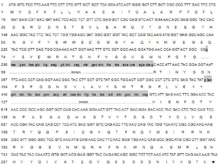
cDNA with two short intervenings by two introns of 56bp and
55bp as indicated in Fig.5 and Fig.6.
Figure 5. Cloning of the ORF sequance. Using the primers F1 and R1for PCR with restriction enzyme cutting site in the 5’
end, clone the ORF for expression in Ecoli. PCR products were
used to construct an expression plasmid and translate into
Ecoli.BL21 (DE3). M: PCR fragment. 1: blank. 2-3: PCR
413
Zhan-Ling, X. et al. Xylanase gene from Fusarium sp.
Figure 6. Nucleotide sequence of the Xyn8 gene and its deduced amino acid. DNA sequence of two introns is shown in lower-case letters and grey boxes. The bold letters in boxes, ATG and TAA, indicate the start codon and stop codon.
DNA sequence analysis of the xyn8 gene
The predicted peptide consisted of 230 amino acids with
the theoretical M.W. of 25.7 kDa. Amino acid homology
alignment of the predicted Xyn8 with other four xylanases
from Fusarium oxysporum f. sp., Glomerella graminicola,
Verticillium dahliae, Verticillium albo-atrum was carried out
(Fig. 7). The identities between F. sp. Q7-31 with F.
oxysporum f. sp (AAK 27975.1), Glomerella graminicola
(EFQ30380.1), Verticillium dahliae (ABE02800.1), V.
albo-atrum (XP✡ 003006739.1) were 87%, 78%, 74%, 74%,
respectively.
The phylogenetic analysis of the xylanases included 25
known different amino acid sequences of xylanase and was
complied using the data base☛ Fig.8 . The phylogenetic tree of ☞
the xylanases was then constructed by MEGA 3.1, as shown in
Fig. 9. According to this phylogenetic tree, the Xyn8 is
genetically closely related to the hypothesis protein of
Gibberella zeae (89%), and xylanase F. oxysporum (87%). We
found that the topology of the evolutionary tree of the Xyn8 is
quite similar to that of the other GHF 11 xylanases, so that the
Xyn8 should be classified into the family 11 glycosyl
Figure 7. Comparison of Xyn8 to other microbial xylanases. Abbreviations: Fusarium oxysporum f. sp., Glomerella graminicola, Verticillium dahliae, Verticillium albo-atrum. The conserved and similar amino acids in five xylanases are
indicated by solid and same color boxes, respectively.
Figure 8. Neighbor-joining (NJ) phylogenetic tree using MEGA 3.1
showing the evolutionary relatedness and levels of homology
between the xylanase amino acid sequences. The estimated genetic
distance between sequences is proportional to the lengths of the
horizontal lines connecting one sequence to another. This is an
un-rooted tree. Bootstrap values of the major branch points are shown;
they represent the number of times the group consisting of the
species to the right of that branch occurred among 100 trees. The
sequences are of the following proteins: F.o., Fusarium oxysporum;
G.g., Glomerella graminicola; V.d., Verticillium dahliae; V.a.,
Verticillium albo-atrum; C.s., Cochliobolus sativus; C.c.,
Cochliobolus carbonum; P.t., Pyrenophora tritici-repentis; P.n.,
Phaeosphaeria nodorum; G.z., Gibberella zeae; Sch.s.,
Scheffersomyces stipitis; H.g., Humicola grisea; Scl.s., Sclerotinia
sclerotiorum; B.f.,Botryotinia fuckeliana; A.t., Aspergillus terreus;
T.s., Talaromyces stipitatus; P.m., Penicillium marneffei; P.f.,
Penicillium funiculosum; M.o., Magnaporthe oryzae; P.c.w.,
Penicillium chrysogenum Wisconsin; S.m., Sordaria macrospore;
P.a., Podospora anserine; A.c., Aspergillus clavatus; N.c.,
415
Zhan-Ling, X. et al. Xylanase gene from Fusarium sp.
Figure 9. Analysis of SDS-PAGE (A) and western blot (B) A: M. Protein marker; 1. Un-induced with IPTG; 2, 3, 4, and 5: induced for by IPTG; B: 1, pGEX5x-1+BL21; 2, pGEX5x-1-xyn8+BL21; 3.
Expression of Xyn8 in E. coli
To further test if the cloned sequence truly represents F.
sp. Q7-31. Primers OF and OR were applied to amplify the
peptide encoding cDNA with adequent restriction enzyme sites
so that the cDNA could be inserted into expression vector
pGXE5x-1 in the right reading frame. Because of the presence
of the GST-tag fusion peptide in expression vector pGXE5x-1,
the recombinant protein had a 26 kDa peptide. As shown in
Fig. 9(A), an efficiency of Xyn8 protein expression was
achieved with pGXE5x-1. The recombinant protein showed a
M.W. of about 52 kDa which was close to the theoretical value.
Western blotting and SDS–PAGE showed that the cell
extracts from E. coli BL21CodonPlus(DE3)-RIL harboring
pGXE5x-1-xyn8 exhibited a clear band with a molecular
weight about 52kDa (Fig. 9, A-Lane2- 5, B-Lane 2), which was
a fusion hybrid protein and has the same size as estimated from
the deduced amino acid sequence of the fusion region in
pGXE5x-1-xyn8. The expression of the solubility fusion hybrid
protein could be induced by IPTG. A maximum activity of
2.34U/ mg was obtained from cellular extract of E. coli
BL21-CodonPlus (DE3)-RIL harboring pGXE5x-1-xyn8 (table 3).
These predictions are consistent with experimental data
previously obtained from the GST-Xyn8 fusion protein (7).
Properties of Xyn8
From the pH profile, the pH for optimal activity of Xyn8
was determined to be pH 6.0, with 50% of maximum activity
being retained between pH 5.8 and pH 9.0 (Table 3). The
apparent optimal temperature for enzyme activity at pH 6.0
was 40 °C. Crude xylanase from Q7-31 was used to evaluate its
biochemical properties.
DISCUSSION
The Qinghai–Tibet Plateau is located in east Asia and
represents a unique permafrost environment, being a result of
high elevation caused by land uplift. However, its
microbiology remains largely unexplored to date. Zhang G
reported that the total 33 analyzed isolates from Qinghai-Tibet
Plateau, 9 isolates related to 8 genera might be new taxa, it
suggested that the Qinghai-Tibet Plateau permafrost region is a
specific ecologic niche that accommodates an original
microbial assemblage (11, 27). Yu-Qin Zhang (26) reported
there was abundant actinobacterial species diversity in the soil
samples from the Qinghai–Tibet plateau, including
xylanase-producing microorganisms (2010). No xylanase-xylanase-producing
fungi have been reported in the Qinghai–Tibet plateau. In this
study, we isolated and identified a xylanse-producing strain of
Fusaurium sp.Q7-31 from a morchella fruiting body in the
Qinghai–Tibet plateau. The ITS rDNA sequence of Fusaurium
sp.Q7-31 showed high identity with Fusarium oxysporum,
there are reports on xylanses produced by Fusarium
oxysporum(), while the gene Xyn8 from Fusaurium sp.Q7-31 is
deferent from other reports. The gene Xyn8 from Fusaurium
sp.Q7-31 encodes a GHF11 xylanse.
Xyn8 has relatively the highest sequence identity to Xyl5
(87%) from F. oxysporum (5), but the properties of these two
enzyme are distinct. Xyl5 resulting from differential splicing of
the third intron, while Xyn8 resulting from differential splicing
of the second intron.
The cloning of a novel gene depends largely on the
combination of DNA library construction and the screening of
genes. Various methods have been developed to amplify the
process, such as screening of an expression DNA library or
genomic DNA library with antibodies or detecting specific
enzyme activity of the target protein; however, all the present
methods are still time-consuming and laborious.
In this work, cloning the cDNA and the gene of the Xyn8
from F. sp Q7-31 were achieved by using three steps of PCR.
The first step was to use degenerated primers for amplification
of cDNA. The second step was to obtain the full-length cDNA
by a modified 5, RACE approach. The third step was to obtain
the introns sequence in genomic DNA corresponding to the
full-length cDNA. It has been reported that the xylanases can
be divided into two major families based on their 3D
structures, GHF 10 and GHF 11. From the prediction of tertiary
structure, we found that the 3D structure of the Xyn8 of F. sp.
Q7-31 was mostly similar to some xylanases of GHF 11.
Therefore, we concluded that Xyn8 belonged to the family 11
glycosyl hydrolases.
The favorable properties of Xyn8, such as high activity
over a wide temperature range and so on, make this xylanase
promising for various applications in the biofuel industries.
ACKNOWLEDGEMENTS
Project was supported by the china National Science
Foundation (30860048). This research was supported by
Professor Ge Rili and Liu Yujiao who both come from Qinghai
University. We also should thank the station master Shen
Zhixin.
REFERENCES
1. Alconada TM, Martínez MJ. (1994). Purification and characterization of an extracellular endo-1,4-beta-xylanase from Fusarium oxysporum f. sp. melonis. FEMS Microbiol Lett. 15;118(3):305-10.
2. Bradford, M.M. (1976). A rapid sensitive method for the quantitation of microgram quantities of protein utilizing the principle of protein-dye binding. Anal. Biochem. 72: 248–254.
417
Zhan-Ling, X. et al. Xylanase gene from Fusarium sp.
to family F/10 Aug 7;302(3-4):191-5. Carbohydr Res.
4. Christakopoulos, P.; Nerinckx, W.; Kekos, D.; Macris, B.; Claeyssens, M. (1996). Purification and characterization of two low molecular mass alkaline xylanases from Fusarium oxysporum F3. Journal of Biotechnology. 51: 181-189.
5. Esperanza Gómez-Gómez, M.C.; Isabel, G.; Roncero. (2001). Molecular characterization of a novel endo- -1, 4-xylanase gene from the vascular wilt fungus Fusarium oxysporum. Current Genetics. 40: 268-275. 6. Esperanza Gómez-Gómez, M.C.; Ruiz-Roldan, A.; Di Pietro, M.I.G.;
Roncero.; Hera, C. (2002). Role in Pathogenesis of Two Endo- -1, 4-xylanase Gene from the Vascular Wilt fungus Fusarium oxysporum. Fungal Genet Biol. 35, 213-222.
7. Fu, D.D.; Li, A.J.; Xie, H.; Wu, M.C. (2006). Purification and properties of xylanase from Aspergillus usamii. Food. Sci. 27, 116–120.
8. Gómez-Gómez E, Isabel M, Roncero G, Di Pietro A, Hera C. (2001) Molecular characterization of a novel endo-beta-1,4-xylanase gene from the vascular wilt fungus Fusarium oxysporum. Curr. Genet. 40 (4), 268-275.
9. Gómez-Gómez E, Ruíz-Roldán MC, Di Pietro A, Roncero MI, Hera C. (2002) Role in pathogenesis of two endo-beta-1,4-xylanase genes from the vascular wilt fungus Fusarium oxysporum. Fungal Genet Biol. Apr;35(3):213-22.
10. Jorge I, de la Rosa O, Navas-Cortés JA, Jiménez-Díaz RM, Tena M. (2005) Extracellular xylanases from two pathogenic races of Fusarium oxysporum f. sp. ciceris: enzyme production in culture and purification and characterization of a major isoform as an alkaline endo-beta-(1,4)-xylanase of low molecular weight. Antonie Van Leeuwenhoek. Jul;88(1):48-59.
11. Gaosen Zh., Xiaojun M., Fujun N., Maoxing D., Huyuan F., Lizhe A. and Guodong Ch. (2007). Phylogenetic diversity of bacteria isolates from the Qinghai-Tibet Plateau permafrost region. Canadian J. Microb. 12. Laemmli, U.K. (1970). Cleavage of structural proteins during the
assembly of the head of bacteriophage T4. Nature. 227: 680–685. 13. Miller, GL. (1959). Use of dinitrosalicylic acid reagent for determination
of reducing sugars. Anal. Chem. 31: 426-428.
14. Ruiz Roldan, M.C.; Di Pietro, A.; Roncero, M.I.G. (1997). Purification and characterization of an acidic endo- -1,4-xyalnase from the tomato vascular pathogen Fusarium oxysporum f.sp. lycopersici. FEMS Microbiol Lett. 148: 75-82.
15. Ruiz Roldan, M.C.; Di Pietro, A.; Huertas Gonzalez, M.D.; Roncero M.I.G. (1999). Two xylanase genes of the vascular wilt pathogen Fusarium oxysporum are differentially expressed during infection of tomato plants. Mol. Genet. 261: 530-536.
16. Sambrook, J.; Fritsch, E.F.; Maniatis, T. (1989). Molecular Cloning: a Laboratory Manual, seconded. Cold Spring Harbor Laboratory Press, Cold Spring Harbor, New York.
17. Sambrook, J.; Fritsch, E.F.; Maniatis, T. (2001). Molecular Cloning: A Laboratory Manual. Cold Spring Harbor Laboratory Press, Cold Spring Harbor, New York.
18. Sapag, A.; Wouters, J.; Lambert, C. (2002). The endoxylanases from family11: computer analysis of protein sequences reveals important structural and phylogenetic relationship. J. Biotechnol. 95, 109–131. 19. Sengupta, S.; Jana, M.L.; Sengupta, D.; Naskar, A.K. (2000). A note on
the estimation of microbial glycosidase activities by dinitrosalicylic acid reagent. Appl. Microbiol. Biot. 53: 732- 735.
20. Shao, W.; Wiegel, J. (1992). Purification and characterization of a thermostable xylosidase from Thermoanaerobacter ethanolicus. J. Bacteriol. 174, 5848–5853.
21. Van Burik, J.A.; Schreckhise, R.W.; White, T.C.; Bowden, R.A.; Myerson. D. (1998). Comparison of six extraction techniques for isolation of DNA from filamentous fungi. Med. Mycol. 36: 299- 303. 22. Vallander, L.; Eriksson, K. E. L. (1990). Production of ethanol from
lignocellulosic materials: State of the art. Adv. Biochem. Eng. Biotechnol. 42: 63-95.
23. Verma, I.M. (1977). Reverse transcriptase-The Enzymes. Academic Press, New York.
24. Walton, J.D. (1994). Deconstructing the plant cell wall. Plant Physiol. 104: 1113-1118.
25. Yang, G.L.; An, Q.; Li, J.; Lin, W.; Liu, L.; Lin, X. (2007). Genotyping of Trichophyton rubrum by analysis of ribosomal-DNA intergenic spacer regions. Mycopathologia. 164: 19- 25.
26. Yu-Qin, Zh.; Hong-Yu, L.; Jie, Ch.; Li-Jie, Y.; Wei, S. (2010). Diversity of culturable actinobacteria from Qinghai-Tibet plateau, China. Antonie van Leeuwenhoek, 98, : 2, s: 213-223
27. Zhang, G.; Niu, F.; Ma, X.; Liu, W.; Dong, M.; Feng, H.; An, L.; Cheng, G. (2007). Phylogenetic diversity of bacteria isolates from the Qinghai-Tibet Plateau permafrost region. Can J Microbiol. Aug;53(8):1000-10.