http://www.uem.br/acta ISSN printed: 1806-2636 ISSN on-line: 1807-8672
Doi: 10.4025/actascianimsci.v37i3.26535
Bacterial diversity in bovine rumen by metagenomic 16S rDNA
sequencing and scanning electron microscopy
Raphael Barbetta de Jesus1,2, Wellington Pine Omori1,2, Eliana Gertrudes de Macedo Lemos1,3
and Jackson Antônio Marcondes de Souza1,2*
1
Faculdade de Ciências Agrárias e Veterinárias, Universidade Estadual Paulista, Via de Acesso Professor Paulo Donato Castellane, s/n, 14884-900, Jaboticabal, São Paulo, Brazil. 2
Departamento de Biologia Aplicada à Agropecuária, Faculdade de Ciências Agrárias e Veterinárias, Jaboticabal, São Paulo, Brazil. 3
Departamento de Tecnologia, Faculdade de Ciências Agrárias e Veterinárias, Jaboticabal, São Paulo, Brazil. *Author for correspondence. E-mail: [email protected]
ABSTRACT. The bacterial diversity by 16S rDNA partial sequencing and scanning electron microscope (SEM) of the rumen microbiome was characterized. Three Nellore bovines, cannulated at the rumen, were utilized. Liquid and solid fractions from the rumen content were processed for the extraction of metagenomic DNA and later 16S rDNA amplicons were utilized to construct the WGA library for further clone sequencing. Data were analyzed by MEGA and MOTUR (University of Michigan) softwares. Approximately 97.96% of operation taxonomic units (OTUs) were related to Bacteriodetes phylum and 2.04% of sequences were affiliated to Firmicutes phylum. In the case of Bacteriodetes, the great part of sequences (47.96%) was attributed to Prevotella genus. Bacteroidetes phylum was predominant in rumen content and the Prevotella genus was the most abundant, including diverse species related to this taxon. The bacterial morphological diversity associated to plant fibers was detected by SEM and showed its role in plant biomass deconstruction beyond the detection of microbiological interactions that involved protozoa.
Keywords: bacterial community, bacterioidetes, Nellore, Prevotella, taxonomic affiliation.
Diversidade bacteriana em rúmen bovino por análise de sequências do 16S rDNA
metagenômico e microscopia eletrônica de varredura
RESUMO. O estudo foi realizado para caracterizar a diversidade bacteriana por meio do sequenciamento parcial do 16S rDNA total e microscopia eletrônica de varredura (MEV) do microbioma ruminal. Foram utilizados três bovinos da raça Nelore, canulados no rúmen. As frações líquidas e sólidas do conteúdo ruminal foram processadas para extração de DNA metagenômico. Em seguida, amplicons 16S rDNA foram utilizados na construção da biblioteca WGA para posterior sequenciamento dos clones. Os dados foram analisados pelos softwares MEGA e MOTHUR (The University of Michigan). Aproximadamente 97,96% das UTOs foram relacionadas ao filo Bacteroidetes e apenas 2,04% das sequências foram afiliadas ao filo Firmicutes. Para o filo Bacteroidetes, grande parte das sequências (47,96%) foi atribuída ao gênero
Prevotella. O filo Bacteroidetes foi predominante no conteúdo ruminal e o gênero Prevotella foi o mais abundante, incluindo diversas espécies relacionadas a esse nível taxonômico. A diversidade morfológica bacteriana associada às fibras vegetais foi detectada por MEV, evidenciando seu papel na desconstrução da biomassa vegetal, além da detecção de interações microbiológicas que envolvem protozoários.
Palavras-chave: comunidade bacteriana, bacteroidetes, Nelore, Prevotella, afiliação taxonômica.
Introduction
The demand for greater efficiency in the production of ruminants is limited by the lack of information on rumen microbiota which damages the effective improvement of fermentative digestion. If the symbiotic relationship between the host and microorganisms is one of the most relevant factors in taking the best advantage from the diet, or rather, a greater efficiency in the use of energy and proteins by the animals, the understanding of the structure of this microbial community and its factors is mandatory for
the development of new production technologies (Martins et al., 2007; Martins et al., 2006).
The rumen environment is colonized by thousands of microorganisms which belong to the three dominions (Woese et al., 1990), namely: Bacteria (bacteria), Archaea (archeae) and Eucarya (fungi and protozoa). Bacteria are highly diversified and abundant within the dominions in the rumen and represent approximately 95% of total microbiota (Flint et al., 2008).
such as isolation, nutritional characterization and cell numbering (McSweeney & Mackie, 2012). However, these traditional techniques provide only a limited access to the diversity of microorganisms. They were actually complemented by more precise molecular techniques and shorter analysis duration (McSweeney & Mackie, 2012). Further, 16S rRNA gene sequencing is one of the basic genetic markers for the characterization of prokaryote microorganism community and estimates the phylogenetic richness and classification used in the quantification and taxonomy of the microbiome including non-cultivable microorganisms (Makkar & Cameotra, 2002).
Since several genetic and environmental aspects contribute towards the co-evolution between the ruminant and its rumen microbiota, the importance of the genetic heritage of Nellore cattle and its role as supplier of food resources, current analysis characterizes the rumen’s bacterial diversity by the partial sequencing of total 16S rDNA and Scanning Electron Microscopy (SEM).
Material and methods
The experiment was conducted in an area of the Faculty of Agrarian and Veterinarian Sciences (FCAV-UNESP), Jaboticabal, São Paulo, Brazil and developed in the Laboratory of Applied Genetics (LGA) and the Laboratory of the Biochemistry of Microorganisms and Plants (LBMP). All experimental stages were developed according to ethics in animal experimentation of the Brazilian College of Animal Experimentation and approved by the Committee for Ethics in the Use of Animals of the FCAV (protocol 017621/11).
Three Nellore bulls, castrated, cannulated in the rumen, approximately 24 months old, with mean weight 350 kg, were used. Diet comprised a mixture of ground hay Tifton 85 (Cynodon spp) (70%) and concentrate (30%) made of soybean meal (13.1%), ground corn (15.7%), urea and ammonium sulfate (1.2%), given once a day at 7h00 am. Mean daily ingestion of dry matter was approximately 1.7% of the animal’s live weight; water was given ad libitum and diet adaptation period was 31 days. A sample of approximately 500 g was retrieved from the middle region of the rumen from each animal one hour before feeding and after manual homogenization of the rumen, by rumen cannula.
Samples from each animal were fractioned in a liquid and solid fraction for total DNA extraction after prior processing of the collected material given below:
The microorganisms adherent to the solid fraction were separated as follows: 30 mL of a solution with NaCl 0.85% and containing 0.2% polysorbate were added to 10 g of the solid fraction of the rumen content. The sample was vigorously homogenized for 1 minute and then centrifuged for 10 minutes at 1,000 x g,
at 4°C. The supernatant was transferred to a new tube kept in ice. The sediment was prepared by the same washing process previously described. The second supernatant was transferred to the tube kept in ice whilst the sediment was again re-suspended with the solution previously described, with the addition of glass beads, vigorously homogenized and centrifuged as before. The third supernatant was centrifuged at 7,000 x g for 30 minutes, at 4°C, and the sediment re-suspended in 3 mL of NaCl 0.85%.
The microorganisms in the liquid fraction were obtained as follows: 500 mL of the rumen liquid were centrifuged at 27,000 x g for 30 minutes, at 4°C, and the supernatant disposed of. The precipitate was washed in 40 mL of NaCl 0.85% and centrifuged as previously described; the sediment was re-suspended in 40 mL of NaCl 0.85%.
Total DNA was extracted with 250 mg of each fraction with extraction kit FastDNA® SPIN Kit for
Soil (MP Biomedical, Santa Ana, California, USA). DNA samples were verified in agar gel 0.8% and analyzed by spectrophotometry (Thermo Scientific, NanoDrop™ 1000, Loughborough, Leicestershire, UK) to assess quality and amount. Biological samples and metagenomic DNA extraction were undertaken separately in triplicate till equimolar amounts of each sample were obtained for PCR.
Partial amplicons of 16S rRNA of two samples from rumen contents were produced by PCR with degenerated oligonucleotide initiators: 27 F (5’ AGA GTT TGA TCM TGG CTC AG 3’) and 1492 R (5’ GGY TAC CTT GTT ACG ACT T 3’) (Lane, 1991). PCR reaction employed 20 ng of total DNA in a reaction comprising 1.25 mM of MgCl2, 0.2 mM of
dNTPs, 1 U of enzyme Taq DNA polymerase (Invitrogen); buffer solution for PCR reaction [1x] (Invitrogen) and 10 pmol of each oligonucleotide initiators. The reaction occurred in a thermocycler (MJ Research, Inc., model PTC-100TM, Watertown, Massachusetts, USA) with the following conditions: 95°C for 5 minutes, 30 cycles with denaturation at 95°C for 30 seconds, pairing at 52°C for 30 seconds, extension at 72°C for 2 minutes and final extension at 72°C for 10 minutes.
The amplicons were cleansed with kit Wizard® SV
Gel and PCR Clean-Up System (Promega) and a clone library of 16S rRNA fragments was constructed by kit pGEM®-T Easy Vector System (Promega). The
Biosystems) with kit DNA Sequencing-Big Dye Terminator Cycle Sequencing Ready ABI Prism 3.
Further, 16S rRNA sequences were submitted to GenBank, with access number KJ650099-KJ650196. All sequences were compared to those deposited in the GenBank by Megablast and the sequences with the greatest score were used for the multiple alinement in ClustalW available in BioEdit. Distance matrix was calculated with DNADIST of PHYLIP and a dendrogram was built with MEGA 5.0 (Tamura et al., 2011) by the Neighbor-Joining method (Saitou & Nei, 1987), substitution matrix Jukes and Cantor (Jukes & Cantor, 1969), bootstrap (Felsenstein, 1985), for 1,000 replicates and option for complete deleting. Rarefaction curve and richness estimators Chao1 (Chao, 1987) and ACE (Chao & Lee, 1992) were obtained in MOTHUR (Schloss et al., 2009), whereas Shannon-Weaver and Simpson diversity indexes (Magurran, 1988) were obtained with 97% similarity to classify the Operation Taxonomic Units (OTUs).
Samples of rumen contents, conditioned in falcon-type flasks (50 mL) were used for scanning electron microscopy (SEM). They contained a solution of glutaraldehyde 3% in a potassium phosphate buffer at 0.1 M, pH 7.4, for 48 hours for fixing. They were later post-fixed in a solution of osmium tetroxide 1% for 4 hours and washed in the same buffer solution. Dehydration occurred in a gradual serial of ethyl alcohol 30, 50, 70, 80, 90 and 100%; three times in the later, and remained immersed in each during 20 minutes. The drying of the material was undertaken in a critical point drier with CO2; they were fixed and
metalized with approximately 35 nm of gold-palladium. The samples analyzed by electron micrography with SEM JEOL JSM 5410 at 15 kV (Santos, 1996).
Results and discussion
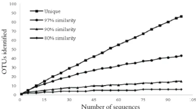
As Figure 1 shows, 43, 15 and 6 distinct OTUs were discovered respectively at 97, 90 and 80% similarity in the evaluated rumen contents, taking into consideration conditions applied in MOTHUR analysis. Rarefaction graph revealed that the sampling effort on orders and phyla (respectively 90 and 80% similarity) was satisfactory, since curve reached the asymptote. Sequences at the genus level (97%) failed to reveal the total presence of the bacterial community evaluated. More sequences could have been obtained to amplify the genus spectrum represented by the community. The above was confirmed by ACE richness estimators (109) by which sequences are grouped into rare and abundant according to frequency; by Chao1 (80) that analyzes the number of absent species; by the Shannon-Weaver diversity index (3.430), an index, which is sensitive to species richness and relative abundance; and by Simpson (0.034) in
which the closer it is to zero, the greater is the diversity in the environment under analysis.
OTUs ident
ified
Number of sequences
Figure 1. Rarefaction curve of cattle rumen contents showing the number of Operation Taxonomic Units (OTUs), taking into account the total number of sequences, with 97%, 90% and 80% of genetic similarity.
Partial sequences of gene 16S rDNA were compared with data bank RDP II by RDPquery. The 98 sequences belonged to the dominion Bacterium, of which approximately 97.96% were related to the Bacteroidetes phylum and only 2.04% of the sequences to the Firmicutes phylum. The predominance of the phyla corroborates data given by several authors who reported that the most relevant identified phyla in bovine rumen are the Bacteroidetes and Firmicutes phyla (Callaway et al., 2010; Chen et al., 2011; Li et al., 2014).
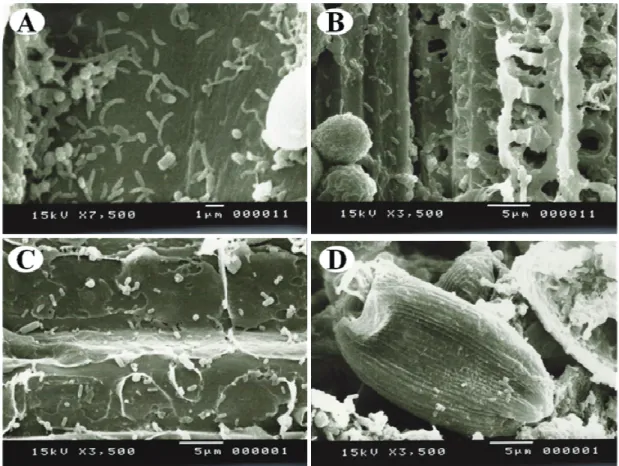
Most sequences (47.96%) of the Bacteroidetes phylum belonged to the genus Prevotella, or rather, gram-negative, obligatory anaerobic, non-sporulating bacteria, without any motility and shaped as pleomorphic rods (Shah & Collins, 1990). The high percentage of Prevotella, mainly the species Prevotella ruminicola, Prevotella brevis, Prevotella bryantii and Prevotella albensis (McSweeney & Mackie, 2012), revealed the genus’s abundance in the rumen (Bekele et al., 2010; Stevenson & Weimer, 2007). The above-mentioned bacteria showed great variability in genetic and phenotypic features (Avguštin et al., 1997)(Figure 2). Important differences were also identified among the four species with regard to the capacity of polysaccharide degradation (Matsui et al., 2000).
Diversity analysis by total 16S rDNA sequencing of the rumen contents was a very relevant approach since it comprised microorganisms which would not be accessible by traditional microbiologic tools. Moreover, a visual analysis by SEM assessed the richness of microbiological forms in the bovine rumen (Figure 2) and thus amplifying further the access capacity for the diversity of the rumen environment.
A great variety of Prevotella species with enzyme activities for carboxy-methylcellulase and xylanase has been isolated from the rumen (Avguštin et al., 1997).
proteolithic activity in the rumen and has an important role in making nitrogen available for other microorganisms from the degradation of protein derived from the host’s feed (Wallace et al., 1997). These species are perhaps the most important ammonia-producing bacteria in the rumen.
Figure 2 demonstrates diversified bacteria composed of bacteria, fungi and protozoa (Figura 2). Bacteria of the genera Bacteroides, Prevotella, Ruminobacter, Fibrobacter, Methanobrevibacter, Methanomicrobium and
Clostridium (spores) are rod-shaped; Butyrovibrio, Selenomonas and Lachnospira are curved rod bacteria; cocci indicate the presence of Ruminococcus, Streptococcus
and Megasphaera. Spiral microorganisms, similar to spirochaetes, have been frequently detected in the rumen and are represented by the genus Treponema
(Stewart et al., 1997).
Albeit surety is not possible, the form standard reported is a relevant indication of species diversity. Several bacteria are decomposers of cellulose, pectin and starch and they are involved in the deconstruction of vegetal biomass ingested by the animal. The bacteria associated with decomposing vegetal fiber are reported (Figure 2). Perforations caused by cellulite activity and the exposure of sap-conducting vessels may be perceived. Prevotella isolates have the shape represented
in Figure 2, whereas Ruminococcus isolates have the pattern shown in Figure 2 (Stewart et al., 1997).
Aerobic yeasts and fungi, which are able to grow in anaerobic conditions, may colonize the rumen, including the species of the genera Aspergillus, Mucro and Sporormia (Orpin & Joblin, 1997). Anaerobic saprophyte fungi, in the main zoosporic fungi, belong to the division Eumycotina, subdivision Mastigomycina and class Chytridiomycetes (Orpin & Joblin, 1997). Thali, sporangia and zoospores of rumen anaerobic fungi may be represented by the shapes under analysis.
Besides fungi and bacteria, the rumen has bigger organisms, measuring 5-250 μm, or protozoa. Ciliated protozoa of the subclass Trichostomatia are the most important (Williams & Coleman, 1997). The protozoa may interact with the archaeas, bacteria and fungi in the rumen (Figure 2). Many protozoa have archaeas adhered to their external surface. The employment of 16S rRNA by Lloyd et al. (1996) confirmed several ectosymbiontes belonging to archaeas, especially metanogenic and hydrogen producers (Williams & Coleman, 1997). Ciliated protozoa are generally attracted by colonies of fungi in which predatory and metabolic interactions among eukaryote microorganisms may be observed (Williams & Coleman, 1997).
Although synergistic and mutualistic relationships between the different groups of prokaryote and eukaryote microorganisms are beneficent to the animal and the microbial community, there is a delicate equilibrium between the individual populations that make up the community. Each population contributes metabolically towards the transformations of the substrates in fermentation products and the population balance may be adjusted by subtle variations in the animal’s diet. An efficient equilibrium corresponds to the animals’ healthy conditions.
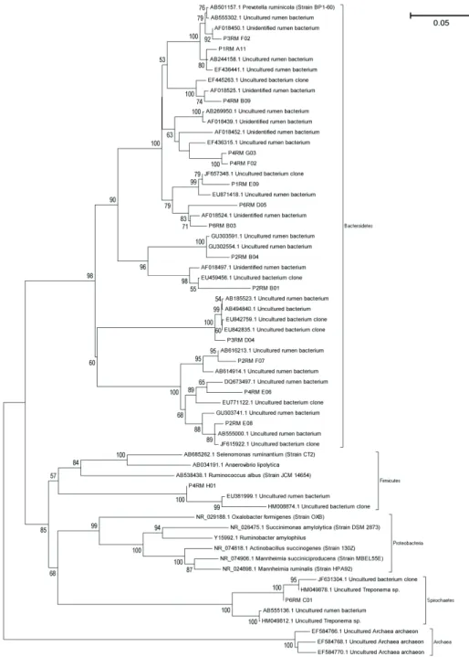
Reinforcing and complementing information produced by phylogenetic classification in the RDP data base, the dendrogram reveals (Figure 3) that almost all the sequences belong to the phylum Bacteroidetes. Moreover, there were genetic differences among these sequences that revealed a great diversity of species related to the phylum, most of which described as non-cultured bacteria. The great number OTUs of non-cultivable organisms identified in current assay reinforce the relevance in studying the rumen.
The information produced in current analysis is primary and primordial for the understanding of the ruminal bacterial composition. The development of new methods and technologies is highly promising in future research in genomics, proteomics, metabolomics, transgenics and immunology among other possible types of knowledge that may be obtained from such a rich and scantily known rumen environment.
Conclusion
The phylum Bacteroidetes was identified as predominant in bovine rumen contents by partial 16S rDNA sequences. The genus Prevotella was the most abundant in this phylum, with several species related to this taxonomic level. SEM also showed the biodiversity of prokaryotes and eukaryotes in full metabolic activity with regard to the degradation of the vegetal biomass.
Acknowledgements
The authors would like to thank the Brazilian Council for Scientific and Technological Development (CNPq) for its funding. Thanks are also due to the Pro-Rectory for Research (PROPe) of the UNESP.
References
Avguštin, G., Wallace, R. J. & Flint, H. J. (1997). Phenotypic diversity among ruminal isolates of
Prevotella ruminicola: proposal of Prevotella brevis sp. nov., Prevotella bryantii sp. nov., and Prevotella albensis
sp. nov. and redefinition of Prevotella ruminicola.
International Journal of Systematic Bacteriology, 47(2), 284-288.
Bekele, A. Z., Koike, S. & Kobayashi, Y. (2010). Genetic diversity and diet specificity of ruminal Prevotella revealed by 16S rRNA gene-based analysis. FEMS Microbiology Letters, 305(1), 49-57.
Callaway, T., Dowd, S., Edrington, T., Anderson, R., Krueger, N., Bauer, N., ... Nisbet, D. (2010). Evaluation of bacterial diversity in the rumen and feces of cattle fed different levels of dried distillers grains plus solubles using bacterial tag-encoded FLX amplicon pyrosequencing. Journal of Animal Science, 88(12), 3977-3983.
Chao, A. (1987). Estimating the population size for capture-recapture data with unequal catchability.
Biometrics, 43(4), 783-791.
Chao, A. & Lee, S.-M. (1992). Estimating the number of classes via sample coverage. Journal of the American statistical Association, 87(417), 210-217.
Chen, Y., Penner, G. B., Li, M. & Oba, M. (2011). Changes in bacterial diversity associated with epithelial tissue in the beef cow rumen during the transition to a
high-grain diet. Applied and Environmental Microbiology, 77(16), 5770-5781.
Felsenstein, J. (1985). Confidence limits on phylogenies: an approach using the bootstrap. Evolution, 35(4), 783-791.
Flint, H. J., Bayer, E. A., Rincon, M. T., Lamed, R. & White, B. A. (2008). Polysaccharide utilization by gut bacteria: potential for new insights from genomic analysis. Nature Reviews Microbiology, 6(2), 121-131. Jukes, T. H. & Cantor, C. R. (1969). Evolution of protein
molecules. In H. N. Munro (Ed.), Mammalian protein metabolism (Vol. 3, pp. 21-132). New York: Academic. Lane, D. J. (1991). 16S/23S rRNA sequencing. In S. E. &
M. Goodfellow (Eds.), Nucleic acid techniques in bacterial systematics (pp. 125-175). New York: John Wiley and Sons.
Li, Y., Ma, S., Zhang, X., Huang, S., Yang, H., Zhao, F., Yi, W., Yang, X., ... S. & Yi, X. (2014). Evaluation of bacterial and archaeal diversity in the rumen of Xiangxi yellow cattle (Bos taurus) fed Miscanthus sinensis or common mixed feedstuff. Annals of Microbiology, 64(3), 1385-1394.
Lloyd, D., Williams, A.G. & Arman, R. (1996). Intracellular prokaryotes in rumen ciliate protozoa: detection by confocal laser scanning microscopy after in sito hybridization with fluorescent 16S rRNA probes. European Journal of Protistology, 32(4), p. 523-531.
Magurran, A. E. (1988). Ecological diversity and its measurement. Princeton: Princeton University Press. Makkar, R. & Cameotra, S. (2002). An update on the use
of unconventional substrates for biosurfactant production and their new applications. Applied Microbiology and Biotechnology, 58(4), 428-434.
Martins, A. S., Vieira, P. F., Berchielli, T. T., Prado, I. N. & Paula, M. C. (2007). Degradabilidade in situ e observações microscópicas de volumosos em bovinos suplementados com enzimas fibrolíticas exógenas.
Revista Brasileira de Zootecnia, 36(6), 1927-1936. Martins, A. S., Vieira, P. V., Berchielli, T. T., Prado, I. N.
& Garcia, J. A. S. (2006). Eficiência de síntese microbiana e atividade enzimática em bovinos submetidos à suplementação com enzimas fibrolíticas.
Revista Brasileira de Zootecnia, 35(3), 1194-1200.
Matsui, H., Ogata, K., Tajima, K., Nakamura, M., Nagamine, T., Aminov, R. I. & Benno, Y. (2000). Phenotypic characterization of polysaccharidases produced by four Prevotella type strains. Current Microbiology, 41(1), 45-49.
McSweeney, C. & Mackie, R. (2012). Commission on Genetic Resources for Food and Agriculture. Micro-organisms and ruminant digestion: State of knowledge, trends and future prospects. Background Study Paper (FAO), 61, 1-62.
Saitou, N. & Nei, M. (1987). The neighbor-joining method: a new method for reconstructing phylogenetic trees. Molecular Biology and Evolution, 4(4), 406-425.
Santos, J. M. (1996). Microscopia eletrônica de varredura aplicada às ciências biológicas. Jaboticabal: Funep.
Schloss, P. D., Westcott, S. L., Ryabin, T., Hall, J. R., Hartmann, M., Hollister, E. B., ... Robinson, C. J. (2009). Introducing mothur: open-source, platform-independent, community-supported software for describing and comparing microbial communities.
Applied and Environmental Microbiology, 75(23), 7537-7541.
Shah, H. & Collins, D. (1990). Prevotella, a new genus to include Bacteroides melaninogenicus and related species formerly classified in the genus Bacteroides.
International Journal of Systematic Bacteriology, 40(2), 205-208.
Stevenson, D. M. & Weimer, P. J. (2007). Dominance of Prevotella and low abundance of classical ruminal bacterial species in the bovine rumen revealed by relative quantification real-time PCR. Applied Microbiology and Biotechnology, 75(1), 165-174.
Stewart, C., Flint, H. & Bryant, M. (1997). The rumen bacteria. In P. N. Hobson & C. S. Stewart (Eds.), The rumen microbial ecosystem (pp. 10-72): Blackie Academic and Professional.
Tamura, K., Peterson, D., Peterson, N., Stecher, G., Nei, M. & Kumar, S. (2011). MEGA5: molecular evolutionary genetics analysis using maximum likelihood, evolutionary distance, and maximum parsimony methods. Molecular Biology and Evolution, 28(10), 2731-2739.
Wallace, R., Onodera, R. & Cotta, M. (1997). Metabolism of nitrogen-containing compounds The rumen microbial ecosystem (pp. 283-328): Springer.
Williams, A. G. & Coleman, G. S. (1997). The rumen protozoa. In P. N. Hobson & C. S. Stewart (Eds.), The rumen microbial ecosystem (pp. 73-139). Netherlands: Blackie Academic and Professional.
Woese, C. R., Kandler, O. & Wheelis, M. L. (1990). Towards a natural system of organisms: proposal for the domains Archaea, Bacteria, and Eucarya.
Proceedings of the National Academy of Sciences, 87(12), 4576-4579.
Received on February 7, 2015. Accepted on May 31, 2015.