Joseph Khalil
Socket Shield and Immediate Implantation
Universidade Fernando Pessoa
Joseph Khalil
Socket Shield and Immediate Implantation
Universidade Fernando Pessoa
Joseph Khalil
Socket Shield and Immediate Implantation
Joseph Khalil
Trabalho apresentado à Universidade Fernando Pessoa como parte dos requisitos para obtenção do grau de
Abstract
Introduction - After tooth extraction, the alveolar bone undergoes a remodeling process, wich leads to horizontal and vertical bone loss. These resorption processes complicate dental rehabilitation, particularly in connection with implants. Various methods of guided bone regeneration have been described to retain the original dimension of the bone after extraction. Most procedures use filler materials and membranes to support the buccal plate and soft tissue, to stabilize the coagulum and to prevent epithelial ingrowth. It has also been suggested that resorption of the buccal bundle bone can be avoided by leaving a buccal root segment (socket-shield technique) in place, because the biological integrity of the buccal periodontum remains untouched. This method has also been decribed in connection with immediate implant placement.
Objective - This literature review aim enumerate and describe the different treatments and tissue reactions after tooth extraction, immediate and delayed implantation. The socket-shield technique, the evolution in tooth extraction and immediate implantation with high esthetic results due to the preservation of hard and soft tissues by leaving a buccal root segment in place.
Materials and methods - For this purpose a research has been done and data was obtained from on-line resources: Medline, Pubmed, Scielo, Bireme, Bon, books and specialized magazines which was conducted between January 2016 and May 2016. A number of articles have been obtained in English and French ,published between 1997 and 2015 . The key words used were implantology, dental implant, hard/soft tissue, tooth extraction, immediate implantation, delayed implantation, socket-shield.
Conclusion - In socket-shield technique, there were neither functional nor aesthetic changes in soft and hard tissues. It’s already a routine practice in the arsenal of high-aesthetic immediate implantology and should be used when indicated. Although this technique is quiet promising, we should be aware of the incoming publications about a
larger follow up and the predictability of leaving a fragment inside the socket after an extraction.
Resumo
Introdução - Após extração dentária o osso sofre um processso de remodelação que consequentemente conduz à perda óssea horizontal e vertical. Este processo de reabsorção óssea condiciona a reabilitação dentária particularmente quando esta é implanto-suportada.
Para preconizar a manutenção da dimensão óssea após exodontia têm sido descritos na literatura vários métodos de regeneração guiada. Recorrem-se a preenchimentos com materiais e membranas de modo a criar suporte na tábua óssea e tecidos moles. Desta forma, estabilize-se o coágulo e evite-se a recessão epitelial.
Foi também sugerido que um método a utilizar para evitar a reabsorção óssea é deixando um segmento radicular no local de modo a que consigamos manter o periodonto intacto (técnica socket-shield). Este método pode ser aplicado concomitantemente com implante imediato.
Objectivo - Esta revisão bibliográfica procura enumerar e descrever diferentes técnicas, tratamentos e reações tecidulares após a extração dentária com recurso à implante imediato ou mediato.
A preservação dos tecidos moles e duros é garantida através da colocação prévia de um segmento radicular no local, da evolução da técnica cirúrgica de exodontia assim como a implantologia imediata com elevada satisfação estética.
Materias e metodos - Com o objectivo de descrever esta técnica foram efectuadas pesquisas em bases de dados como: Scielo, Medline, Bireme, Pubmed, Bon, livros e também revistas da especialidade, entre os meses de Janeiro e Maio de 2016. Um número de artigos obtidos em inglês e francês publicados entre 1997 e 2015 .
As palavras-chave utilizadas foram : implantologia, implante dentário, tecido duro e tecido mole, exodontia, implante imediato, implante mediato, socket-shield.
Conclusão - Na técnica socket-shield não houve alteração estética nem funcional nos tecidos duros e moles. Esta, é já prática corrente na técnica implantar imediata associada ao sucesso estético e deverá, portanto, ser utilizada quando assim for indicada.
Apesar desta técnica ser altamente promissora, devemos mantermo-nos atentos às publicacões relativas ao follow-up e à possibilidade de deixar fragmentos dentro do socket após extração dentária.
Acknowledgments
I would like to thank my dissertation supervisor, Mestre Dr. Jorge Pereira, for his input, advice and orientation to this important subject, an evolution in immediate implantology.
I am very grateful for the support and motivation from family and friends.
I would like to thank my children and love, Gabriella and Alexandre, for being so patient and understanding with me, along this year.
Finally I would like to thank my wife Charlotte for her support,attention, patience and love for our lovely family assuming my long absence.
INDEX
Page
Index of Figures……… Index of Tables……….. INTRODUCTION……… 1 DEVELOPMENT………. 3Materials and Methods……… 3
I.
IMMEDIATE IMPLANTATION………..
41. Original concept……… 4
2. Study of post extractional healing in anterior maxillary………... 5
i. Healing of tooth extraction socket……… 5
ii. Bone healing around implants……….. 6
a. The horizontal bone resorption………. 6
b. Vertical proximal resorption………. 9
c. Vertical vestibule palatal resorption………. 9
iii. Healing inside the peri-implant defect………. 11
3. Evolution of the immediate implantation concept……… 12
a. Position and size of implants………. 13
b. Principles of successful immediate implantation.. 14
4. From immediate implantation to delayed implantation………… 15
i. Clinical situations, indications and patients selection….. 15
ii. Bone environments………... 16
II. DELAYED IMPLANTATION………..
191. Alveolar preservation……… 20
i. The different techniques………... 21
ii. Different times of management of soft tissue…………... 23
3. Management of bone defect………. 24
i. Guided bone regeneration technique………... 24
ii. Bone graft………. 25
a. Different sources………... 25
b. Bone apposition graft……… 26
III. SOCKET-SHIELD TECHNIQUE IN IMMEDIATE
IMPLANTATION………
281. The new technique to avoid buccal bone resorption………. 28
i. Description and definition……… 28
ii. Clinical concept of ridge preservation with modified socket-shield technique………. 29
2. Indication and procedure for ridge preservation………... 30
3. Socket-shield technique to support the bucco-facial tissues at immediate implant placement………... 31
4. Socket-shield technique to replace the conventional immediate implant placement………. 34
i. Preservation of hard and soft tissues………. 35
5. Histological, clinical, volumetric observations after separation of the buccal tooth segment………. 36
6. Socket-shield technique using bone trephine……… 38
IV. CONCLUSION……… 41
Index of Figures
Figure 1: Size representation of vestibular (buccal) and lingual bone (Araujo, 2006) 7
Figure 2: Representation of different points of measure (Araujo, 2005) 10
Figure 3: Cross section of implantation site after 3 months of healing (Araujo, 2005). 13
Figure 4: Different types of bone defects (Zuck, 2009) 18
Figure 5: Guided bone regeneration (Nevins, 2010). 25
Figure 6: Socket-shield technique and digital impression (Hürzeler, 2010) 29
Figure 7: The step by step procedure illustrated on a model (Glocker et al., 2014) 31
Index of Tables
Table 1: Comparative tabulation of procedure to manage the effects of post-extraction resorption adjunct to implant therapy (Gluckman, 2015)
INTRODUCTION
Immediate implant placement is a successful treatment option following tooth removal. Although the success rates for both immediate and delayed implant techniques are comparable, the literature reports that one can expect there to be recession of the buccal/facial gingiva following immediate implant placement, however this recession and ridge collapse can pose an aesthetic disaster in areas such as the anterior maxilla.
The socket-shield technique provides a promising treatment adjunct to better manage these risks and preserve the post-extraction tissues in aesthetically challenging cases. The principle is to prepare the root of a tooth indicated for extraction in such a manner that the buccal / facial root section remains in-situ with its physiologic relation to the buccal plate intact, to prevent the expected post-extraction socket remodeling and to support the buccal/facial tissues.
The choice of the theme of this thesis is the result of a long reflection. The author intended, considering his passion for oral surgery and implantology. Immediate implantation is a basic solution in order to face frequent situations of edentulism in anterior sectors, and to provide adequate and esthetic rehabilitation, to avoid eventual complications on functional, esthetic and psychological levels.
The objective of this study, through a literature review, is to deepen the dentist’s knowledge about the immediate implantation after tooth extraction, the reaction of hard and soft tissues and the post-extractional bone resorption. The socket-shield technique is an evolution in extraction and immediate implantation, in order to avoid the buccal bone resorption and to preserve the integrity of the periodontum with possible immediate prosthetic rehabilitation.
The research has been done on internet by consulting articles in 5 databases, on proposed subject with recourse to keywords.
Tooth loss is of multifactorial etiology and the indications for immediate implantation are multiple depending on the localization of the extracted tooth and the esthetic requirements.
The goal of this technique is to obtain immediate rehabilitation on anterior sectors; dental extraction, immediate implantation and immediate loading with high esthetic results due to the preservation of hard and soft tissues by leaving a buccal root segment in place.
With this technique, we can avoid buccal bone resorption in case of immediate implantation. But we should wait for more follow-up of this technique, because it is a recent practice.
DEVELOPMENT
Materials and Methods
The analysis and preparation of this bibliographic review were based on the scientific material duly published in books, articles and publications. The bibliographic research was conducted via online using the Medline, Scielo, Bireme, Pubmed, Bon search bases.
The key words used in the research are: implantology, dental implant, hard/soft tissue, tooth extraction, immediate implantation, delayed implantation, socket-shield.
A number of articles have been obtained in English and French published between 1997 and 2015. 35 were used which where relevant and useful for this bibliographic review. The selection was made after reading the abstract.
I.
IMMEDIATE IMPLANTATION
1.
Original conceptExtraction and immediate implantation (EII) consist on tooth extraction and immediate placement of the implant in the same surgical process. If this technique was not initially recommended by Branemark, the innovation of materials and design have enabled the frequent use, however there are special indications. This concept is initialized in 1976 by Schulte and Heimke.
At the beginning, we tried to be similar to the morphology of the root with similar implant diameter positioned on the same axis.
Different objectives in immediate implantology:
- reduce the duration of the treatment and the number of operations - minimize the post extraction bone resorption
- benefit from high osteogenic healing potential of the extraction site - minimize the drilling steps
- improve the integration of the future crown for the esthetic and functional purpose.
There are many discussions about some of these objectives because many studies prove some implant failures. To explain, we have to understand the post extractional healing. We are interested specially in the anterior sector where esthetic is the most important in preservation of soft tissues in the socket shield technique.
2. Study of post extractional healing in anterior maxillary
i. Healing of tooth extraction socket
The extraction of the tooth imposes bone modifications during the next 12 months but most of dimentional variations occurs in the first 3 months of healing.
The crestal vetibulo palatal bone resorption is about 50%, and the most resorbed part is the vestibular table specially in the anterior sector.
After 3 months of healing the bone level on mesial and distal adjacent teeth of the extraction site are almost unchanged (Schropp et al., 2003).
After extraction the vestibular and palatal table are at the same level (coronal to apical). However low thickness, histological quality and the different vascularisation of the vestibular table will result in a position of this table approximatively 2,2 /0,2 mm more apical than the palatal table after 3 months of healing.
After extraction of the tooth, the vestibular table loose its ligamentary vascularisation ( and the periostal vascularisation in case of gingival flap). The importance of resorption of the vestibular table is caused by less vascularisation of the coronal part from the intracrestal source. But only this don’t justify the difference of resorption between vestibular and palatal table. Fascicular bone is present in higher proportion in the vestibular table, but this bone is vital if it’s attaches to the periodontal ligament of the tooth. And because of vestibular table is more thin, we understand why it’s more interested by horizontal and vertical resorptions. Light bone resorption appears on the palatal table (Araujo et al., 2005). The regenerated bone level in the middle of the tooth socket will never reach the level of the proximal faces of adjacent teeth.
Concerning the gingival flap, it seems that periostal lift-off increase the vestibular and horizontal resorption around natural teeth and the edentulous ridge. For this reason some clinicians use techniques in “Flapless” (without flap).
Influence of this flap on the volumetric modifications of the tooth socket seems low and non proved by all studies, specially studies of Araujo and Lindhe (2009). Certes, Fickl and al. (2008) found that 0,7 mm supplementary resorption occurs after 4 months in case of periostal flap. Blanco (2006), after a study on dog, confirmed that the bone resorption of the vestibular wall is lower without flap (0,8mm) than with flap (1,4mm). But for Araujo and Lindhe who also do a study on dog, they note that after 6 months of post extraction healing(Araujo, Lindhe, 2009), there is no volumetric difference between the group with flap technique and the group without flap. However we must note that these two studies concerns the mandibular premolars that bone healing is different from the maxillar.
Realizing a flap may delay the healing process, but in long term, the healed bone level is almost the same.
All alveolar modifications described concerns external variations and are almost totally stables after three months but bone formation processes occurs at the same time inside the socket. The healing process fills the socket with mature bone between the third and the twelfth month, the crestal modifications are almost only qualitative; we must know that immature bone covered by thin cortical lay will be transformed to lamellar bone (Araujo, 2005).
ii. Bone healing around implants
If esthetic benefits and time saving in immediate implantation are evident, the bone volume preservation is not evident.
a) The horizontal bone resorption
The crestal bone on the vestibular part seems more resorbed (until 56%) than palatal or lingual part (30%) (Botticelli, 2004). Some authors like Araujo in 2005 have studied this bone resorption around sockets without implants and related the same results than sockets
with immediate implantation: the cortical bone in the vestibular side is resorbed around 50% more than the palatal side.
Repeated in multiple articles like truth, the study of Araujo in 2006 must be analysed; six beagle dogs are selected, exactly their third premolar and first molar in both sides of the mandibule, after flap elevation, the distal roots are extracted and an implant (4,1mm) is immediately placed. Two months later, we reproduce the same experience in the other side and the animals sacrified one month later.
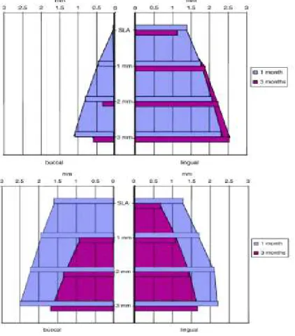
Multiple studies are realized but we are specially interested about the thickness of vestibular and lingual bone (three measures at 1,2 and 3mm from the point SLA). The SLA point corresponds to the most coronal level of the rough surface of the implant.
Figure 1: Size representation of vestibular (buccal) and lingual bone; the premolar side in the upper part, and the molar site in the lower part (Araujo, 2006).
After one month in the molar site, the thickness of the vestibular bone is more important than in the premolar site.
After three months, in the premolar site, there is no bone at the SLA point and 1 mm on vestibular side. On lingual the measures are similar after one month. In the molar site, there is no bone only at the point SLA. The bone resorption occurred on the vestibular and lingual side. Measures are realized on sockets without implants: 3,8 / 0,3 mm average for premolars and 5,8/0,2 mm for molars.
All implants placed were 4,1 mm diameter with different GAPS after implantation between bone and implants. GAP was more important in the molar sites but all healed after four months. Modifications are observed: in the premolar site on vestibular side there is a bone loss and loss of the BIC (Bone Implant Contact). Although the bone GAP is more important in the molar site, there was a similar thickness of the vestibular bone at the moment of implantation.
The decrease of thickness of the vestibular alveolar wall is more pronounced on molars than premolars and more on the vestibular part.
On premolars, the thickness of the lingual wall stayed unchangeable between four and twelve weeks. A bone formation occurred and is suggested by the bone loss on the vestibular side.
Another affirmation by the authors: when implant placed against a thin bone wall, the risks of the bone loss resorption are important. It seems that the small space between the implant and the alveolar wall don’t prevent the bone resorption and the risk of exposition of implant with large diameter after healing. But their radical conclusion was that placement of implant after tooth extraction was a failure in preservation of bone volume.
Other studies were interested on the horizontal bone loss like study of Chen and coll (2007). Thirty trans-gingival implants were placed with Bio-Oss® (1), and with Bio-Oss®
and membrane (2), or without. The results were: loss of 15,8 /16,9%, 20/21,9% and 48,3/9,5% .
An horizontal bone resorption is unavoidable but can be reduced by regenerating techniques. The positions of the implant in the alveolus have a major impact on the preservation of the vestibular wall and it’s better to place the implant more palatal and to fill the bone GAP with bone substitute when necessary. A safety distance of 2 mm must be maintened between the external part of the vestibular cortical bone and the implant. Chen and his team concluded the use of bio-material reduce the horizontal resorption but not the vertical resorption of the vestibular wall (resorption caused by the small thickness of this vestibular wall).
Some techniques can reduce the horizontal bone resorption, but the immediate placement of an implant in the alveolus cannot prevent this resorption during the post extractional healing phase.
b) Vertical proximal resorption
The vertical bone resorption on mesial and distal level of immediate implants placed after extraction is very low during the healing period (four month) (Botticelli et al., 2004). It’s an average of 0, 2/0,7 mm on mesial, and 0,5/0,9 mm on distal.
In fact, the periodontal ligament of the teeth bordering the implantation site has a supplementary vascular supply, and prevents the vertical resorption of the proximal bone boarding the immediate and delayed implantation. The direct surrounding has an influence on the proximal bone resorption.
c) Vertical vestibulo-palatal resorption
Vertical and horizontal resorptions are certainly related. To continue with Araujo, we will analyse his 2005 study.
In this study, five beagle dogs are used, particularly their third and fourth mandibular premolars. After flap elevation, the mesial root is removed and an implant (4,1 mm) is placed with the healing abutment. Three months later, dogs are sacrified. Different measures are realized, particularly the reduction of the height of cortical bones after implant placement in the socket; in another empty socket and around a tooth close to the implanted site and another far away.
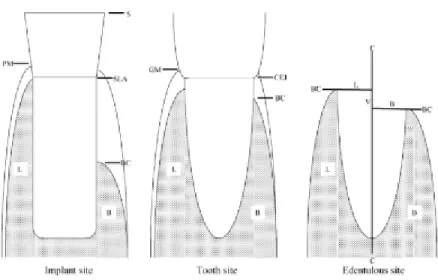
Figure 2: representation of different points of measure (Araujo, 2005) S: the coronal point of the implant
SLA: point 2,8 mm from S BC: crestal bone
GM: Gingival level CEJ: junction enamel-cement
In the implant site, the distance between point SLA and BC at the vestibular side is 2,6/ 0,4 mm and 0,2/0,5 at the lingual side. Far away in the dental side, the average distance between CEJ point and BC is 0,8/0,1 mm on vestibular side and 0,7/0,2 mm on lingual side. For the proximal dental sites, we measure 1,8/0,7 mm vestibular, and 0,8/ 0,1 mm lingual.
They conclude that vertical resorption is more pronounced on vestibular side than the lingual in all the sites (particularly the extraction site).
The immediate placement of implant in the socket cannot prevent the vertical marginal bone resorption.
However this vertical resorption seems to be under the influence of the vestibular cortical bone, but also of the peri-implantal defect.
iii. Healing inside the peri-implant defect
The shape difference between the natural dental root and the implant, as well as the difference between the implant diameter and the socket results in a peri-implantal defect.
During the first three months of healing, an opposition of bone tissue occurs inside the defect in parallel with the external bone resorption.
The healing process of this defect is studied by Paolantonio (Paolantonio et al., 2001). After six months of healing, the contact percentage between implant and bone is equivalent for implant placed immediately after extraction (64,8%) and others on healed bone (62,3%). Immediate implantation doesn’t compromise the osteo-integration in presence of the peri-implantal defect. It seems that bone defect with three walls is filled by remodeling bone without ingrowth of epithelial tissue and the horizontal component must be less than 2 mm. This reduction of the defect occurs without using of membrane or filling materials. However the bone defect can be filled spontaneously four month if it measured less than 2 mm (Boticelli et al., 2004), (Paolantonio et al., 2001). Studies of Evans and Chen (2008) demonstrate the utility of bone filling with collagenic membrane to reduce the resorption.
The extraction and immediate implantation do not prevent horizontal and vertical resorption, but some parameters have their influence: position of the implant and crestal level, the distance between implant and the vestibular cortical bone, and the thickness of bone.
3. Evolution of the immediate implantation concept
Bone healing during the osteo-integration period, around the immediate implants, results in horizontal resorption stable in frequency and value. This resorption presents the same characteristics on an empty socket without implant, and an immediate placement of implant after extraction doesn’t prevent this horizontal resorption. A marginal vertical resorption is observed systematically and depends on intra-operative bone conditions, and the implant position. For a long time, we thought that implantation after extraction could preserve the bone loss, but few studies prove that it has not been the case.
However it is interessant to note some points in the protocols with direct influence on the bone resorption. A literature review (Bousquet et al., 2011) on immediate implantation after extraction and the conservation of bone volume showed conflicting results due to the difference between the analysed parameters:
- the diameter of the implant: sometimes the diameter of implant is large and reduce the defect between implant and bone; sometimes defect is large and there is necessity of bone filling (with or without membrane).
- Surface and shape of implants
- topography of the socket : the integrity of the vestibular wall has a direct impact on the results.
- localisation of implants: bone tissue has not the same constitution in the maxillary or the mandibule, anteriorly or posteriorly.
- the selection criteria of patients (smoking, hygiene, cause of extraction…)
- evaluation of results : some measures concern all the periondontum, others concern only the bone level. We must note that a thick periodontum can hide an important bone resorption.
- position of the implant in the extraction site: primary in the results of bone resorption. It is not the same position in all the studies, and they didn’t use a tomographic section to evaluate the position of the implant in the bone or the thickness of the vestibular bone after healing.
a) Position and size of implants
The position of implant remains a key point. While the thickness of the vestibular cortical bone is important, why many studies were not concentrated on this point? On the anterior segment, the tooth are closed to the cortical bone, then if implants are placed exactly in the extraction site, the security thickness is not respected. When the team of Araujo concluded a failure of the immediate extraction in the preservation of bone volume (Araujo et al., 2005) we can discuss two aspects:
- the implant diameter: the diameter of the implants used in the study is 4,1 mm and the sockets of beagles dogs measured 3,6 mm; this provokes an over drill with negative effects on the preservation of bone mass.
- Position of the implant : placed more vestibular as illustrated in this cross section :
Figure 3: Cross section of the implantation site after three months of healing (Araujo et al., 2005)
To benefit from all advantages of the extraction and immediate implantation, you have to place a small diameter implant more palatal and use bone filling materials when necessary. This successful technique adopted by Araujo and team in 2011 (Araujo et al., 2011). In these conditions, this concept presents more advantages like possibility of placement of temporary crown. In fact, on the anterior sector, many authors recommend immediate implant placement to guide the healing of soft tissues and preserving of gingival architecture (Jemt, 1997).
The immediate implantation concept has been developed after multiple studies explaining that placement of the implant in the same position of the extracted tooth was not an ideal solution. Large implants were used to hold on the cortical bone and minimize the defect between implant and vestibular cortical bone. Finally, we realized that this can be traumatic to the vestibular cortical bone and may expose the surface of the large implant, as well as in a case of small diameter implant the defect may be filled with or without bio-materials, it depends on the size of the defect during the healing period. The position and the diameter of the implant are modified (small diameter implant and palatal position) and associated with bone filling methods.
b) Principles of successful immediate implantation
The extraction and immediate implantation concept is based on different principles to perform best results:
- atraumatic extraction of the tooth
- optimize the positioning of the implant in the socket
- obtain primary stability: by drilling beyond the apex of the socket minimum 3 mm (Antoun et al., 2007) with less drill of the implant site in the apical part to increase the insertion stability of the implant. This implant must have the shape, size and surface adapted on the received area.
- regenerate the bone in the defected areas, and maintain a tight contact between implant and alveolar walls with filling bone materials and membrane if necessary.
- obtain an hermetic flap repositioning in the regeneration techniques.
4. From immediate implantation to delayed implantation
i. Clinical situations, indications and patient selections
The success of the extraction and immediate implantation technique depends not only on the respect of indications, and good patient selections; the pre-implant analysis is important and we should be interested particularly on the cause of extraction, and on biotype of tissues and alveoli.
In fact, the cause of extraction may result a contraindication of extraction and immediate implantation (case of periodontal infections and acute endodontic infections).
However there are classical indications: traumatologic, endodontic failure and periodontitis with slow evolution at terminal stage, replacement of persistent temporary teeth replacement of fractured implant
Also the biotype must be known because has influence on the prognosis: thin biotype induces less stability, the bone is thin and bone regeneration must be done.
There are difficult clinical cases with possible complications, A thick biotype has good prognosis and is suitable for extraction and immediate implantation (Touati, 2010).
The destruction of the alveolar site during the tooth extraction and a severe interproximal bone loss without inter-dental papilla prevents this technique. For an accurate assessment, a cone beam must be realized it’s important to explore around the tooth to evaluate if bone
remains intact, partially, or totally absent. This will inform about possible future papilla, and thickness of soft tissues.
Elian (Elian et al., 2007) has established the indications of extractions and immediate implantations depending on the tooth socket:
- in class 1, hard and soft tissues are sufficient, and socket is intact.
- in class 2, small bone loss, but gingiva is in normal level.
- in class 3, important gingival and bone loss.
Tarnow (Tarnow et al., 2003) suggests extraction and immediate implantation only in class 1 and 2.
Each clinical situation must be analysed, the bone situation after extraction will guide to extraction- immediate implantation or to delayed implantation after a healing period.
ii. Bone environments
To take good decisions, the nature of bone defect must be analysed. The potential of bone repairing is related to presence of bone cells close to the site; it is interesting to classify the bone defects according to number of walls around the implant (Zuck, 2009).:
- bone environment with five walls: ideal situation, implant is totally surrounded by bone with optimal repairing potential. This category also concerns the remaining small gaps (1 to 2 mm) or more, but with limit at same level of the implant; in this case, implant is well placed with spontaneous repairing through “jumping effect”. It consists in differenciation of mesenchymal stem cells of the peri-implant blood clot under action of growth factors and morphogenic proteins of the environment bone walls. If there is a small gap, it is preferable to fill with autogenous bone if possible.
- bone environment with four walls: one part of the implant is not covered by bone tissue. This is caused by previous situation (periodontal infection) or during the surgical phase. In this categorie, there are coronal defects (limited to the third of the implant length), localized fenestrations, presence of coronal GAP more than 3 mm (on vestibular side with marginal edge apicaly relative to the implant top). Each defect must be studied specifically to choose the best therapies, but the bone healing is never spontaneous. We can realize a guided bone regeneration, autogenic bone placement or other substitute, and always hermetic sutures.
- bone environment with three walls: this includes coronal defects with exposition of more than 1/3 of the length of the implant, fenestrations, remaining of marginal GAP more than 3 mm with implant exposition of one or more proximal face in addition to the vestibular part. These cases are difficult and usually, absence of primary stability. This is preferable to defer the implantation and go through a bone graft.
In favorable cases we can go through guided bone regeneration, bone filling or connective tissue graft. Each case must be well studied and hermetic site must be realized. In case of fenestrations with the coronal part of the intact wall, there is no risk of resorption of soft tissues, but for some defects in anterior esthetic sector, the immediate implantation is impossible. But it is possible to manage the soft tissues during the extraction phase.
- bone environment with two or one wall: primary stability is impossible to obtain; we must go during preliminary important bone management, like fixing of autogenic bone bloc taken from a second intra oral site.
Figure 4: different types of bone defects (Zuck, 2009)
Bone resorption is similar if implant is placed immediately or in a second operative step. The immediate implantation doesn’t compromise the osteo-integration, provide time saving for patients preferring frequently rapid processing. However this concept can be established in specific cases. Without respect of contra indications, the result will be a failure. Unfortunately, sometimes post extractional bone conditions prevent the extraction and immediate implantation. We will be in obligation of realizing a delayed implantation.
II.
DELAYED IMPLANTATION
An implant can be placed in two different circonstances: directly after extraction of the unpreserved tooth, or on edentulous healed site since more or less longtime. When the tooth is present, it is difficult to take decisions of treatment sequences; the decision can be definitive during the operation after tooth extraction. With absence of good conditions, the implantation must be delayed, then we go to classic cases with all indications and protocols recommended by Per-Ingvar Branemark.
We make the tooth extraction, and wait for a complete healing of tooth socket; in the cases of tooth infections, we wait for the resolution of the infectious problem. After eight weeks, the socket reparation will be almost complete, and after three months, we consider that more than 2/3 of thickness reduction of the crestal bone is achieved (Schropp, et al., 2003).
After extraction , it is possible to use bone filling materials with or without membrane to prevent the bone resorption, and improve the area of future implant placement.
The result must be analysed: is there enough hard bone and soft tissue, or we must increase?. Different possibilities to optimize the bio-esthetic success:
- first we must manage the mucosal tissue, the bone defect is managed after at the same time of the implant placement.
- the three steps are dissociated: we have a mucosal step, bone step and finally implantation.
1. Alveolar preservation
In order to prevent first months bone resorption, and flap opening (if possible case), we can go through technique of socket recovering with connective tissue graft (Stimmelmayr et al., 2009); it associates crestal preservation to realize filling by the natural opening of the socket, non invasive and alveolar recovering. Iglhaut and coll. was the first using connective tissues graft with epithelium to recover the post extractional socket (Iglhaut et al.,2006). These pedicles permit better vascularization of the graft and thickness of vestibular soft tissues. This is basic for formation of esthetic emergence profile, support papillas and prevent shrinkage of attached gingiva .
A study of twenty-eight filling is realized; a bone bloc taken from retro molar area is crushed, mixed to bio-oss and the patient’s blood. An atraumatic extraction of the tooth and supra periostal tunnels realized to permit positioning of pedicles epithelium and connective tissue. The mixture is placed in the tooth socket, and bio-guide membrane is placed in palatal direction on the bone graft. The epithelium and connective tissue graft taken from the palate are placed. The implantation will be five or six months after bone filling (a removable denture is placed). Non invasive implantation realized after crestal incision. (shifted to palatal) and sulcular incisions. Operating area closed and a period of three to five months is respected before the second surgical step and temporary charge on the implants. Finally twenty-five implants posed after crestal preservation but in five cases, a vestibular filling was necessary at the same time of the implantation. The success rate of this technique is 92,9% successfully compared to lateral filling techniques : 92% to 100%. (Chiapasco et al., 2006).
This method also realized with bio-oss allow pre-compensation of post-extractional resorption by proceeding oversize and crestal preservation. The graft limits the soft tissue’s invagination in the first weeks after extraction. In fact, it protects the blood clot. The gingival cells proliferates faster than the bone cells, the gingival invades the socket and change the healing of bone tissues. The graft optimize the bone healing, maintain the marginal gingiva , preserve bone volume and create keratinized gingiva . In case of filling
after healing of bone tissues, we must realize a flap and open the periosteum of the alveolar process resulting a new resorption. The presence of pedicle epithelium and connective tissue permits better vascularization and better integration rate of the graft compared to connective tissues only.
2. Management of soft tissues
Each prosthetic technique on the anterior sector pass through necessity of esthetic results. Soft tissues in addition to hard tissue have a significant role in both quantitative and qualitative terms. Some periodontal biotypes are easy, others need more precautions and more treatments before receiving implants. We must observe the residual keratinized tissue before placing the implant in order to see if we have to increase or not. If this keratinized mucosa doesn’t appear essential to obtain osteo-integration and functional success (Wennstrom et al., 1994), we admit about 3 mm in heightto optimize the esthetic result, limit the risk of tissue recession, and improve the dental plaque control (Berglundh et al., 1992). There are different techniques of manipulation of peri-implants soft tissues described before in parodontology and adapted in implantology. They focused of improving the quantity of tissues. We will discuss the different techniques for alveolar preservation and management of soft tissues. This is a large subject but, we will see the most common, however some of these techniques can be used during the post extractional implant placement.
i. The different techniques
- the technique of connective tissue or envelope technique (Langer et al.,1985). The connective tissues taken from the palate is placed under the flap moved coronaly to fill the small gaps of the vestibular outline of the edentulous ridgeor to optimize the quantity (and the quality) of the mucosa. This can be realized before or during the implant operation.
- pedicles graft for vestibular thickening or roller technique (Abrams, 1980) . With this method, we slide under the vestibular flap the connective tissue prepared from the palate or the crestal side, in case of wide crest.
- modified flap for vestibular thickening (Martinez et al., 2004 ).This non invasive technique permits with thin biotype the increasing of vestibular thickness relative to the connective tissues localized on the alveolar crest. Concerning the quality of soft tissues, there are many processes to protect the appropriate mucosa or to correct an unfavorable mucosa. When we are in an appropriate situation with keratinized mucosa covering the vestibular and palatal crest (at least 5 mm), a technique without flap can be realized. We prevent all risks of tissue modifications. Without going into the “flapless” implantation, we must know that this technique can be realized in specific and appropriate situations (wide crest, absence of bone concavity…). When enabling conditions, we can realize extraction and immediate implantation, without flap in the same operation to preserve natural tissues. In other appropriate situations, when vestibular is almost 4 mm, we have to realize a planning for adaptation of residual architecture in determining the location of the first incision, which is primordial to the new positioning (Martinez et al., 2004) of tissues. The choice of the other components is also important (neck of implant, anatomy of the adapted healing abutment). They influence on the shape of tissues, and give a new peripheral architecture to the soft tissues. Frequently, we realize a crestal incision with vestibular moving of the keratinized mucosa. After few weeks of maturation, the new shape of the mucosa will give an optimized tissue aspect. In situations without keratinized mucosa on the vestibular side, we have to realize an important tissues supply. Frequently we go to epithelium and connective tissue graft taken from the palate. This technique described by Bjorn in 1963 can be realized during or before the implant placement (alternatively with the graft of pedicles connective tissue). If keratinized mucosa is present, but small, we can realize a shifted incision with keratinized mucosa moved apically (Martinez et al., 2008). We must notethat pedicles graft allow better vascularization with better prognosis than simple tissue graft.
ii. Different times of the soft tissues management
It’s logical to assume that soft tissues management may be realized at different stages of the implant treatment: before implant placement, during the implant placement, during the second surgical step (healing abutment placement) or lately during the maintenance step. The most frequent stages: before and during implant placement, with different objectives:
before implant placement: we preserve the bone tissues and obtain a favorable biotype.
- the alveolar preservation: in order to prevent gingival invagination in the bone compartment, an epithelium and connective tissue graft can be realized. This protects the blood clot, optimize bone healing, and preserve the soft tissue volume creating keratinized gingiva .
- connective tissue supply: esthetic result depends on the proportion of the crown in height and width. The situation of papilla and gingiva and their symmetry, are important factors considered to optimize esthetic results. In case of recession, there is necessity of recovering by connective tissue graft. Because gingiva around the tooth is more vascularized than around the implant, and success of recovering depends largely on vascularization , it is preferable to realize this operation before extraction of the tooth.
the day of the implant placement: we look for creating and thickening of peri-implantal tissues.
- thickening of peri-implantal tissues: stability of the marginal gingiva depends on two anatomic parameters: presence of bone tissue under the peri-implantal gingiva, and the thickness of the gingiva . In order to improve the situation of the marginal gingiva , we can, when insufficient gingiva , increase the thickness by connective tissue graft or rolled technique described before.
During the maintenance phase: it is the moment to correct the defects of mucosa like recessions. The frequent method is combination of palatal graft with flap repositioned
laterally (Nelson, 1987) but this technique go through unpredictable results. It seems that soft tissues management is optimal when managed as soon as possible. Indeed tissues are in their original position and still have possibility to re-operate later. However association of implantation and management of soft tissues is certainly an effective medical procedure. The type of our clinical objective determines the best moment to manage peri-implantal soft tissues. If the muco gingival surgery of peri-implantal tissues brings solutions to integration, and stability of tissues, it cannot correct the errors of implant treatment; mainly the size and position of implant. Because of principal impact on the long term result of the implant reconstitution, management of soft tissues is now an indissociable step during the implant treatment.
3. Management of bone defects
i. Guided bone regeneration technique
Guided bone regeneration is realized in implantology since years 1980. This technique comes through the guided regeneration tissue technique; indicated for treatment of bone defects associated or not to the surgical implant placement. The basic principle is to manage a space between bone defect and soft tissues to promote remodeling. The size of this space is equivalent to the regenerated bone volume. It is based on the clot stability and the principle of cells selection enabling bone formation; epithelial and conjunctival cells are excluded from the site due to a membrane enabling osteoblastes responsible of the bone neoformation. There are two types of membrane: absorbable preventing a second surgical operation with low mechanical quality; and non absorbable armed with titanium. In all cases, isolation is essential (flap with hermetic sutures) associated to follow up visits and good buccal hygiene.
This technique can be associated with a bone filling. Without bone substitute, membrane is away from the bone defect; but in the two cases, the membrane must be well adapted (covering all bone defects, away from roots of adjacent teeth). In implantal tissue reconstruction, indications of guided bone regeneration: local increasing of the alveolar
crest, bone dehiscence after implant placement and peri-implant fenestrations (Martinez et al., 2008).
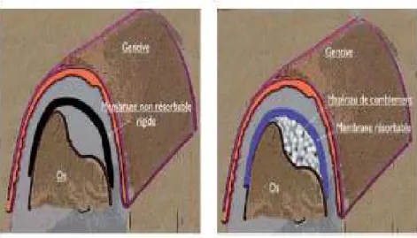
Figure 5: Guided bone regeneration with bone substitute (right)/without bone substitute (left) (Nevins, 2010)
If until now hermetic site is essential to avoid a failure, we try to obtain a membrane that may be exposable. This may be the case of Dynamatri (keystone dental , Voiron, France), that can permit more predictable results during the extraction and immediate implantation and prevent graft techniques (Nevins, 2010).
ii. Bone graft
a) Different sources
Xenografts: the origin is natural, not human (usually bovine). These grafts are treated in
without rejecting risk, but can potentially cause an immune response. The bio-oss frequently used in implantology is produced from bovine bone.
Synthetic materials: in order to prevent risks of contamination, there is synthetic materials
(ceramics, calcium carbonate…) steriles, healthy and well tolerated, absorbables or not.
Allograft: the origin is from a human person, but not the one who receives the graft. Like
xenograft and synthetic materials, they don’t require a second surgical operation on the same patient.
Autogenic bone: this bone remains the gold standard, and is used preferably when it is possible. There are different benefits: high osteogenic potential, osteo-inductive and/or osteo-conductive, with rapid cicatrisation. In case of insufficient quantity, it must be associated with synthetic materials. Bone tissue is taken mostly from intra-oral sites of the same person receiving the graft; there are two operating sites which is the only disadvantage in this technique. Then the bone is crushed to be used as thin particules, in case of guided bone regeneration; but it can be used in high volume in the grafts. We will see the collection bone areas in the apposition graft.
b) Bone apposition graft
When bone defect is too important for guided bone regeneration success, we go through apposition graft. The indications of onlay grafts: important bone less of external cortical bone, high bone resorption and advanced combined crestal defect. Donor sites can be extra orals (iliac crest, tibia or parietal bone) when there are important defects and high resorptions of alveolar crests requiring large bone quantity. This is clear significant disadvantages: long recovery period, frequent pains, and necessity of general anesthesia. Collection sites remain frequently intra-orals: grafts can be trabecular, corticals or cortico-trabeculars. Trabecular grafts contain more osteogenic cells than cortical bone grafts, but contain more morphogenic proteins (BMP) essentials to the bone formation. Chin symphysis and mandibular ramus are the primary prelevation sites, and sometimes, from
zygomatic arch, and coronoid process. We will not detail protocols and incisions, but we must get attention on anatomic obstacles. This technique will increase the intervention time uncomfortable and more risks of infection. In general, maxillary bone apposition graft has best prognosis. But for successful result, the bone has to be well fixed without mobility, protected from bacterial infiltrations through hermetic flap and sutures. Implant placement will be after four months.
Guided bone regeneration technique and bone graft improve the bone site by filling the bone defects. Each technique has specific indications to achieve success and optimal positioning of implants. These techniques and specially the guided bone regeneration technique can be used at different moments of the treatment, and can be associated to other soft tissue operations (Martinez et al., 2008, Princ et al., 2008). There are many techniques to maintain and increase soft tissues, but the bone quantity below is essential to preserve. We try to see the better to conserve bone tissues or recreate bone.
When it is absent for a specific purpose: positioning of the implant where we want, not where we can, because prosthesis remain our guide. Many solutions are presented with protocols and with different times of management. Selecting a technique is related to our objectives, we can prevent the defects if we go early to the concerned critical cases, and we still have possibility of re-operating in case of failures. There are different possibilities with management of hard and soft tissues at same time, or at different moments. With the post-extractional implantation, the guided bone regeneration remains successful. This success doesn’t need more demonstration (Chen et al., 2007). Analysing bone defect to choose the technique remains essential for success. We have seen before the adapted protocol based on the number of bone walls around the implant, this can be also adapted for bone defect of post-extractional tooth socket. The same reasoning for a healed site; we must go through these techniques to prevent recessions, dehiscences and implant failures. They often require special cares. We must be adapted on their evolution, old techniques although remains practicable (envelope technique and rolled technique).
III.
SOCKET
SHIELD
TECHNIQUE
IN
IMMEDIATE
IMPLANTATION
1. The new technique to avoid buccal bone resorption
i. Description and definition
Several methods have been described to avoid the negative effect of an extraction like immediate implant (Boticcelli, 2004), (Araujo et al., 2006), barrier membranes (Lekovic et al., 1997) although the most suitable technique advocated to preserve the volume of the socket is the ridge preservation (Araujo, 2009). Lately, a new technique is being described as an option to perfom an immediate implant without the negative consequences of the bone remodeling after an extraction (Hürzeler, 2010) , and the rationale behind this technique is preserving a tooth fragment that will avoid the post extraction resorption. Although this technique is quiet promising, we should be aware of the incoming publications about a larger follow up of this technique, and the predictability of leaving a fragment inside the socket after an extraction (Baumer et al., 2013).
The socket shield technique is a recent method to avoid buccal bone resorption when immediate implants are performed. We should wait for new literature about this technique with larger follow up before applying it on our daily practice .
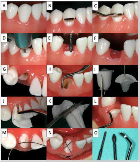
Background to avoid tissue alterations of the ridge tooth extraction, the socket shield technique was first introduced in 2010 by Hürzeler. It was suggested that instead of extracting the whole tooth, the buccal aspect of the root could be left intact to preserve the buccal plate of bone, and prevent post-extraction resorption , at the same time an immediate implant is placed this would lead to an optimal stable esthetic result after the final delivery of the restauration. To extract the tooth while keeping the buccal aspect intact, a fissure bur is used to cut the tooth medio-distaly, then the lingual aspect of the tooth is extracted leaving a socket where the implant is to be placed.
ii. Clinical concept of ridge preservation with modified socket shield technique
With the root submergence technique (RST), submucosal root retention can virtually eliminate bone resorption (Salama et al., 2007). Based on this concept, the retention and stabilization of the coronal and buccal bundle bone and the retention of the periodontal membrane by retaining a coronal tooth fragment (so called “Socket shield”), including adequate blood supply, can be expected.
To ensure complication-free healing, special attention should be paid to wound stabilization: stabilization of the clot with a criss-cross suture is optimized by placing a collagen cone with integrated collagen membrane, such as collagen sponge with integrated membrane into the tooth socket. Depending of the individual treatment plan of a patient, there is the option to either wait for two to six months to allow for the formation of new bone, followed by implantation, or to leave the site without subsequent second procedure.
2. Indications and procedure for ridge preservation
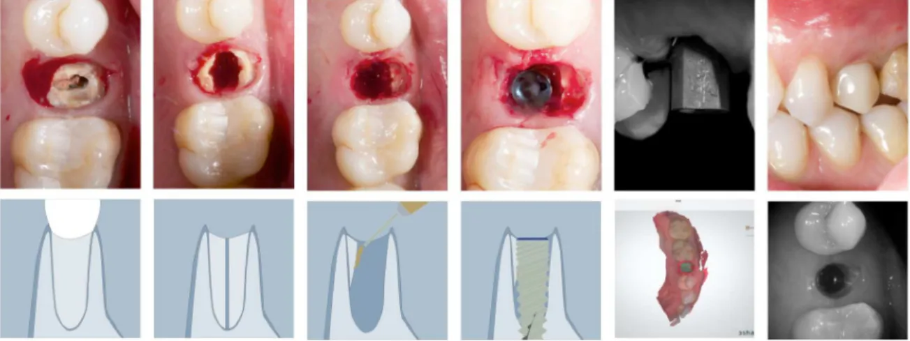
Potential indications of such techniques include their use as part of the (delayed) late implantation approach or the optimization of pontic support in crown-bridge reconstructions, or to improve the prosthesis base for removable dentures. As general contra indications, the usual restrictions for oral surgical procedures (biphosphonate medication, immuno-suppression, radiation therapy, anticoagulation,…) . Local contra-indications include an absent buccal lamella, which develops for instance after vertical root fractures or periodontis. A step by step illustration of the proposed procedure using a model illustrating the given in the figure.
Firstly the hopeless tooth is split supragugivally (B) and the crown fragment is carefully dislocated and removed using a suitable instrument (C). The root is separated vertically in a ration between 1/3 and 2/3 (D). The smaller buccal root fragment is retained and the larger lingul root fragment is removed in a manner that spares bone and soft tissue to the greatest possible extend. The height of the buccal socket shield is reduced to the level of the bone (E,F), and the gingiva overlying the retained buccal root fragment is tunneled by 2mm (H) to allow the insertion of the collagen cone (I, L) into the tooth socket and placement of the membrane part of the collagen cone under the buccal mucosa.
Finally, the collagen cone is secured with a criss-cross suture (M, N).
After the procedure, patients rinse with 0,2% chlorexidine mouthwash two or three times daily for one minute over a period of at least ten days. During this time mechanical oral hygiene is avoided in the affected area and only restarted after the follow-up examination, and suture removal after ten days. Anti-inflamatory drugs are prescribed as needed. Typically no antibiotics are prescribed. Each patient was informed verbally and in writing about the treatment, and the materials used as well as the associated pre and post-operative risks and gave their written consent to the use of the collected data and photos.
3. Socket shield technique to support the bucco-facial tissue at immediate
implant placement
Tooth loss and subsequent ridge collapse continue to burden restorative implant treatment. Careful management of the post-extraction tissues is needed to preserve the alveolar ridge. Instead of surgical augmentation to correct a ridge defect, the socket shield technique offers a promising solution. As the root submergence technique retains the periodontal attachment and maintains the alveolar ridge for pontic site development, retention of a prepared tooth root section as a socket shield prevents the recession of tissues bucco-facial to an immediately placed implant.
The retention of the bucco-facial root section at immediate implant placement achieved osteo-integration without resorptive response of the ridge bucco-facial to the implant (Hürzeler, 2010). The technique offers a viable solution when managing the post extraction ridge and its complications associated with immediately placed implants. Prior to the socket shield technique the implant surgeon conventionally, was to select between an immediate placement protocol with an augmentation of the jump gap, with or without bulking of the bucco-facial soft tissues, or a delayed approach with additional surgical intervention to correct an existing ridge defect (Baumer, 2013) overbuilding the ridge buccal/facial to the implant by guided bone regeneration and soft tissue augmentation can only partly compensate. A wealth of literature supports these ridge management techniques, but an amount of shrinkage with healing is to be expected. Moreover, healing is not without complication by infection and complete failure with a worse outcome is possible. Alternatives are thus desired and the benefits of the socket shield technique can be appreciated.
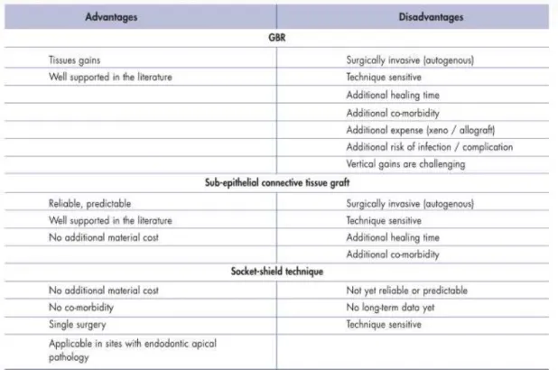
Table 1: Comparative tabulation of procedure to manage the effects of post-extraction resorption adjunct to implant therapy (Gluckman 2015).
First reported in 2010, the socket shield technique had progressed from concepts introduced in the 1950 s that the retention of a tooth limits tissue alterations following extraction. The submergence of tooth roots was introduced originally to preserve alveolar ridge volume beneath removable full prostheses. Malmgren and coworkers had also more than three decades ago reported successful tissue regeneration around submerged tooth roots (Malmgren et al., 1984). Thereafter submerging a tooth root for pontic site development has become a well-documented treatment. Salama and coworkers reported on preserving of the alveolar ridge when developing pontic sites (Salama et al., 2007). This technique typically decoronates the tooth at the bone crest or preferably 1 mm above it so as to preserve the supra crestal fibers with epithelial and connective tissue attachment. By comparison, ridge preservation techniques may reduce the amount of ridge resorption but cannot prevent the loss of interdental bone and papillae.
Preservation of supracrestal fibers however can better develop pontic sites by in turn preserving the papillae, and thus it has been shown that the retention of part of the tooth, its fibers and reticulate vascularity interconnected with bundle bone, eludes the physiological remodeling of an extraction socket and the alveolar crest. These delicates tissues can be preserved, bundle bone, buccofacial plate and overlying keratinized mucosea (Filippi et al., 2001). It can be postulated that retention of part of the tooth as a socket shield eludes the body from mealizing the tooth has been extracted and circumvents the normal events of physiological healing that would resorb the alveolar socket.
The resorption of a post-extraction socket is the direct result of trauma to the bone periodontal ligament-tooth complex. Bundle bone born from a functionally loaded periodontal ligament is lost following extraction and sees an almost certain recession of residual buccofacial tissue (Gluckman, 2015).Complete maintenance of ridge volume after tooth extraction with preservation techniques utilizing currently available materials as a primary is not yet possible (Baumer, 2015).
However as started before, the retention of tooth roots in the alveolar process can preserve the ridge tissues. Histologically, this was demonstrated by Hürzeler and coworkers
(Hürzeler, 2010). Their report confirmed the retains attachment of the socket shield to the buccal plate via a physiologic periodontal ligament free of any inflammatory response. The buccal plate crest showed an absence of osteoclastic activity – an absence of active remodeling. The coronal soft tissue demonstrated a physiologic junctional epithelium also free of any inflammatory response. The clinical outcome of Hurzeler and coworkers report presented the successful osseointegration of an implant placed simultaneous to the socket shield technique, and a restoration with prosthetics indistinguishable from the adjacent maxillary central incisor. Whilst the authors reported preservation of the buccofacial tissues, it should be noted that absolute preservation has not yet been shown. The authors later reported a mean of 1 mm horizontal loss after final restoration, Chen and coworkers reported 0,72 mm of buccal resorption (Baumer, 2015),(Chen, 2013). In spite of the histological and clinical finding to date and the prospects of the socket shield technique, to safety apply a newly introduced treatment in everyday practice data from long-term clinical studies are required and at present this data is not yet available. Only one case series with a two year or more follow up of a significant number case exists in the literature (Siormpas et al., 2014). However that technique differed significantly. The authors had prepared the implant osteotomy directly through the intact tooth root and thereafter prepared what they termed the “root membrane”. That said the study is a significant contribution to literature on these techniques. Most have also deviated from the original protocol. The modified (proximal) socket shield reported by Kan and Rungcharassoeng had the input gap grafted with a xenograft material, the facila soft tissues augmented (Kan, 2013). In their report, the methodology further differs by sectioning the socket shield into mesial and distal sections for the purpose of the papillae preservation by a modified socket shield sectioned in a similar manner (Chevel, 2014).
4. Socket Shield technique to replace the conventional immediate implant
placement
Implantology invisible frontal region demands extreme precision due to the high aesthetic requirement of patients. Years of age immediate implant placement was considered main approach for preserving bone volume after tooth extraction due to its close relation to the
tissues to the periodontal ligaments. Therefore a risk of losing vestibular bone height and respectively soft tissue which is unacceptable from aesthetic point of view. One of the methods used in order to avoid bone loss is the so called “socket-shield” technique published by Hürzeler and coworkers(hürzeler et al., 2010).
Comparison of the results gained after immediate implant placement by conventional and socket shield techniques for a period of two years, twenty-six titanium screwed implants placed in post-extraction socket with conventional immediate implantation and socket-shield technique, xenogenic bone-graft material, PRGF, individualized factory titanium interface, press-ceramic e-max, metal-ceramic. Sector X-rays and intraoral photos for determining bone and soft tissue loss after immediate implant placement. It was discovered minimal from functional point of view but unacceptable from aesthetic point of view vertical bone loss of the vestibular lamella at conventional implant placement. To compare in socket shield technique, there were neither functional nor aesthetic changes in soft and hard tissues. Socket shield technique is already a routine practice in the arsenal of high-aesthetic immediate implantology and should be used when it is indicated.
i. Preservation of hard and soft tissues
There is a dramatic remodeling of hard and soft tissues after Tooth extraction. Data reported in clinical studies indicate that an overall reduction in the horizontal dimensions occurred following tooth extraction and that the resorption of the buccal part of the ridge was more pronounced than the lingual part (Pierokovski et al., 1967, Schropp 2003). Similar observations were also made on histological evaluation in an animal experiment by Lindhe and Araujo (2005). In this way morphology of the healed alveolar ridge following tooth extraction is almost always presenting with discrepancy in bone height between the two bone plate of the alveolar ridge, lingual and buccal. It is known that physiologic processes taking place immediately after tooth extraction up to the end of the first week include increasing the number of osteoclasts on the inner surface of the socket walls indicates that the bundle bone, which is closely related with the periodontal tissue is being resorbed. Anatomically buccal bone plat of the teeth is thinner than lingual or palatal. There