1. Associate member of the BSCVS, Heart surgeon of PROCAPE-UPE, Invited professor of the University of Pernambuco – UPE. 2. Full member of the BSCVS, Heart surgeon of PROCAPE-UPE, Invited professor of the University of Pernambuco – UPE. 3. Full member of the BSCVS, Full Professor of the University of Pernambuco – UPE; Heart surgeon of PROCAPE-HUOC. 4. Full member of the BSCVS, Professor Adjunto of the University of Pernambuco – UPE, Cardiothoracic surgeon of PROCAPE-HUOC. 5. Full member of the BSCVS, Professor Adjunto of the Federal University of Pernambuco – UFPE, Cardiothoracic surgeon of PROCAPE-HUOC.
Work carried out in Hospital Jaime da Fonte (HJF), Recife, PE. Correspondence address:
Alexandre Motta de Menezes. Rua das Pernambucanas, 167 (Hospital Jayme da Fonte - Cecordis), Graças - Recife - PE. CEP: 52011-010 Phone: 81-3221-2222 (com) 81-8833-9099 (cel).
E-mail: [email protected]
Alexandre Motta de MENEZES1, Frederico Pires de VASCONCELOS2, Ricardo de Carvalho LIMA3, Mário Gesteira COSTA4, Mozart Augusto Soares de ESCOBAR5
Article received in December 7th, 2006 Article accepted in June 6th, 2007
RBCCV 44205-888
Aspectos técnicos na esqueletização da artéria torácica interna com bisturi ultra-sônico
Technical aspects in skeletonization of the internal
thoracic artery using an ultrasonic scalpel
Abstract
Objective: To describe the technique and evaluate the immediate results of using an ultrasonic scalpel in the skeletonization of the internal thoracic artery for coronary artery bypass grafting surgery.
Methods: From January 2000 to October 2006, 188 patients were submitted to coronary artery bypass grafting with the internal thoracic artery skeletonized using an ultrasonic scalpel. Seventy-one patients (37.8%) were women. The patients’ ages varied from 28 to 81 years old. The entire internal thoracic artery was exposed opening the endothoracic fascia using scissors as close as possible to the arterial adventitia. An ultrasonic scalpel was used to transect and coagulate all the intercostal branches, thereby minimizing the use of metallic clips.
Results: The skeletonized internal thoracic arteries presented with excellent flow, obviating the need for
intraluminal manipulation for vasodilatation. In the immediate postoperative period, two patients were found to have temporary left-sided diaphragmatic paralysis. There were no sternal wound infections in this series. The dissection can be performed in approximately 33 minutes however with more experience this time may be reduced.
Conclusion: This technique facilitates and shortens the internal thoracic artery skeletonization procedure and does not cause arterial spasms. Cauterization of the collateral branches with an ultrasonic scalpel is efficient and the use of metallic clips is almost unnecessary. It is a procedure that is easy to reproduce and may be recommended as the first-choice technique for the dissection of the internal thoracic artery.
an ultrasonic scalpel, stressing the technical reproducibility and the results obtained.
METHODS
In the period from January 2000 to October 2006, 188 consecutive patients were submitted to CABG using skeletonized ITA grafts dissected using an ultrasonic scalpel. Seventy-one (37.8%) patients were female and 117 (62.2%) male. Their ages varied from 28 to 81 years old (mean 59.4 ± 9.5 years old) with 57 (30.3%) patients older than 65. Relevant preoperative data such as diabetes mellitus and chronic obstructive pulmonary disease are listed in Table 1.
INTRODUCTION
The utilization of the internal thoracic artery (ITA) is common in coronary artery bypass grafting surgery (CABG) [1,2]. The utilization of ITA for the anterior descending artery is associated to higher patency and greater survival than the use of saphenous vein grafts [3,4]. This arterial conduit can be used pediculated, skeletonized or as a free graft. Its use as a free graft or pediculated requires direct dissection by electrocautery, however, this technique implies much injury to the retrosternal region that may result in serious complications, including sternal infection, respiratory dysfunction, thoracic pain and paralysis of the diaphragm [5-7].
The ITA skeletonization technique has been shown to preserve residual sternal flow, reducing dissection injuries and, consequently, reducing the incidence of infection [8]. Thus, the use of skeletonized ITAs has been praised as a form of decreasing postoperative morbidity [9]. The classical technique of ITA skeletonization consists in the dissection of the artery using scissors and occluding intercostal branches using metal clips.
Higami et al. [10] and Lamm et al. [11] introduced the ultrasonic scalpel for skeletonized dissections of the ITA, with the objective of decreasing the time of dissection and injuries caused in the conventional technique.
The objective of this work is to report the experience of the authors with skeletonized dissection of the ITA utilizing
Resumo
Objetivo: Descrever a técnica e avaliar os resultados imediatos da utilização do bisturi ultra-sônico nas esqueletizações da artéria torácica interna, na cirurgia de revascularização do miocárdio.
Método: Foram operados com essa técnica 188 pacientes submetidos à cirurgia de revascularização do miocárdio, no período de janeiro de 2000 a outubro de 2006. Setenta e um (37,8%) pacientes eram do sexo feminino. A idade variou de 28 a 81 anos. A técnica utilizada na dissecação consistiu em expor toda artéria torácica interna, abrindo-se a fáscia endotorácica com tesoura o mais próximo possível da adventícia da artéria. Com o bisturi ultra-sônico é feita a secção dos ramos colaterais e sua respectiva hemostasia, dispensando-se o uso de “clips” metálicos na artéria torácica interna.
Resultados: As artérias torácicas internas esqueletizadas
com bisturi ultra-sônico apresentaram fluxos excelentes, não sendo necessárias manipulações intraluminais para vasodilatação. No pós-operatório imediato, dois pacientes apresentaram paralisia temporária da hemicúpula diafragmática esquerda. Não houve infecção do esterno nesta série. O tempo de dissecação foi de aproximadamente 33 minutos, mas com o aumento da experiência esse tempo pôde ser reduzido.
Conclusão: Essa técnica facilita e abrevia o procedimento da esqueletização da artéria torácica interna, não promove espasmos e a cauterização dos ramos colaterais com o bisturi ultra-sônico é eficiente, dispensando o uso de “clips” metálicos. É um procedimento de fácil reprodução, podendo ser recomendado para sua realização de maneira preferencial.
Descritores: Revascularização miocárdica. Ponte de artéria coronária. Artérias mamárias.
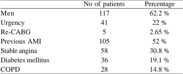
N = 188. CABG = coronary artery bypass grafting surgery; AMI = acute myocardial infarction; COPD = chronic obstructive pulmonary disease
Table 1. Preoperative data
Men Urgency Re-CABG Previous AMI Stable angina Diabetes mellitus COPD
No of patients 117
41 5 105
58 36 28
Percentage 62.2 %
22 % 2.65 %
The pulmonary arterial pressure was continuously monitored using a Swan-Ganz catheter. Additionally, routine monitoring was performed of the mean arterial and central venous pressures by cardioscope, the urinary outflow and pulse oximetry. A thermal mattress was used to assist the cooling or heating of patients during the surgery. Tranexamic acid (Transamine) was administered at a dose of 50 mg/kg for all patients after anesthetic induction. In patients submitted to cardiopulmonary bypass (CPB), anoxic arrest of the heart was achieved with administration of cold blood cardioplegia at the aortic root; topical ice was not used in the pericardial cavity. All the patients were prescribed oral diltiazem for the first six postoperative months. The study was approved by the Ethics Research Commission of the institution and complied with the norms of the declaration of Helsinki.
Ultrasonic scalpel
The Ultracision Harmonic scalpel (Ethicon Endo-Surgery, Cincinnati, OH) was used with a 5-mm HS2 manual applicator and curved or hook-type blades. The scalpel produces lengthwise motions at 55,500 cycles/sec or 55.5 kHz with heat emission of between 50ºC at 60ºC at the diamond tip during coagulation.
Dissection of internal thoracic artery
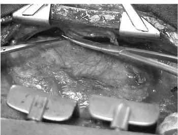
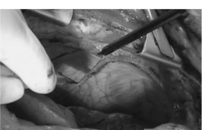
Median longitudinal sternotomy was performed with rigorous hemostasis, which facilitated ITA dissection; electrocautery and bone wax were used always when necessary for hemostasis. After dissection of the mediastinal pleural reflection and opening of the endothoracic fascia using delicate scissors and clamps, as close as possible to the adventitious layer of the ITA, the entire artery and satellite veins are accessed (Figure 1). Artery displacement and exposure of the intercostal vessel are carefully achieved using scissors or a fine spatula (sculptor) – Figure 2. We use level 3 USS power. The intercostal branches are coagulated for 2 to 3 seconds and sectioned as far from the arterial wall as possible (at least 1 mm from the ITA) (Figure 3), so that adjusted protein coagulation of the branches is possible and not to damage the ITA trunk [10]. We adopted the hook-type blade and considered level 3 ideal, because it presented good coagulation with a slower cut. Coagulation of venous vessels was avoided. The ITA was dissected from its distal bifurcation in the phrenic muscle and superior epigastric artery to its upper portion near to the first rib (Figure 4). After ITA dissection, gauze soaked in papaverine solution was wrapped around the entire artery.
After systemic heparinization at a dose of 4 mg/kg weight and before establishing CPB, the distal part of the ITA was occluded using a clip and sectioned. After sectioning, the
free blood flow without resistance was measured over one minute using a 100mL container and the arterial pressure of the patient was maintained at around 70 mmHg. After flow measurement, hemostasis of the graft was performed using metal clips, when necessary.
Fig. 1 – Opening of the endothoracic fascia using scissors, exposing the artery and satellite veins
RESULTS
At the beginning of the series, there was a case of an injury involving cutting the ITA which was then excluded from the study. Blood loss during dissection was very low. Significant vasospasms did not occur and intraluminal manipulations for ITA vasodilatation were unnecessary. It was observed that coagulation of venous vessels using the USS is not very efficient and that it is important to avoid handling these vessels. In a few ITA, occlusion of intercostal branches using clips was necessary due to bleeding.
The time of dissection ranged from 22 to 50 min (mean
33 ± 4.9 min). The mean free blood flow was 98.7 mL/min (± 27.5 mL/min). The diameter of the treated coronary vessels varied between 1.5 and 3.0 mm.
In five patients (2.6%), associated procedures were performed including aortic valve replacement (1.06%), mitral valve replacement (0.53%), left ventricular aneurysmectomy (0.53%) and the Bentall-de Bono procedure (0.53%). In 181 patients (96.27%), the left ITA was anastomosed in the anterior descending coronary artery. The two internal thoracic arteries were utilized in four (2.12%) patients. The radial artery was used in eight (4.25%) patients. The left ITA was used sequentially in six (3.31%) individuals. In this series, eighteen (9.6%) patients were operated on without the use of CPB.
Two (1.06%) patients presented with temporary hemidiaphragmatic paralysis. There were no occurrences of infection in the sternotomy (superficial, mediastinitis or osteomyelitis). One patient, who was submitted to CABG associated with aortic valve replacement, was re-operated in the immediate postoperative period due to blood dyscrasia. One hospital death occurred due to left ventricular insufficiency of a patient operated during the acute phase of myocardial infraction.
DISCUSSION
Recent studies have demonstrated that collateral circulation to the sternum is preserved when skeletonization of the ITA is used. The division of collateral braches utilizing clips and scissors preserves the sternal collateral perfusion through intercostal and muscle branches [12]. The use of electrocautery for the pediculated dissection of the ITA may destroy any possibility of maintaining the sternal collateral circulation [8,13] and it may be responsible for the higher infection rate in diabetic patients [9]. A comparison between the use of pediculated and skeletonized ITAs has been carry out by several authors and recently summarized by Athanasiou et al. [6] in a meta-analysis that emphasizes the main advantages of skeletonization, such as a lower sternal infection rate and longer grafts thus simplifying management of sequential grafts. The free blood flow can be considered satisfactory when compared to other studies [6], stressing that there was no intraluminal manipulation of vessels.
Even though used little in this study, the utilization of both ITAs (2.12%) has proved beneficial over the long term when compared to the use of only one ITA, chiefly in young patients [14-16]. However, the utilization of both ITAs in diabetic patients has been associated to a higher incidence of sternal infection [17,18]. Recent studies have demonstrated that, with the skeletonization of both ITAs, there is a reduction in this incidence, without differences in Fig. 3 – Coagulation and sectioning of the intercostal veins with
the ultrasonic scalpel
REFERENCES
1. Green GE, Swistel DG, Cameron AA. Bilateral internal thoracic artery surgery: 17-year experience. Eur Heart J. 1989;10(Suppl H):57-60.
2. Loop FD, Lytle BW, Cosgrove DM, Stewart RW, Goormastic M, Williams GW, et al . Influence of the internal-mammary-artery graft on 10-year survival and other cardiac events. N Engl J Med. 1986;314(1):1-6.
3. Singh RN, Sosa JA, Green GE. Long-term fate of the internal mammary artery and saphenous vein grafts. J Thorac Cardiovasc Surg. 1983;86(3):359-63.
4. Okies JE, Page US, Bigelow JC, Krause AH, Salomon NW. The left internal mammary artery: the graft of choice. Circulation. 1984;70(3 Pt 2):I213-21.
5. Keeley SB. The skeletonized internal mammary artery. Ann Thorac Surg. 1987;44(3):324-5.
6. Athanasiou T, Crossman MC, Asimakopoulos G, Cherian A, Weerasinghe A, Glenville B, et al. Should the internal thoracic artery be skeletonized? Ann Thorac Surg. 2004;77(6):2238-46.
7. Lehtola A, Verkkala K, Järvinen A. Is electrocautery safe for internal mammary artery (IMA) mobilization? A study using scanning electron microscopy (SEM). Thorac Cardiovasc Surg. 1989;37(1):55-7.
the morbimortality between the utilization of only one ITA or of both [19-23]. In our series, no cases of infections (osteomyelitis or mediastinitis) were observed even though electrocautery and bone wax were used for sternal hemostasis, when necessary.
Higami et al. [10] presented histological results of tissue submitted to USS, demonstrating its safety when used at a distance of more than 1 mm from the vessel; less than 1 mm may cause slight injuries in adjacent tissues. With coagulation resulting from collagen molecule degradation, the emanated heat is around 50ºC to 60ºC. In this study, microscopic analyzes were not performed although they are necessary to better evaluate the method. Lehtola et al. [7] and Yoshikai et al. [24] demonstrated, by electronic microscopy, that endothelial injuries of the ITA did not occur when USS was used. Experience derived from endoscopic surgery shows that the method is safe in vessels of up to 5.0 mm. Our experience was with vessels smaller than 3.0 mm, agreeing with the observations reported in the user’s manual provided by the manufacturer of the USS. One limitation of the USS is its utilization in venous vessels; its use in this type of vessel or in surgical injuries provoked during at dissection should be avoided.
Definitively adopted in endoscopic surgery, the USS was an important technological breakthrough for modern surgery. In conventional surgery, however, we feel the need for a more delicate instrument in respect to the applicator and the blades. Nevertheless, it is possible to skeletonize the ITA with the USS causing the minimum amount of injury and without the necessity of using metal clips, that in spite of their qualities can at any moment make the surgical process more difficult (collateral vessel injuries or excess of clips in the region at be anastomosed). The final aspect of ITAs skeletonized using the USS is very similar to a classically skeletonized artery, however, the time required for dissection seemed shorter to us, as initial occlusion of the ITA branches was unnecessary. Also the minimal surgical injury that we observed in the retrosternal region by macroscopy should be stressed.
In this series, only two patients, due to the return of symptoms suggestive of angina, were re-studied by cinecoronariography in the late postoperative period which demonstrated that the ITAs were patent. The postoperative cinecoronariography was not performed in a systematic way in this study thus this number is not statistically significant. However, authors such as Higami et al. [25] have demonstrated, by angiographic studies carried out in the postoperative period, that there are no significant differences related to the dissection technique in relation to graft patency. Diaphragmatic paralysis and a cutting injury of one graft occurred at the beginning of our series.
CONCLUSION
8. Parish MA, Asai T, Grossi EA, Esposito R, Galloway AC, Colvin SB, et al. The effects of different techniques of internal mammary artery harvesting on sternal blood flow. J Thorac Cardiovasc Surg. 1992;104(5):1303-7.
9. Bical O, Braunberger E, Fischer M, Robinault J, Foiret JC, Fromes Y, et al. Bilateral skeletonized mammary artery grafting: experience with 560 consecutive patients. Eur J Cardiothorac Surg. 1996;10(11):971-5.
10. Higami T, Maruo A, Yamashita T, Shida T, Ogawa K. Histologic and physiologic evaluation of skeletonized internal thoracic artery harvesting with an ultrasonic scalpel. J Thorac Cardiovasc Surg. 2000;120(6):1142-7.
11. Lamm P, Juchem G, Weyrich P, Schütz A, Reichart B. The harmonic scalpel: optimizing the quality of mammary artery bypass grafts. Ann Thorac Surg. 2000;69(6):1833-5.
12. de Jesus RA, Acland RD. Anatomic study of the collateral blood supply of the sternum. Ann Thorac Surg. 1995;59(1):163-8.
13. Seyfer AE, Shriver CD, Miller TR, Graeber GM. Sternal blood flow after median sternotomy and mobilization of the internal mammary arteries. Surgery. 1988;104(5):899-904.
14. Lytle BW, Blackstone EH, Loop FD, Houghtaling PL, Arnold JH, Akhrass R, et al. Two internal thoracic artery grafts are better than one. J Thorac Cardiovasc Surg. 1999;117(5):855-72.
15. Carrel T, Horber P, Turina MI. Operation for two-vessel coronary artery disease: midterm results of bilateral ITA grafting versus unilateral ITA and saphenous vein grafting. Ann Thorac Surg. 1996;62(5):1289-94.
16. Pick AW, Orszulak TA, Anderson BJ, Schaff HV. Single versus bilateral internal mammary artery grafts: 10-year outcome analysis. Ann Thorac Surg. 1997;64(3):599-605.
17. Cosgrove DM, Lytle BW, Loop FD, Taylor PC, Stewart RW, Gill CC, et al. Does bilateral internal mammary artery grafting
increase surgical risk? J Thorac Cardiovasc Surg. 1988;95(5):850-6.
18. Grossi EA, Esposito R, Harris LJ, Crooke GA, Galloway AC, Colvin SB, et al. Sternal wound infections and use of internal mammary artery grafts. J Thorac Cardiovasc Surg. 1991;102(3):342-6.
19. Gurevitch J, Paz Y, Shapira I, Matsa M, Kramer A, Pevni D, et al. Routine use of bilateral skeletonized internal mammary arteries for myocardial revascularization. Ann Thorac Surg. 1999;68(2):406-11.
20. Uva MS, Braunberger E, Fisher M, Fromes Y, Deleuze HP, Celestin JA, et al. Does bilateral internal thoracic artery grafting increase surgical risk in diabetic patients? Ann Thorac Surg. 1998;66(6):2051-5.
21. Calafiore AM, Vitolla G, Iaco AL, Fino C, Di Giammarco G, Marchesani F, et al. Bilateral internal mammary artery grafting: midterm results of pedicled versus skeletonized conduits. Ann Thorac Surg. 1999;67(6):1637- 42.
22. Matsa M, Paz Y, Gurevitch J, Shapira I, Kramer A, Pevny D, et al. Bilateral skeletonized internal thoracic artery grafts in patients with diabetes mellitus. J Thorac Cardiovasc Surg. 2001;121(4):668-74.
23. Bical OM, Khoury W, Fromes Y, Fischer M, Sousa Uva M, Boccara G, et al. Routine use of bilateral skeletonized internal thoracic artery grafts in middle-aged diabetic patients. Ann Thorac Surg. 2004;78(6):2050-3.
24. Yoshikai M, Ito T, Kamohara K, Yunoki J. Endothelial integrity of ultrasonically skeletonized internal thoracic artery: morphological analysis with scanning electron microscopy. Eur J Cardiothorac Surg. 2004;25(2):208-11.