UNIVERSIDADE DA BEIRA INTERIOR
Ciências
Synthesis and characterization of heterocyclic
rings linked to sugar moieties
Marta Moniz Santos
Dissertação para obtenção do Grau de Mestre em
Bioquímica
(2º ciclo de estudos)
Orientador: Prof. Doutor Maria Isabel Guerreiro da Costa Ismael Co-orientador: Prof. Doutor Arnaud Tatiböuet
iii
Para os meus pais
Para a minha irmã
v
Agradecimentos
Em primeiro lugar gostaria de agradecer aos meus orientadores, a Professora Doutora Maria Isabel Guerreiro da Costa Ismael e ao Professor Doutor Arnaud Tatiböuet, pela orientação dispensada ao longo do trabalho.
Gostaria também de agradecer à Universidade da Beira Interior e ao Institut de Chimie Organique et Analitique, por terem permitido o uso das instalações e de reagentes, e por toda a ajuda prestada
E por último à minha família, em especial ao meus pais e à minha irmã, e amigos pela ajuda, apoio e sobretudo compreensão, demonstrada ao longo deste ano de trabalho.
vii
Resumo
Este trabalho teve como objectivo a construção de cadeias ramificadas regio- e estereosselectivas, a parir da D-ribose e D-xilose, para sintetizar de N-óxidos de tio-imidatos derivados de açúcares.
Foram usados três métodos de síntese, dois deles a partir de açúcares, a ribose e a xilose, e o terceiro método foi iniciado por lactonas, nomeadamente a -valerolactona e a D-(-)-pantolactona. A síntese a partir da D-ribose deu origem a um N-óxido de tio-imidato, num rendimento global de 48%. A D-ribose, em acetona, reagiu durante uma hora com H2SO4
concentrado, adicionado a 0ºC, originou o composto 2,3-O-isopropilideno-β-D-ribofuranose (1), com os grupos hidroxilo em C-2 e C-3 protegidos, num rendimento de 95%. Este composto em MeOH, reagiu com NaBH4 durante uma hora e a 0ºC, de seguida evaporaram-se os
solventes e adicionou-se t-BuOH/H2O (3:2) e deixou-se reagir durante mais cinco minutos, e a
0ºC adicionou-se NaIO4, e deixou-se reagir mais doze horas. Esta é uma reacção de clivagem
oxidativa, e o composto obtido, num rendimento de 48%, foi 2,3-O-isopropilideno-L-eritrose (2). A oxima 3 foi obtida a partir deste, por reacção com NH2OH.HCl em piridina e peneiros
moleculares durante doze horas, composto com isomeria geométrica (E e Z), sendo o rendimento desta reacção de 68%. A reacção seguinte foi a protecção dos grupos hidroxilos restantes, com TBDMS.Cl e piridina, foram obtidos dois compostos, o composto di- 4 e mono-sililado 5, ambos com isomeria E e Z. Nesta reacção, que durou doze horas, o rendimento foi de 24% para o composto 4 e de 35% para o composto 5. A partir do composto 5 ao reagir, em dois passos diferentes, com NCS e DMF durante quatro horas, e depois com EtSH e Et3N
durante mais doze horas, consegue-se introduzir o grupo SEt no C-1, levando ao composto 6, num rendimento de 27%. A desprotecção do composto 6 foi conseguida pela reacção deste com TBAT e THF, durante doze horas, originando o composto 8, sendo o rendimento da reacção de 38%. O fecho do anel, para dar origem ao N-óxido de tio-imidato 9, foi feito com PPh3, DEAD e THF anidro. Esta reacção foi feita sobre refluxo, e teve um rendimento de 4%.
A actividade do composto 9 foi testada pelo método do radical DPPH, e com uma concentração de 12500µg/ml a % de actividade anti-oxidante obtida foi de 32,42.
Usando D-xilose, como produto inicial, e reagindo com H2SO4 concentrado em MeOH durante
3h e sobre refluxo, conseguiu-se a protecção do hidroxilo primário, dando origem ao composto 14, em ambas as formas, piranose e furanose. A protecção dos restantes grupos hidroxilo foi efectuada com adição de NaH, BnBr, nBuNI4 em DMF, a 0ºC. Esta reacção durou
quarenta e oito horas, e o rendimento foi de 30% para a forma piranose e 30% para a forma furanose. A desprotecção do grupo primário foi conseguida pela adição de AcOH, a 0ºC, ao composto 15, seguidamente adicionou-se dioxano e H2SO4 1M e deixou-se reagir durante 16
horas sobre refluxo, dando origem ao composto 16 com um rendimento de 16%. A partir deste obtém-se a oxima 17, por reacção de sódio com MeOH, durante dez minutos, e de seguida,
adicionou-se NH2OH.HCl e o composto 16, e deixou-se reagir por doze horas sobre refluxo.
Esta reacção teve um rendimento de 36%, e o rendimento global foi de 35%.
A % actividade anti-oxidante, para uma concentração de 17000µg/ml, foi de 24,83.
Partindo das lactonas, e em relação à -valerolactona obteve-se apenas o ácido hidroxâmico correspondente 7, pois as protecções dos grupos hidroxilo não foram bem sucedidas. Para obter o composto 7, fez-se reagir MeOH, NH2OH.HCl e KOH, de seguida filtrou-se, e
adicionou-se a -valerolactona, e deixou-se reagir durante doze horas. Em relação à D-(-)-pantolactona, a obtenção do ácido hidroxâmico correspondente não foi conseguida, devido a uma reciclização do composto.
Todos os compostos obtidos foram isolados e purificados por cromatografia em coluna. A análise da sua estrutura foi efectuada por espectroscopia de Infravermelho, de Ressonância Magnética Nuclear (RMN) de protão (1H RMN) e de carbono treze (13C RMN). Assim como por
espectrometria de massa. Foram também determinados os pontos de fusão (dos compostos sólidos) e os poderes rotatórios específicos dos compostos isolados.
Foi realizada a determinação da actividade antioxidante para o composto 16 utilizando o método do radical DPPH (2,2-difenil-1-picril-hidrazilo), baseado na capacidade que este radical tem em reagir com doadores de hidrogénio para conhecimento da sua actividade antioxidante.
Os restantes compostos analisados apresentam actividade antioxidante residual.
Palavras-chave
ix
Abstract
The aim of this work was the synthesis of several compounds namely by the regio- and stereoselective branched-chain construction starting from D-ribose or D-xylose which led to the synthesis of thio-imidate N-oxides sugar derivatives. The synthesis of compounds was made by several reactions, either starting from two different sugars or by two different lactones.
Starting from D-ribose and making it react with H2SO4 in acetone, it’s easy to convert it to
2,3-O-isopropylidene-β-D-ribofuranose (1). From this compound, and by reaction with NaBH4
and NaIO4 in MeOH, 2,3-O-isopropylidene-L-erythrose (2) was obtained. The conversion into
the aldoxime (3) was achieved using hydroxylamine hydrochloride in pyridine. The hydroxyl groups were protected with TBDMSCl in pyridine, giving the di- and mono-silylated compounds, respectively, compounds 4 and 5. The introduction of the SET group was achieved in a two step reaction by using NCS in DMF, and then by the addition of EtSH and Et3N, leading to the obtainment of compound 6. The de-O-silylation was performed using
TBAT in THF. The ring-closing allowed the obtainment of the thioimidate N-oxide (9), and it was performed using PPh3 and DEAD in anhydre THF. The global yield of all these reactions
was 42%. When starting from D-xylose, and using H2SO4 in MeOH, the primary hydroxyl group
was protected, giving compound 14, in both the pyrano and furano form. The protection of the remaining hydroxyl groups was performed using NaH, BnBr and nBu4NI in DMF. The
deprotection of the primary hydroxyl group was achieved by using H2SO4 and diocane, in
AcOH. The oxime 17 was obtained using Na, NH2OH.HCl in MeOH, in a global yield of 35%. The
lactones used were -valerolactone and D-(-)-pantolactone. When starting from the latter it wasn’t possible to obtain correspondent hydroxamic acid, but the same was not true to -valerolactone, with hydroxamic acid 7 obtained in 87%. The following steps were to protect the hydroxyl groups in this compound. However, these reactions weren’t successful.
The obtained compounds were isolated and purified by column chromatography. The characterization of compounds was made by NMR analysis (1H NMR and 13C NMR). The anti oxidant activities was also evaluated, by the DPPH method, for some obtained compounds.
Keywords
xi
Index
Chapter 1 ... 1
General Introduction ... 1
Carbohydrates ... 2
Representations of Carbohydrate Structure ... 6
Glucosinolates ... 8
Hydrolysis of glucosinolates ... 8
Chemo-preventive activity of glucosinolates ... 9
Thioimidate N-oxides ... 9
Synthesis of thioimidate N-oxides ... 11
Hydroxyl group protection... 13
Acetals ... 13
Silyl ether protecting groups ... 14
O-Acylation ... 14 Benzoylation ... 15 Methylation ... 15 Benzylation ... 15 Aldoxime formation... 16 Mitsunobu... 16 Oxidative cleavage ... 17 Conversion to a thiohydroximate... 17 De-O-silylation ... 18
Primary hydroxyl deprotection ... 18
Antioxidant activity ... 18
Free radicals ... 19
Reactive oxygen species (ROS) ... 19
Reactive nitrogen species (RNS) ... 20
Oxidative stress ... 20 Lipid peroxidation ... 21 Protein oxidation ... 22 DNA oxidation ... 22 Antioxidants ... 22 Antioxidant systems... 23
Relation between oxidative stress and pathologies ... 24
Measurement of antioxidant activity ... 25
DPPH method ... 25
Chapter 2 ... 27
Experimental Part ... 27
General Methods ... 27
Antioxidant activity... 45
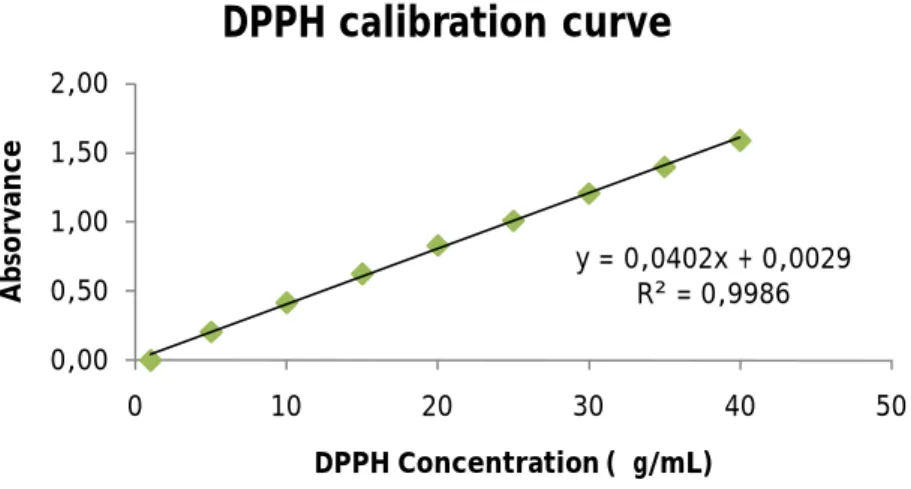
DPPH Calibration curve construction ... 45
Compound samples absorbance measurements ... 45
Chapter 3 ... 49
Results Discussion ... 49
Chapter 4 ... 59
Conclusions ... 59
xiii
Schemes list
Scheme 1 - The general structure of a thioimidate N-oxide 1 Scheme 2 – Aldoses (left) and ketoses (right) 2
Scheme 3 – (D)-Glyceraldehyde (left) and (L)-Glyceraldehyde (right) 2
Scheme 4 – Tetroses 3
Scheme 5 – D-mannose (right) is C-2 epimer of D-glucose (left) 3
Scheme 6 – The intermolecular reaction 4
Scheme 7 – The intramolecular reaction 5
Scheme 8 – Cyclic hemiacetals: Furanose (up) and pyranose (down) 5
Scheme 9 – Cyclization of glucose 6
Scheme 10 – D-glucopiranose is more stable in the configuration on the left 6
Scheme 11 – Fischer representation of D-glucose 7
Scheme 12- Haworth representation of D-glucose 7
Scheme 13 – “Chair” form of D-glucose 7
Scheme 14 - Glucosinolates skeleton 8
Scheme 15 - Glucosinolates structure and glucoraphenin desulfation B resulting in
a cyclized TIO 10
Scheme 16 – Two different routes to prepare a TIO 10
Scheme 17 - Synthesis path starting from D-ribose 12
Scheme 18 - Synthesis starting from a lactone 12
Scheme 19 - Synthesis starting from D-xylose 13
Scheme 20 – Hydroxyl group protection with isopropylidene 14
Scheme 21 – Protection with TBDMS.Cl 14
Scheme 22 – O-acylation of hydroxyl groups 15
Scheme 23 – Benzoylation of hydroxyl groups 15
Scheme 24 – Methylation of primary hydroxyl group 15
Scheme 25 – Benzylation of hydroxyl groups 16
Scheme 26 – Aldoxime formation 16
Scheme 27 – Mitsunobu reaction 17
Scheme 28 – Oxidative cleavage 17
Scheme 29 – Conversion to a thiohydroxamate 18
Scheme 30 – De-O-silylation 18
Scheme 31- Primary hydroxyl group protection 18
Scheme 32 - Hydroxyl group protection with isopropylidene 49
Scheme 33 – Oxidative cleavage 49
Scheme 34 – Aldoxime formation 50
Scheme 35 – Protection with TBDMS.Cl 50
Scheme 37 – De-O-silylation 52
Scheme 38 – Ring-closing 52
Scheme 39 – Hydroxamic acid formation 53
Scheme 40 – Protection with TBDMS.Cl 53
Scheme 41 – Protection with Ac2O and benzoyl chloride 54
Scheme 42 – Conversion to a hydroxamic acid 54
Scheme 43 – Primary hydroxyl group protection 55
Scheme 44 – Protection with benzyl bromide 55
Scheme 45 – Deprotection of the primary hydroxyl group 56
xv
Table list
Table 1 – Antioxidant activity of compound (9) 46
xvii
Acronims List
Ac2O – acetic anhydride
AcOH – acetic acid BnBr – benzyl bromide DCM - dichlorometane
DEAD – diethyl azodicarboxylate DMF – dimethylformamide DMSO – dimethylsulfoxide d6 DPPH – 2,2-diphenyl-1-picrylhidrazyl EA – ethyl acetate eq - equivalents Et3N – triethylamine EtSH – ethanethiol GL - glucosinolates MeOH – methanol
nBu4NI – tetrabutyl ammonium iodide
NCS – N-chlorosuccinimide
NMR – nuclear magnetic resonance MS – molecular sieves
PE – petroleum ether PPh3 – triphenylphosphine
TBAT – tetrabutylammonium difluorotriphenylsilicate TBDMSCl – tert-Butylchlorodimethylsilane
THF – tetrahydrofuran TIO – thioimidate N-oxide TLC – thin layer chromatography
1
Chapter 1
Introduction
General Introduction
Sugars or saccharides are the most abundant bio-molecule on the planet. They are important in a number of biological roles and a major component in the human diet. Glucosinolates are a class of naturally occurring thioglycosides that play numerous important roles in living organisms. They are sulfur-containing secondary metabolites that display a structural homogeneity based on a hydrophilic β-D-glucopyrano unit, an O-sulfated anomeric (Z)-thiohydroximate function connected to a rather hydrophobic side chain, the only structural variant, in which 120 different combinations have already been identified in the vegetable kingdom. They are present in various vegetables, namely the Cruciferae family. The glucosinolates are hydrolyzed by an enzyme called myrosinase (thioglucoside glycohydrolase E.C. 3.2.3.147), the only identified glycohydrolase able to break an anomeric carbon-sulfur bond [1]. As a defense mechanism the plant uses the relationship between the enzyme and the substrate, by production of bio-active compounds with a large activity spectrum, which include anti-fungi, anti-bacterial and insecticide activity. The principal degradation products of the glucosinolates are the isothyocianates, which are known for their efficacy as chemo-preventive agents. Glucosinolates can be extracted from vegetable sources and then refined by chromatography procedures, but the chemical synthesis approach seems to be a more general and efficient way to get access to glucosinolates in pure form. Functional groups are described as specific groups of atoms within molecules that are responsible for the characteristic chemical reactions of those molecules. The word moiety is often used as a synonym of functional group but, according to the IUPAC definition, a moiety is a part of a molecule that may include functional groups as substructures. The N-oxide thioimidate function is rare, and little can be found in literature about it [2].
Carbohydrates
Carbohydrates are polihydroxy aldehydes, polihydroxy ketones or compounds that, by hydrolysis, can be transformed into the previous compounds. Their general formula is Cx(H2O)y
and contain ketones groups and aldehyde groups, but they exist mainly as hemi-aketals or aketals. The simplest carbohydrates are the ones that can be hydrolyzed into more simple compounds, and they are denominated monosaccharides; the ones that can be hydrolyzed in 2 monosaccharids molecules are called disaccharides; and the ones, that by hydrolysis, originate many monosaccharides molecules are polysaccharides. Monosaccharides can be subdivided: if they contain an aldehyde group, they are called aldoses; and if they are formed by a ketone group, they are ketoses; and depending on the number of carbon atoms, the monosaccharide is called triose (3 carbon atoms), tetrose (4), pentose (5), etc. As an example, an aldohexose, is as monosaccharide with 6 carbon atoms that contains an aldehyde group.
Scheme 2 – Aldoses (left) and ketoses (right)
Glyceraldehyde, which is and aldotriose, is one of the simplest monosaccharides and exists in 2 enantiomeric forms. The middle carbon atom in glyceraldehyde is chiral, that is it bears four different substituents, and consequently has non-superimposable stereo isomers. Glyceraldehyde possesses enantiomeric (mirror-image) forms.
Scheme 3 – (D)-Glyceraldehyde (left) and (L)-Glyceraldehyde (right)
If the secondary OH is on the right we have D-glyceraldehyde, and vice-versa. For longer chains the D/L distinction is based on the orientation of the secondary OH furthest from the C=O, C5 in hexose. In this context D/L refers only to the configuration about this carbon atom and does not specify the optical activity of the sugar; the latter is denoted by +
3 (dextrorotatory) or – (levorotatory). N chiral centers yield 2N isomers: tetroses have 2 chiral
centers, which yield 4 stereoisomers.
Scheme 4 – Tetroses
D-erythrose and L-erythrose are enantiomers, L-threose and D-erythrose are diastereomers and L-threose and D-threose are also enantiomers. D-glucose and D-mannose, which differ by the orientation of a –O at a single chiral center are called epimers.
Scheme 5 – D-mannose (right) is C-2 epimer of D-glucose (left)
D-glyceraldehyde L-glyceraldehyde
Carbohydrates have been so far represented in the linear form, but in reality the linear form is a minor species. In solution carbohydrates are usually closed rings. Ring closure occurs by attack of a secondary alcohol on the carbon of the electron deficient C=O. This attack can occur on either face of the planar CHO with the result that the –OH group that is created at C1 can be oriented in either of two directions (if the attack is on the left structure the –OH created will point to the left and vice-versa). The two forms that are formed are called anomers and the C bearing the C=O is the anomeric carbon. When the newly created –OH has the same orientation as the –OH that did the attacking (the two –OH’s is cis) it’s called the α-anomer, otherwise it’s the β-anomer. Usually the –OH group that does the attacking is located on C5 and a 6-membered ring (pyranoside) is formed. When the ring is closed another chiral center is formed, so the ring form has twice as many isomers as the linear form. The attack by the C4 -OH is less common, and leads to a 5-membered ring (furanoside). An important result of the formation of the cyclic hemiacetal is that the hydroxyl group created on C1, bearing the original aldehyde function can be oriented in either of two ways, so a new chiral center is created. Thus the α and β anomers described are geometric forms of each other. They can be crystallized separately and can be distinguished by a measurement of optical activity. In the case of glucose, both α and β forms are dextrorotatory, that when dissolved in water leads to the production of the mixture: α-glucopyranoside 40%, open form 0,1%, and β-glucopyranoside 60%, a phenomenon called mutarotation, because the optical activity changes from that of the pure form observed immediately on dissolution to that of the equilibrium mixture hours later. Aldehydes and ketones react with alcohols to form hemiacetals. When the reaction is intermolecular the equilibrium is unfavorable and the amount of hemiacetal present is very small. However, when the aldehyde group and alcohol group are contained in the same molecule the intramolecular reaction is much more favorable and the hemiacetal is the predominant species present at the equilibrium.
5 H O O O H H O
Scheme 7 – The intramolecular reaction
Because monosaccharides contain both and aldehyde group and alcohol group they exist predominantly in the form of cyclic hemiacetals (5- or 6-membered rings).
Scheme 8 – Cyclic hemiacetals: Furanose (up) and pyranose (down)
In the case of the cyclization of glucose there are five hydroxyl groups that might react with the aldehyde group. However 5- and 6-membered rings are much more stable than others.
Scheme 9 – Cyclization of glucose
Scheme 10 – D-glucopiranose is more stable in the configuration on the left
Representations of Carbohydrate Structure
There are several ways to draw the structure of carbohydrates. The simplest is the method introduced by Fischer which represents the sugar as a straight chain of carbon atoms with the lowest numbered at the top and the OH’s of the secondary alcohol shown to the right or left. The horizontal lines are to be visualized as projecting out of the page; the vertical lines project into the page so that the carbon backbone has the overall profile of a banana, the top and bottom of which lies behind the plane of the page. The assignment of D or L depends on the orientation of the OH in the penultimate carbon and the orientation of the other OH’s are relative to this one. As sugars exist mainly in the ring form, the Fischer representation is converted to the ring formation by drawing a “box” connecting the C bearing the keto function to the penultimate carbon.
7
Scheme 11 – Fischer representation of D-glucose
An alternative is the Haworth representation which attempts to convey more three-dimensional information. The carbohydrate is drawn as a hexagon (or pentagon). The OH’s are shown and lie either above or below the plane if the hexagon.
Scheme 12- Haworth representation of D-glucose
However the most realistic representation of the structure would show that the ring form is not planar. The predominant forms in solution are both “chair” forms, but a small percentage of the sugar molecules are present in the boat form [3].
Glucosinolates
The glucosinolates are a class of secondary metabolites found in fifteen botanical families of dicotyledonous plants. Glucosinolates can be subdivided into three major classes, depending on the nature of their side chains, which may be derived from aliphatic, indolyl, or aralkyl α-amino acids. The skeleton of glucosinolates consists of a thioglycosides link to the carbon of a sulphonated oxime. The R group (side chain) and the sulphate group have anti stereochemical configuration. The R group is derived from amino acids and is highly variable. It can be aliphatic, aromatic or heterocyclic. The sulphate group imparts strongly acidic properties and thus the glucosinolates occur in nature as anions counterbalanced by a cation. The cation is usually potassium, being one of the most abundant cations in plant tissues.
Scheme 14 - Glucosinolates skeleton [1]
Generally, the concentration of GL’s is higher in the seed, as opposed to the low levels find in the leaf, stem and root. Concentrations differ according to tissue type, physiological age, plant health and nutrition. Studies have shown that myrosinases are localized in vacuoles of specialized plant cells, called myrosin cells. Thus the two components of the system are separated until autolysis or tissue damage brings them into contact. The precise localization of glucosinolates is not known, but they have been reported to be stored in vacuoles.
Hydrolysis of glucosinolates
The enzymes catalyzing the hydrolysis of glucosinolates are known as myrosinases. The complexity of the myrosinase-glucosinolate system indicates an important role in the life
Methylene substituted chain Β-D-glucopyranosyl moiety
9 for nutrients like nitrogen and sulphur, while the products of hydrolysis may have important roles in the plant defense system against insect, fungi and microorganism infections.
Glucosinolates behave like bio-precursors to produce electrophilic isothiocianates, compounds that display a diversified and marked biological activity. The myrosinase-assisted hydrolysis cleavage of GL releases a labile aglycon chain which is converted into isothiocyanate through a Lossen-type rearrangement. When crushed plant tissue or seeds containing glucosinolates are added to water, myrosinases catalyze the hydrolytic cleavage of the thioglucosidic bond, giving D-glucose and a thiohydroximate-O-sulphonate (aglycone). The latter compound rearranges nonenzymatically with release of sulphate to give one of several possible products. The predominant product is dependent on the structure of the glucosinolate side chain and the presence of protein co-factors that modify the action of the enzyme. The most frequent fate of the unstable aglycone is to undergo rearrangement spontaneously via a proton independent Lossen rearrangement with a concerted loss of sulphate to yield an isothiocyanate, or a competing proton dependent desulphuration yielding a nitrile and elemental sulphur. Some glucosinolates also give rise to the formation of thiocyanates. [4]
Chemo-preventive activity of glucosinolates
They have the ability to block the carcinogenic potential of many particularly dangerous substances that can damage cell DNA, leading to cell damage that allows the growth of tumours. Glucosinolates stimulate our immune systems, and accelerate the elimination of carcinogens from our bodies, depriving them of a longer more destructive stay. In short, these vegetables prevent carcinogenic substances from causing the type of genetic damage that leads to the onset of cancer and the growth of cancerous tumours in the human body.
Thioimidate N-oxides
The thioimidate N-oxide function is a rare and original function, of which little is known. Glucoraphenin A displays a unique behavior: the desulfo-counterpart B is readily converted into a cyclic thioimidate N-oxide C, and that can be explained by intramolecular concerted Michael addition of the thiohydroximate moiety of the desulfo-counterpart onto the vinyl sulfoxide acceptor.
-sulfatase (E.C.3.1.6.1)
A B
C
Scheme 15 - Glucosinolates structure and glucoraphenin desulfation B resulting in a cyclized TIO [4]
J.Schleiss et al designed preparative synthetic methods as a prerequisite to evaluate the chemical potential and reactivity scope of this rare function. The formation of thioimidate N-oxide can be controlled by avoiding direct attack to the electrophilic carbon. They chose to introduce the thiohydroximate function in one end of the chain and the activable group at the other end, and they also used to ways to convert the thiohydroximate into a thioimidate N-oxide: halociclization, under the conditions developed by Grigg et Jäger, and using as the activable group an alkenyl; and nucleophilic substitution, under the conditions of the Mitsunobu reaction and using an hydroxyl as the activable group. [6-9]
halociclization
11 The compounds obtained from the enzymatic desulfation of the sugar ring were unstable and degradation occurred at room temperature. Using a carbohydrate template and performing halociclization showed compounds with some instability when kept at room temperature for a few days. When performing nucleophilic substitution the reaction had a low yield. Converting 2,3-O-isopropylidene-L-erythrose into the aldoxime, in a 4 step sequence, gave acess to the thiohydroximate and then de-O-sylilation with TBAT. To close the ring two methods were used, one involved a mesyl activation of the primary alcohol prior to application of basic conditions to induce intramolecular ciclization and the second method uses the Mitsunobu procedure. Then, starting from a group of lactones which were converted into hydroxamic acids, and protected in the form of bi-O-silylated derivatives, these compounds originated nitrile oxides, following a protocol developed by Carreira et al (formation of the triflated hydroximates, then condensation with ethanethiol, under basic conditions). To prevent the hydrolysis of the cyclic TIO into and N-hydroxylactam, a mild neutral reaction protocol should be used.
Synthesis of thioimidate N-oxides
Three paths of synthesis were tested to obtain TIO. The first path starts from a carbohydrate template. Starting from D-ribose, the latter can be converted to 2,3-O-isopropylidene-L-erythrose (1), which can be converted to the aldoxime (6), and in a four step sequence the thiohydroximate (8) was achieved. De-O-silylation was performed using TBAT, and then closing of the ring was performed using the Mitsunobu procedure:
H2SO4 conc acetone 1) NaBH4, MeOH, 0ºC 2) NaIO4, ta (1) (2) NH2OH.HCl pyridine TBDMSCl pyridine, rt (3) (4) (5) 1) NCS, DMF 2) EtSH, Et3n TBAT THF, rt (9) (8) (6) PPh3, DEAD anydre THF, reflux
Scheme 17 - Synthesis path starting from D-ribose
In the second path the depart product is a lactone. -valerolactone was converted to a hydroxamic acid (7), which was readily protected by either TBDMSCl or Ac2O or benzoyl
chloride. KOH, MeOH (7) (10) (11) (12) benzoyl chloride pyridine TBDMSCL, Imidazole DMF Ac2O pyridine
13 This synthesis path wasn’t further explored, and so we moved to the third path. Starting from D-xylose, the anomeric carbon was protected by a methyl group (14), and then protection to the others groups was performed using benzyl bromide (15). Deprotecting the anomeric carbon and then ring opening allowed the access to the aldoxime (18).
NaH, BnBR, nBu4NI DMF (15) (17) H2SO4 conc MeOH, reflux AcOH H2SO4 1M, dioxane (14) (18) Na, NH2OH.HCl MeOH, reflux
Scheme 19 - Synthesis starting from D-xylose
Hydroxyl group protection
Monossacharides contain several hydroxyl groups and to execute certain reactions in a specific location the protection of these groups is necessary. To protect a group the following characteristics are required: a) easily and selectively on; b) stable under the reaction conditions, and; c) easily and selectively off. Carbohydrate may be protected by conversion of hydroxyl groups in acetals, hemiacetals, esters and ethers. Protective groups exist in a big variety, and some are more selective than others, therefore we can achieve the desired compound [10].
Acetals
Acetals are useful in carbohydrate chemistry because they can protect two hydroxyl groups at the same time, and they are often selective for certains sorts of hydroxyl groups. When acetals are derived from aldehydes they generally prefer to be 6-membered rings, that is protection of 1,3-diols. The formation of isopropylidene can be achieved in acid conditions using acetone as solvent, taking as example (1) [11].
H
2SO
4Acetone
(1)
O H O O O H O O H O H O H O H OScheme 20 – Hydroxyl group protection with isopropylidene
Isopropylidene are stable in basic conditions and cleaved by acidic conditions.
Silyl ether protecting groups
The silyl ether family is a large one and they are popular protecting groups because they are readily formed and cleaved under mild conditions and their relatively stability can be adjusted by simply varying the substituents on silicon. Using TBDMS in pyridine allows us to achieve compounds (4) and (5). The hydroxamic acid (7), originated from -valerolactone, was protected using TBDMS and Imidazole in DMF. In both cases the silicon is directly attached to the oxygen.
(4) (5) TBDMS pyridine (7) (10) TBDMS.Cl Imidazole, DMF
Scheme 21 – Protection with TBDMS.Cl
O-Acylation
Using acetic anhydride in pyridine also allows the protection of the hydroxyl groups in compound (7) [12].
15
(11) Ac2O
Pyridine
(7)
Scheme 22 – O-acylation of hydroxyl groups
Benzoylation
When benzoyl chloride reacts with alcohols it originates esters. In this reaction benzoyl chloride in pyridine reacted with compound (7) and originated (12).
Benzoyl chloride Pyridine
(7) (12)
Scheme 23 – Benzoylation of hydroxyl groups
Methylation
Methylation is an easy way to protect a hydroxyl group so that we don’t want to react in the next reactions. It can be done using sulphuric acid in MeOH [3][13].
(14) H2SO4
MeOH
Scheme 24 – Methylation of primary hydroxyl group
Benzylation
Benzylation of (14) allowed the hydroxyl group protection of the remaining groups. Using NaH as a base, BnBr for the introduction of the benzyl group and nBuNI4 as a catalyst, in DMF
NaH, BnBr, nBuNI4
DMF
(14) (15)
Scheme 25 – Benzylation of hydroxyl groups
Aldoxime formation
An aldoxime is an oxime produced in the condensation reaction between an aldehyde and hydroxylamine, and exists always as two stereoisomers. When compound (2) was allowed to react with hydroxylamine hydrochloride in pyridine and with molecular sieves it originated the aldoxime (3) [15]. Compound (16) reacted with Na and hydroxylamine hydrochloride in MeOH and so we obtained aldoxime (17).
NH2OH.HCl Pyridine, MS (2) (3) O H O O O O H N O H O O (17) Na, NH2OH.HCl MeOH (16) O H O O O H O O H N O H O BnO OBn BnO OBn OBn BnO OBn
+
Scheme 26 – Aldoxime formation
Mitsunobu
The mitsunobu reaction is a reaction in which an alcohol reacts with PPh3 and DEAD in THF.
17
(8) (9)
PPh3, DEAD
THF
Scheme 27 – Mitsunobu reaction
Oxidative cleavage
When NaBH4 and NaIO4 in MeOH react with compound (1) first occurs a reduction which is
followed by a oxidative cleavage, originating compound (2) [18][19].
NaBH4, NaIO4
MeOH
(1) (2)
Scheme 28 – Oxidative cleavage
Conversion to a thiohydroximate
The thiohyroximate is generated in a two step sequences, in which the intermediary is not isolated. Starting from compound (5), and using NCS, that is a mild oxidant, in DMF, these substances are allowed to react for 4 hours. After that EtSH and Et3N are added and the
(6) 1. NCS, DMF 2. EtSH, Et3N (5) O H N O O S O H N O O SMDBTO SMDBTO
Scheme 29 – Conversion to a thiohydroxamate
De-O-silylation
Using TBAT in THF allows the de-O-silylation of compound (6) originating compound (8).
TBAT THF
(6) (8)
Scheme 30 – De-O-silylation
Primary hydroxyl deprotection
When compound (15) is allowed to react with AcOH and dioxane in H2SO4 it originates
compound (16), in which the primary hydroxyl group is deprotected.
AcOH, dioxane H2SO4
(15)
(16)
Scheme 31- Primary hydroxyl group protection
Antioxidant activity [24-28]
19 meaning, including reactions that do not involve oxygen. A reduction is a reaction in which electron gain is involved when an element is oxidized, and so it acts as a reducing agent, because it donates electrons to another substance, causing the reduction. In other hand, when an element is reduced it acts as an oxidizing agent, because it accepts electrons causing its oxidation [20].
Free radicals
A radical (often, but unnecessarily called a free radical) is an atom or group of atoms that have one or more unpaired electrons. Radicals can have positive, negative or neutral charge. They are formed as necessary intermediates in a variety of normal biochemical reactions, but when generated in excess or not appropriately controlled, radicals can cause damage on a broad range of macromolecules. Radicals have extremely high chemical reactivity, which explains not only their normal biological activities, but how they inflict damage on cells. [21]
Free radicals can be produced by various ways, such as:
- Enzymes: in redox reactions in the electron transport chain in mitochondria, endoplasmatic reticulum and plasmatic and cell membrane; as intermediaries during drug desintoxication, prostaglandin synthesis and during platelet and leucocyte activation.
- Environmental factors: light, UV radiation or ionizing radiation.
- Non-enzymatic processes: through self-oxidation of many substances (polyunsaturated fatty acids, hemoglobin, myoglobin, catecholamines), than can be stimulated by metallic ions (iron and copper) with redox capacity (ionizing or UV radiation and by photo-activated pigments).
Reactive oxygen species (ROS)
Oxygen has two unpaired electrons in separate orbitals in its outer shell. This electronic structure makes oxygen especially susceptible to radical formation, and so they represent the most important class of radical species generated in living systems.
Sequential reduction of molecular oxygen (equivalent to sequential addition of electrons) leads to formation of a group of reactive oxygen species. Molecular oxygen (O2) has a unique
electronic configuration and is itself a radical. The addition of one electron to dioxygen forms the superoxide anion radical (O2-). The hydroxyl radical (OH) is the neutral form of the
hydroxide ion, and it has a high reactivity, making it a very dangerous radical with a very short half-life in vivo. Adittionaly peroxyl (ROO) radicals, also derived from oxygen, can also
Oxygen-derived radicals are generated constantly as part of normal aerobic life. They are formed in mitochondria as oxygen is reduced along the electron transport chain. Reactive oxygen species are also formed as necessary intermediates in a variety of enzyme reactions. Examples of situations in which oxygen radicals are overproduced in cells include:
- White blood cells, such as neutrophils specialize in producing oxygen radicals, which are used in host defense to kill invading pathogens.
- Cells exposed to abnormal environments, such as hypoxia or hyperoxia generate abundant and often damaging reactive oxygen species.
- Ionizing radiation, is well known to generate oxygen radicals within biological systems.The damaging effects of radiation are higher in well oxygenated tissues than in tissues deficient in oxygen.
Reactive nitrogen species (RNS)
Nitric oxide (NO) is a small molecule that contains one unpaired electron on the anti-bonding
2πy* orbital and is, therefore, a radical. It is generated in biological tissues by specific nitric
oxide synthases, and is an abundant reactive radical that acts as an important oxidative biological signaling molecule in a large variety of diverse physiological processes, including neurotransmission, blood pressure regulation, defense mechanisms, smooth muscle relaxation and immune regulation.
Oxidative stress
ROS as well as RNS are products of normal cellular metabolism and are well recognized for playing a dual role as both deleterious and beneficial species, since they can be either harmful or beneficial to living systems. Beneficial effects occur at low/moderate concentrations and involve physiological roles in cellular responses to noxia. The harmful effect of free radicals causing potential biological damage is termed oxidative stress and nitrosative stress. Oxidative stress results from the metabolic reactions that use oxygen and represents a disturbance in the equilibrium status of pro-oxidant/antioxidant reactions in living organisms. The excess ROS can damage cellular lipids, proteins or DNA, inhibiting their normal function. Because of this oxidative stress has been implicated in a number of human diseases as well as in the aging process. The delicate balance between beneficial and harmful effects of free radicals is a very important aspect of living organisms and is achieved by mechanisms called “redox regulation”. This process protects living organisms from various oxidatives stresses and maintains “redox homeostasis” by controlling the redox status in vivo. How the oxidative process starts is still not clarified, but the HO radical and peroxynitrite
radical (ONOO) are the ones that initiate it. The O2- radical also as an important part, due to
21 H2O2 + O2- OH- + HO + O2
This reaction only occurs in the presence of a transition metal (usually iron), which is reduced by O2- and reacts with H2O2, in a reaction type Fenton:
O2- + Fe3+ O2 + Fe2+
H2O2 + Fe2+ OH- + HO + Fe3+
Once formed, ROS travel to the microcirculation, and due to their high reactivity, act at different cellular structures (DNA and proteins) and also at the cellular membrane (polyunsaturated lipids). When an imbalance between ROS/free radicals formation and antioxidant defenses/organism repair mechanisms is verified, oxidative stress exists and can be disease associated.
Lipid peroxidation
Lipid peroxidation refers to the oxidative degradation of lipids. It is the process in which free radicals "steal" electrons from the lipids in cell membranes, resulting in cell damage. This process exists due to a free radical chain reaction mechanism. Usually it affects polyunsaturated fatty acids, because they contain multiple double bonds. As with any radical reaction, the reaction consists of three major steps: initiation, propagation, and termination.
Initiation: Initiation is the step in which a fatty acid radical is produced. The most notable
initiators in living cells are ROS, such as OH and HO2, which combines with a hydrogen atom
to make water and a fatty acid radical.
Propagation: The fatty acid radical is not a very stable molecule, so it reacts readily with O2,
thereby creating a peroxyl-fatty acid radical. This too is an unstable species that reacts with another free fatty acid, producing a different fatty acid radical and a lipid peroxide, or a cyclic peroxide if it had reacted with itself. This cycle continues, as the new fatty acid radical reacts in the same way.
Termination: When a radical reacts with a non-radical, it always produces another radical,
which is why the process is called a chain reaction mechanism. The radical reaction stops when two radicals react and produce a non-radical species. This happens only when the concentration of radical species is high enough for there to be a high probability of collision of two radicals.
Usually many reactive products are formed, namely lipids radicals and malondialdehyde. These don not only act directly on cell membrane components, but can also infiltrate the blood stream, increasing the blood and plasma levels. This increase indicates damage in the cell membrane of organs or tissues that can be responsible by the triggering of many pathologic processes.
Protein oxidation
When the properties of a protein are altered and when amino acids peroxide formation occurs it can lead to protein degradation, by fragmentation and by cross-linking, which can result in polymerization and inactivation, specifically in proteins with SH and aromatic groups. Protein oxidation induces alteration in the tertiary structure which leads to protein aggregation and amyloids formation. Amino acid oxidation by free radicals leads to alterations in the enzymatic activity, with antioxidant compromise in cells and tissues.
DNA oxidation
DNA damage is one of the most important results of the peroxidative process in vivo. DNA alteration caused by oxidative damage is one of the main causes that trigger carcinogenese, either by proto-oncogenes activation, or by tumor suppressor genes inactivation. Apoptose inhibition may also be associated with oncogenese and with DNA alterations, and can appear as a consequence of free radicals action that leads to an increase of cytoplasmic calcium.
Antioxidants
Antioxidants can be defined as any substance that, present in relatively low concentrations (when compared to the one of the substrate), prevent or slow significantly substrate oxidation. For convenience, antioxidants were divided into two classes, primary or chain-breaking antioxidants and secondary or preventative antioxidants. Secondary or preventative antioxidants are compounds that retard the rate of oxidation. This may be achieved in a number of ways, including removal of substrate or singlet oxygen quenching. Primary antioxidants (AH) when present in trace amounts, may either delay or inhibit the initiation step by reacting with a lipid radical or inhibit the propagation step by reacting with peroxyl or alkoxyl radicals:
L + AH LH + A LOO + AH LOOH + A
LO + AH LOH + A
The antioxidant free radical may further interfere with chain-propagation reactions by forming peroxy antioxidant compounds:
A + LOO LOOA
A + LO LOA
The activation energy of the above reactions increases with increasing A-H and L-H bond dissociation energy. Therefore, the efficiency of the antioxidant increases with decreasing A-H bond strength.
23 such antioxidant is vitamin E. Other anti-oxidants made within the body include the enzymes superoxide dismutase, catalase, and peroxidase.
Antioxidant systems
The production of free radicals is controlled in humans by several antioxidant compounds, some of which may be from and endogenous origin and some can be originated from the diet. Antioxidants can stabilize or deactivate free radicals before they attack the biological targets in the cells. The human body has several control mechanisms of ROS production and mechanisms to limit and reaper tissue damage. The integrated antioxidant system has many components:
- Antioxidants that prevent ROS formation: Cu-ceruloplasmin, Cu-albumin, Fe-transferrin and Fe-myoglobin.
- Antioxidants that remove ROS, preventing chain reactions: enzymes, such as superoxide dismusate, glutathione peroxidase, glutathione redutase, catalase, metaloenzymes; and small molecules, like glutathione, vitamin C, tocopherol, bilirubin, uric acid, carothenoids and flavonoids.
- Repair enzymes: which include DNA repair enzymes.
However, the primary intracellular antioxidant defense is given to enzymatic antioxidants and non-enzymatic antioxidants.
Enzymatic Antioxidants
Three groups of enzymes play significant roles in protecting cells from oxidant stress:
Superoxide dismutases are enzymes that catalyze the conversion of two superoxides into hydrogen peroxide and oxygen. The benefit here is that hydrogen peroxide is substantially less toxic that superoxide. They accelerate this detoxifying reaction roughly 10,000-fold over the non-catalyzed reaction.
O2- + O2- O2 + H2O2
They are metal-containing enzymes that depend on bound manganese, copper or zinc for their antioxidant activity. In mammals, the manganese-containing enzyme is most abundant in mitochondria, while the zinc or copper forms predominant in cytoplasm. Interestingly, they are inducible enzymes, which mean that exposure of bacteria or vertebrate cells to higher concentrations of oxygen results in rapid increases in the concentration of SOD.
Catalase is found in peroxisomes in eucaryotic cells. It degrades hydrogen peroxide to water and oxygen, and hence finishes the detoxification reaction started by superoxide dismutase.
Glutathione peroxidase is a group of enzymes, the most abundant of which contain selenium. These enyzmes, like catalase, degrade hydrogen peroxide. They also reduce organic peroxides to alcohols, providing another route for eliminating toxic oxidants.
Non-enzymatic Antioxidants
Three non-enzymatic antioxidants of particular importance are:
Vitamin E (or tocopherol) is the major lipid-soluble antioxidant, and plays a vital role in protecting membranes from oxidative damage. Its primary activity is to trap peroxyl radicals in cellular membranes.
Vitamin C (or ascorbic acid) is a water-soluble antioxidant that can reduce radicals from a variety of sources. It also appears to participate in recycling vitamine E radicals. Vitamin C also functions as a pro-oxidant under certain circumstances.
Glutathione may be the most important intracellular defense against damage by reactive oxygen species. It is a tripeptide (glutamyl-cysteinyl-glycine). The cysteine provides an exposed free sulphydryl group (SH) that is very reactive, providing an abundant target for radical attack. Reaction with radicals oxidizes glutathione, but the reduced form is regenerated in a redox cycle involving glutathione reductase and the electron acceptor NADPH.
Relation between oxidative stress and pathologies
It is now common knowledge that oxidative stress is related to a wide number of pathologies namely:
- Cancer, because free radicals have initiation and promoting capacity, which are the two fundamental phases of cancer development.
- Cardiovascular diseases, free radicals excess can initiate aterosclerose by damaging the blood vessels.
- Diabetes Mellitus, free radicals contribute to pancreatic cell destruction in insulin-dependent diabetes mellitus.
- Inflammatory disease, free radicals may act indirectly as cellular messengers and trigger inflammatory response.
- Infertility, free radicals initiate lipid peroxidation and peroxide accumulation in the spermatozoid membrane, causing motility and viability reduction.
- Cataracts, superoxide and hydroxyl radicals cause damage to proteins and lipids in cell membranes that accumulate in the crystalline surface, causing opacity. - Aging, due to free radicals production in mitochondria that accumulate with age.
25 - Neurologic diseases, the brain is rich in polyunsaturated fatty acids and iron, and poor in antioxidants, and it’s also surrounded by spinal fluid, and it has none or slim capacity to iron chelation.
- Hepatic disease, high levels of short chain fatty acids increase iron captation by hepatocytes, and so increasing hydrogen peroxide production and free radicals production.
- Pulmonary disease, ROS and other toxic products produced by pulmonary cells and by neutrophilic cells activity, that accumulate in the lungs when pure oxygen is breathed, can possibly contribute to oxidative damage.
Measurement of antioxidant activity
The methods to evaluate antioxidant behavior can be clustered in two categories that reflect antioxidant activity importance in food or human bioactivity. In the case of food systems, the need consists in evaluating antioxidant efficacy in providing food protection against oxidative deterioration. A subcategory involves measuring antioxidant activity in food, especially fruits, vegetables and beverages, but with the goal of determining antioxidant load present in the diet and its antioxidant activity in vivo.
Antioxidants can act by several mechanisms, such as free radical trapping, peroxide decomposition and metallic ions chelation. Therefore, antioxidant activity can be measured and evaluated by various methods that look on different antioxidant activity chemical mechanisms.
According to the chemical reactions used, the antioxidant activity determination methods can be clustered into two classes: hydrogen atoms transfer (HAT) and electron transfer (ET). HAT methods measure the ability of an antioxidant to inactivate free radical by hydrogen atoms donation. ET methods measure the ability of a potential antioxidant to transfer an electron to the reduced radicals, metals or carbonyls.
One of the problems when determining antioxidant activity is that the antioxidant activity is variable depending on the method used. But it’s known that the antioxidant mechanism in many biological matrixes is very complex and many other factors can intervene in such process.
DPPH method
The DPPH method is based on the study of the trapping activity of the stable free radical 2,2-diphenyl-1-pycril-hydrazyl (DPPH), which has a purple coloration, and absorbs at a specific wavelength (515-517nm). When an antioxidant (AH) or a radicalar species (R•) intervene, DPPH is reduced forming diphenylpycril-hydrazine, of yellow coloration, with consequent absortion disappearement, and this can be monitored by the decrease in absorbance. From
the obtained results the percentage of antioxidant activity and the percentage of remaining DPPH are determined.
The percentage of antioxidant activity corresponds to the quantity of DPPH consumed by the antioxidant. The efficient concentration (EC50) is the necessary quantity of antioxidant that
27
Chapter 2
Experimental Part
General Methods
The TLC was used to verify the progress of the reactions and the progress of the product purifications. The TLC sheets (aluminium sheets covered by Silica Gel 60 F254) where revealed
with UV light (λ=254nm) and with immersion in one of the following visualization reagents, and then heated:
- Sugars: 800ml EtOH, 150ml water, 50ml H2SO4
- KMnO4 : 5g KMnO4, 500ml water, 8,5ml AcOH, 33g K2CO3
- Phosphomolibdic acid: solution of 5% or 10% of phosphomolibdic acid in EtOH
The Rf can be calculated by the relation between the height of the elution product and the height of the eluent.
The products were purified by column chromatography in silica gel SI 60 (40-63 µm), and these columns were eluted by gravity with the help of compressed air.
The proton NMR spectra were registered in CDCl3 or DMSO by Bruker Spectrometer (250MHz)
and (400MHz), at the Institut de Chimie Organique et Analytique. The homonuclear correlations (COSY) were done to allow the complete attribution of some signals. The chemical shift (δ) of the signals is indicated in ppm, and the solvent was used as an internal reference. The coupling constants are indicated in Hz (Hertz) and the multiplicity of the signals is indicated by the following abbreviations: s (singlet), d (doublet), t (triplet), q (quadruplet) and m (multiplet).
The 13C NMR spectra were registered at 100MHz in the same spectrometer used for the
proton spectra. The heteronuclear correlations between the proton and the carbon (HSQC) were done to allow the correct interpretation of some signals.
To evaluate the Antioxidant Activity of some compounds, the latter were allowed to react with a stable radical, 2,2-diphenyl-1-picrylhydrazyl (DPPH•) in a methanol solution. The
reduction of DPPH• was followed by monitoring the decrease in absorbance at a characteristic
wavelength (515nm) during the reaction. DPPH• absorbs at 515nm, but upon reduction by an
antioxidant (AH) or a radical species (R•), the absorption disappears.
DPPH• + AH → DPPH-H + A•
DPPH• + R• → DPPH-R
Scheme -
These measurements were performed using a UV-Vis spectrophotometer in the 515nm wavelength, using as positive control trolox (6-hydroxy-2,5,7,8-tetramethylchromam-2-carboxylic acid).
General Procedures
(1) 2,3–O–isopropylidene–β–D–ribofuranose [11][31]
190,19 g/mol C8H14O5
Procedure: To a solution of D-ribose (10g, 66,6mol, 1 eq) in acetone (100ml), was added
H2SO4 (0,25ml, 4,5mmol), at 0ºC. The mixture was stirred for 1h, and then the NaHCO3 was
added, a 0ºC. The solvents were evaporated. The suspension was filtered through celite and with EA, and evaporated. Toluene was added, and the solvents were co-evaporated.
Yield = 95% Rf = 0,7 (EA/PE 9:1) 1 H NMR (400MHz, CDCl3) δ: 1,27 (s, 3H, CH3iPr), 1,43 (s, 3H, CH3iPr), 3,63-3,69 (m, 2H, CH2 -5), 4,34 (t, 1H, J=4Hz, J=2,8Hz, CH-4), 4,52 (d, 1H, J=6Hz, CH-3), 4,76 (d, 1H, J=6Hz, CH-2), 5,35 (s, 1H, CH-1) 13 C NMR (100MHz, CDCl3) δ: 24,7 (CH3iPr), 63,6 (CH2-5), 81,7 (CH2-2), 86,8 (CH2-3), 87,7 (CH-4), 102,8 (CH-1), 112,3 (C(CH3)2)
MS (IS): m/z = 173 [M-OH]+, 213 [M+Na]+, 403 [2M+Na]+
IR : 867, 1036, 1159, 1209, 1376, 1458, 2940, 3369
[ ] = -20 (CHCl3)
IR : 778, 867, 923, 1000, 1036, 1063, 1159, 1210, 1241, 1325, 1376, 1458, 1642, 2941, 2986,
29
(2) 2,3-O-isopropylidene-L-erythrose [18-19][31-32]
160,19 g/mol C7H12O4
Procedure: To a solution of 1 (2g, 10,5mmol, 1eq) in MeOH (20ml), NaBH4 was added (0,598g,
15,8mmol, 1,5eq) and the mixture was stirred for 1h, at 0ºC. The solvents were evaporated, then it was added t-BuOH/H2O (3:2) (18/12ml) and the mixture was stirred for 5min. The
NaIO4 was added (8,9g; 42mmol, 4eq), at 0ºC and the mixture was stirred for 12h. The
resulting mixture was diluted with EA and filtered through celite and with EA. The organic phase was washed with a saturated solution of NaHCO3 (50ml) and a saturated solution of
NaCl, and dried with MgSO4. The suspension was filtered and re-evaporated. The product was
purified by column chromatography (PE/EA 7:3).
Yield = 48% Rf = 0,66 (PE/EA 7:3) 1H NMR (250MHz, CDCl3) δ: 1,27 (s, 3H, CH 3iPr), 1,41 (s, 3H, CH3iPr), 3,91-4,06 (m, 2H, CH2 -4), 4,24 (sl, 1H, OH), 4,51 (d, 1H, J2,3=6Hz, CH-2), 4,79 (dd, 1H, J=6Hz, J=3,2Hz,CH-3), 5,35 (s, 1H, CH-1) 13 C NMR (100MHz, CDCl3) δ: 24,8 (CH3iPr), 26,3 (CH3iPr), 71,9 (CH2-4), 80,1 3), 85,3 (CH-2), 101,8 (CH-1), 112,4 (C(CH3)2) [ ] = +50 (CHCl3) IR : 665, 761, 817, 855, 873, 907, 969, 985, 1042, 1063, 1097, 1161, 1208, 1331, 1375, 1459, 2940, 2085, 3411
(3) 2,3-O-isopropylidene-L-erythrose oxime [15]
E+Z 175,18 g/mol
C7H13NO4
Procedure: To a solution of (2) (3,21g, 0,02mol, 1eq) in pyridine (45ml), the TM (2,5g) and
hydroxylamine hydrochloride (4,2g, 0,06mol, 3eq) were added. The mixture was stirred for 12h. The resulting mixture was diluted with EA and filtered through celite and with EA. The organic phase was washed 3 times with a saturated solution of NaCl and dried with MgSO4,
and then filtered. The solvents were evaporated. Toluene was added and then the solvents were co-evaporated. Yield = 68% Rf = 0,3 (EA/PE 50:50) 1 H NMR (400MHz, CDCl3) δ: 1,37 (s, 6H, (CH3)2iPr-Z), 1,47 (s, 6H, (CH3)2iPr-E), 3,49-3,74 (m, 4H, J1= 5Hz, J2=12Hz, (CH-2)2-E+Z), 4,33-4,36 (m, 1H, CH-3-E), 4,51-4,7 (m, 1H, CH-3-Z), 4,88 (t, 1H, J2-1=J2-3=7,2Hz, CH2-4-E), 5,26 (sl, 1H, CH2-4-Z), 6,94 (sl, 1H, CH-1-Z), 7,44 (d, 1H, J=7,2Hz, CH-1-E) 13C NMR (100MHz, CDCl
3) δ (E+Z mixture): 24,8 & 25,2 & 27,3 & 27,6 (CH3iPr), 61 & 61,9 (CH2
-4), 72 (CH-2-Z), 74,9 (CH-2-E), 78,4 & 78,5 (CH-3), 109,6 & 110,1 (C(CH3)2), 149 (CH-1-E), 151
(CH-1-Z) BP/MP: 46-51 ºC MS (IS): m/z = 176 [M+H]+, 198 [M+Na]+, 214 [M+K]+ [ ] = -76 (CHCl3) IR : 698, 792, 837, 883, 909, 927, 957, 984, 1043, 1077, 1104, 1125, 1161,1218, 1253, 1337, 1377, 1415, 1438, 2937, 2989, 3082, 3209, 3383
31
(4)
N-[tert-butyl(dimethyl)silyl]oxy-1-[(4R,5S)-5-[[tert-butyl
(dimethyl)silyl]oxymethyl]-2,2-dimethyl-1,3-dioxolan-4-yl]methanimine
E+Z 403,7 g/mol C19H41NO4Si2Procedure: To a solution of (3) (2,41g, 13,8mmol, 1eq) in pyridine (22ml), the TBDMSCl
(2,5g, 16,6mmol, 1,2eq) was added. The mixture was stirred for 12h. The resulting mixture was diluted with EA and filtered through celite and with EA. The organic phase was washed with distillated H2O, HCl 1M, a saturated solution of NaCl and dried with MgSO4, and then
filtered. The solvents were evaporated, and then toluene was added to eliminate the pyridine. The solvents were co-evaporated and the product was purified by column chromatography (PE/EA 95:5).
Yield = 24%
Rf = 0,71 (PE/EA 95:5)
1
H NMR (400MHz, CDCl3) δ (E+Z mixture): 0,04 (d, 12H, Si(CH3)2), 0,14 (m, 12H, Si(CH3)2), 0,88
(d, 18H, Si(CH3)3), 0,91 (d, 18H, Si(CH3)3), 1,36 (d, 6H, (CH3)2iPr), 1,49 (d, 6H, (CH3)2iPr), 3,48
(dd, 1H, J1=4,58Hz, J2=11,02Hz, H-4b), 3,61-3,71 (m, 1H, H-4a), 4,24-4,28 (m, 1H, H-3-E),
4,39-4,4 (m, 1H, H-3-Z), 4,71 (dd, 1H, J1=6,79Hz, J2=8,11Hz, H-2-E), 5,24 (dd, 1H, J1=7,26Hz,
J2=4,21Hz, H-2-Z), 7,08 (d, 1H, J1= 4,29Hz, H-1-Z), 7,49 (d, 1H, J1=8,19Hz, H-1-E)
13C NMR (100MHz, CDCl
3) δ (E+Z mixture): 5,18 & 5,21 (Si(CH3)2), 18,35 (C(CH3)2iPr) 25,31 &
25,51 ((CH3)2iPr), 26,08 & 26,16 (SiC(CH3)3), 27,31 & 27,85 ((CH3)2iPr), 61,7 (CH2-4), 62,88
(CH2-4), 72,1 (CH-2-Z), 75,2 (CH-2-E), 78,7 (CH-3-E), 79,11 (CH-3-Z), 109,33 (C(CH3)3), 109,7 (C(CH3)3), 152,2 (CH-1-E), 155,0 (CH-1-Z) MS (IS): m/z = 404 [M+H]+, 426 [M+Na]+ [ ] = -53 (CHCl3) IR : 673, 722, 778, 834, 878, 936, 983, 1007, 1094, 1143, 1216, 1251, 1380, 1471, 2858, 2888, 2930, 2955
(5) 4-O-tert-butyldimethylsilyl-2,3-O-isopropylidene-L-erythrose oxime
E+Z 289,4 g/mol
C13H27NO4Si
Procedure: To a solution of (3) (2,41g, 13,8mmol, 1eq) in pyridine (22ml), the TBDMSCl
(2,5g, 16,6mmol, 1,2eq) was added. The mixture was stirred for 12h. The resulting mixture was diluted with EA and filtered through celite and with EA. The organic phase was washed with distillated H2O, HCl 1M, a saturated solution of NaCl and dried with MgSO4, and then
filtered. The solvents were evaporated, and then toluene was added to eliminate the pyridine. The solvents were co-evaporated and the product was purified by column chromatography (PE/EA 95:5).
Yield = 35%
Rf = 0,313 (PE/EA 95:5)
1H NMR (400MHz, CDCl
3) δ (E+Z mixture): 0,0 (m, 6H, Si(CH3)2), 0,10 (m, 6H, Si(CH3)2), 0,82
(s, 9H, SiC(CH3)3), 0,82 (m, 9H, SiC(CH3)3), 1,30 (s, 6H, C(CH3)2), 1,41 (s, 6H, C(CH3)2), 3,61
(m, 2H, CH-4), 3,61 (m, 2H, CH-4), 4,62 (dd, 1H, J2,3=6,8Hz, CH-3), 4,67 (t, 1H, J3,4=7,2Hz,
CH-3), 5,19 (dd, 1H, CH-2), 5,22 (dd, 1H, CH-2), 6,84 (d, 1H, J1,2=4,8Hz, CH=N), 7,36 (d, 1H,
J1,2=4,8Hz, CH=N)
13
C NMR (100MHz, CDCl3) δ (E+Z mixture): -5.56 (Si(CH3)2), -5.48 (Si(CH3)2), -5.40 (Si(CH3)2),
-5.37 (Si(CH3)2), 18.1 (SiC(CH3)3), 18.2 (SiC(CH3)3), 25.6 (SiC(CH3)3), 25.8 (C(CH3)2), 25.9
(SiC(CH3)3), 25.9 (C(CH3)2), 27.0 (C(CH3)2), 27.1 (C(CH3)3), 27.4 (C(CH3)3), 27.5 (C(CH3)2), 27,5
(SiC(CH3)3), 27,6 (C(CH3)3), 61.4 (CH2-4), 62.5 (CH2-4), 74.7 (CH-2), 74.9 (CH-2), 78.3 (CH-3),
33
(6)
(Z)-4-O-tert-butyldimethylsilyl-2,3-isopropylidene-N-hydroxy-L-erythroimidothioate S-ethyl
349,56g/mol C15H31NO4SSi
Procedure: To a solution of (5) (1,96g, 6,8mmol, 1eq) in DMF ([C]=0,25M), it was added NCS
(1,36g, 10,2mmol, 1,5eq) and the mixture was stirred for 4h. The Et3N (2,8ml, 20,4mmol,
3eq) and EtSH (1,5ml, 20,4mmol, 3eq) were added and the resulting mixture was stirred for 12h. The mixture was diluted with EA, and then the organic phase was washed 4 times with distillated H2O, once with saturated NaCl, dried with MgSO4 and then filtered. The solvents
were evaporated. The product was purified by column chromatography (PE/EA 9:1).
Yield = 27%
Rf = 0,24 (PE/EA 9:1)
1
H NMR (400MHz, CDCl3) δ: 0,05 (s, 3H, SiCH3), 0,05 (s, 3H, SiCH3), 0,87 (s, 9H, (CH3)3C), 1,29
(t, 3H, J=8,0Hz, CH2CH3), 1,37 (s, 3H, OCH3iPr), 1,53 (s, 3H, OCH3iPr), 2,97-3,1 (m, 2H,SCH2),
3,73 (dd, 1H, J4a,4b=10,6Hz, J4b,3=6,2Hz, H-4b), 3,79 (dd, 1H, J4a,4b=10,6Hz, J4a,b=6,0Hz, H-4a),
4,37 (q, 1H, J3,2=6,7Hz, H-2), 9,09 (sl, 1H, N-OH) 13 C NMR (100MHz, CDCl3) δ: -5,2 (SiCH3), -5,1 (SiCH3), 15,1 (CH3CH2S), 18,6 (C(CH3)3), 25,3 (CH3iPr), 25,8 (CH2S), 26,2 ((CH3)3C), 27,1 (CH3iPr), 62,6 (C-4), 77,3 (C-2), 79,1 (C-3), 109,7 (C(CH3)2), 149,8 (C=N) MS (IS): m/z = 350 [M+H]+, 372 [M+Na]+, 388 [M+K]+ IR : 667, 777, 835, 900, 992, 1078, 1161, 1214, 1252, 1378, 1462, 1601, 2857, 2930, 3288
(7) 4-hydroxypentanehydroxamic acid
133,146g/mol C5H11NO3
Procedure: At 0ºC, to a solution of MeOH (50ml) and NH2OH.HCl (17,4g, 0,25mol, 5eq), the
KOH (14g, 0,25mol, 5eq) was added and the mixture was stirred for 10min. The resulting mixture was filtered and then it was added -valerolactone (4,8ml, 49,94mmol, 1eq). The mixture was stirred for 12h, and then the solvents were evaporated. The product was purified by column chromatography (EA/MeOH 9:1).
Yield = 87% Rf = 0,24 (EA/MeOH 9:1) 1H NMR (250MHz, DMSO) δ: 1,04 (d, 3H, J 5-4=6,1Hz, CH3-5), 1,57-1,47 (m, 2H, CH2-3), 1,99 (dd, 2H, J1=7,5Hz, J2=15,023Hz, CH2-2), 3,51-3,57 (m, 1H, CH-4), 4,46 (d, 1H, OH), 8,65 (sl, 1H, NH), 10,34 (s, 1H, NOH) [ ] = +0,7 (CHCl3) IR : 904, 944, 1015, 1047,1085,1127,1181, 1376,1451, 1538, 1634, 1759, 2969, 3194
35
(8) (Z)-2,3-O-isopropylidene-N-hydroxi-L-erythronimidothioate de S-ethyl
235,3g/mol C9H17NO4S
Procedure: To a solution of (6) (0,65g, 1,86mmol, 1eq) in THF (11ml), TBAT (1,41g, 2,6mmol,
1,4eq) was added. The resulting mixture was stirred for 12h. The mixture was diluted with EA, and the organic phase was washed twice with distillated H2O and once with a saturated
solution of NaCl, and then dried with MgSO4. The resulting mixture was filtered and then the
solvents were evaporated. The product was purified through column chromatography (PE/EA 50:50). Yield = 38% Rf = 0,28 (PE/EA 5:5) 1 H NMR (400MHz, CDCl3) : 1,33 (t, 3H, J= 7.6 Hz, SCH3CH2), 1,40 (s, 3H, CCH3), 1,53 (s, 3H, CCH3), 3,11 (m, 2H, SCH3CH2), 3.68 (m, 1H, CH2-4b), 3,79 (m, 1H, CH2-4a), 4,40 (q, 1H, J2,3= J3,4= 4.4, CH-3), 4,89 (d, 1H, J2,3= 6.0, CH-2), 9,40 (s, 1H, NOH) 13 C NMR (100MHz, CDCl3) : 15,1 (CH3CH2), 25,4 (C(CH3)2), 25,6 (CH3CH2), 27,3 (C(CH3)2), 61,7 (CH2-4), 76,9 (CH-3), 76,8 (CH-3), 109,5 (C(CH3)2), 150,8 (C=N) BP/MP: 88-94 ºC MS (IS): m/z = 236 [M+H]+, 258 [M+Na]+, 274 [M+K]+ [ ] = +18 (CHCl3) IR : 670, 699,715, 736, 794, 811, 856, 894, 950, 929, 969, 1001, 1033, 1057, 1086, 1122, 1147, 1162, 1204, 1240, 1273, 1315, 1377, 1408, 1453, 1618, 1688, 2868, 2981, 3034, 3155, 3479
![Table 1 – Antioxidant activity of compound (9) t(min) []=12500µg/mL %antioxidant activity t(min) []=12500µg/mL %antioxidant activity 1 1,176 26,07028352 44 1,084 31,8539008 2 1,173 26,25887974 45 1,083 31,9167662 3 1,164 26,82466838](https://thumb-eu.123doks.com/thumbv2/123dok_br/19171396.941346/64.892.101.746.126.1152/table-antioxidant-activity-compound-antioxidant-activity-antioxidant-activity.webp)
