REVISTA UNIMONTES CIENTÍFICA
Montes Claros, II Congresso Nacional deOncologia da Associação Presente.
II Congresso Nacional de Oncologia da Associação Presente
REVISTA
UNIMONTES CIENTÍFICA
RELATO DE CASO: TUMOR NEUROECTODÉRMICO PRIMITIVO Case report: primitive neuroectodermic tumor
Sthefany Indiara Silva Gomes¹ Silvio Tibo Cardoso Filho¹ Victor Thadeu de Freitas Veloso¹ Vithória Ferreira Mendes² Túlio Marcus Ramos Silveira¹ Jonatan Dantas Neto¹ Débora Mayra de Freitas Veloso¹
Lucas Carvalho³
Abstract: The Ewing's tumor is an aggressive bone neoplasm that representatives approximately 6% of primary bone tumors. It is most frequently diagnosed on the second decade of life, with a mild predominance in men and rare in black population. It belongs to the group of the neoplasia of primitive neuroectodermal cells of the neural crest. It can appear in any bone of the body, however is rare in hands and feet. The rare presentation of Ewing's sarcoma in this patient motivated this case report. The study makes an analysis of a male patient aged 14 years old who searched for medical care because of pain on the left foot. The study approaches the epidemiological evidence and clinical features of the Ewing’s sarcoma, as well as the current therapeutic options.
Keywords: Ewing’s sarcoma; Peripheral primitive neuroectodermal tumors. Autor para correspondência: Sthefany Indiara Silva Gomes
E-mail: [email protected]
¹ Acadêmico do curso de medicina, Universidade Estadual de Montes Claros.
² Acadêmico do curso de medicina, Faculdades Integradas Pitágoras de Montes Claros
³Médico especialista em ortopedia oncológica pelo Hospital da Baleia. Docente da Universidade Estadual de Montes Claros. Medico atuante na instituição ONCOVIDA Montes Claros.
GOMES,S.I.S.;FILHO,S.T.C.;VELOSO,V.T.F;MEND ES,V.F.;SILVEIRA,T.M.R.;NETO,J.D;VELOSO,D.M. F.;CARVALHO,L.
Resumo: O Tumor de Ewing é uma neoplasia óssea de comportamento agressivo que corresponde a cerca de 6% dos tumores ósseos primários. É diagnosticado mais frequentemente na segunda década de vida, possui um leve predomínio no gênero masculino e é raro na população negra. É pertencente ao conjunto de neoplasias de células neuroectodérmicas primitivas migrantes da crista neural. Pode surgir em qualquer osso do corpo, sendo raros os acometimentos de mãos e pés. A apresentação rara do Sarcoma de Ewing neste paciente influenciou o relato deste caso. O estudo faz uma análise de um paciente do sexo masculino de 14 anos que procurou atendimento médico devido a queixas de dor em pé esquerdo. O estudo aborda as evidências epidemiológicas e as características clínicas do Tumor de Ewing, bem como as opções terapêuticas atuais.
REVISTA UNIMONTES CIENTÍFICA
Montes Claros, II Congresso Nacional deOncologia da Associação Presente.
INTRODUCTION
Ewing's Sarcoma (TE) is a tumor of small rounded cells belonging to the spectrum of neoplasms of primitive neuroectodermal cells.¹ In Brazil, the National Cancer Institute (INCA) has no data for this neoplasm, but north American epidemiological studies indicate that this is the most frequent malignant bone neoplasm in the first decade of life and the second most frequent in the second decade, behind only osteosarcoma.²
Pain is a highly prevalent complaint, being initially mild and intermittent, causing the patient to delay seeking medical care. The average delay between the onset of symptoms until the diagnosis has been reported as being 34 weeks. In addition to pain, fever, erythema and edema are also common symptoms.3
The treatment can be performed with several modalities, such as surgery, radiotherapy or chemotherapy, being often associated. With the evolution of chemotherapeutic agents, the survival
and the chances of cure has improved considerably in recent years.3
CASE REPORT
JVSD, 14 years old, male, single, student, leukoderma, born and resident in Montes Claros, sought the health service complaining of pain on the left foot after soccer match with trauma on site. Assisted in the emergency room and referred for outpatient evaluation because of persistent edema for about 4 months and the appearance of firm mass in the second toe about 1 month ago.
He had hyperoxia and weight loss of approximately 3kg in the last month. Radiography was performed on the left foot which showed a permeative lesion, sprinkled, “moth-eaten” aspect and periosteal subtle alterations of the type "onion skin" continued the investigation with the realization of a Nuclear Magnetic Resonance (NMR) which showed bone tumor with impairment of soft parts suggesting biological aggressiveness.
GOMES,S.I.S.;FILHO,S.T.C.;VELOSO,V.T.F;MEND ES,V.F.;SILVEIRA,T.M.R.;NETO,J.D;VELOSO,D.M. F.;CARVALHO,L.
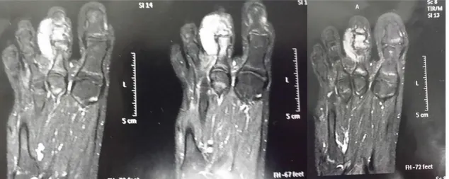
Figure 1: NMR of left foot showing a lesion in 2nd toe. It is remarkable the involvement of soft parts, suggesting biological aggressiveness, being characteristic
of bone tumor.
A biopsy was requested which showed fragments of connective tissue infiltrated by undifferentiated malignant neoplasm, highly mobile, consisting almost entirely of small cells with rounded nuclei, fine chromatin containing discrete nucleoli and sparse cytoplasm of undefined limits.
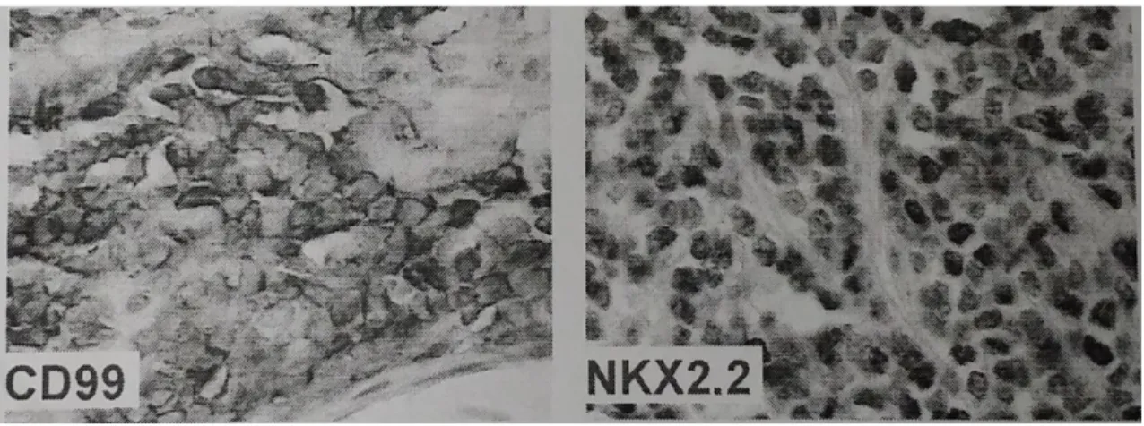
In the immunohistochemistry the detection system based on polymers
was used in which positivity was observed for CD99 protein MIC - 2 (p30/32) clone 12E7, S-100 polyclonal protein, NKX2.2 , homeobox protein, transcription factor gene, clone 74.5A5. It was concluded that the cell morphology associated with the immunohistochemical pattern suggested Primitive Neuroectodermal Tumor (PNET)/Ewing's Sarcoma.
REVISTA UNIMONTES CIENTÍFICA
Montes Claros, II Congresso Nacional deOncologia da Associação Presente.
Figure 2: Immunohistochemistry
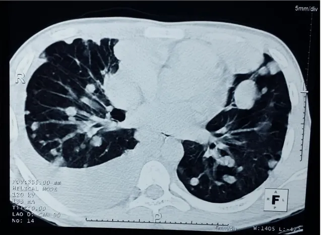
In the possession of the anatomopathological results, a bone scintigraphy was requested, as well as a computed tomography scan of the chest and abdominal ultrasound, whose results showed the presence of pulmonary nodules bilaterally, being that to the right was associated to
moderate pleural effusion, as well as an increase of the osteoblastic activity of the skull (front), of the costal arches on the left (3º and 7º), in the thoracic spine (T6, T8, T12), in the sacrum, on the pubis projections and the greater trochanter of the left femur that suggest neoplastic involvement.
GOMES,S.I.S.;FILHO,S.T.C.;VELOSO,V.T.F;MEND ES,V.F.;SILVEIRA,T.M.R.;NETO,J.D;VELOSO,D.M. F.;CARVALHO,L.
Figure 3: Computed tomography of the chest showed nodular formations of varying sizes in both lungs, and on the right, moderate pleural effusion.
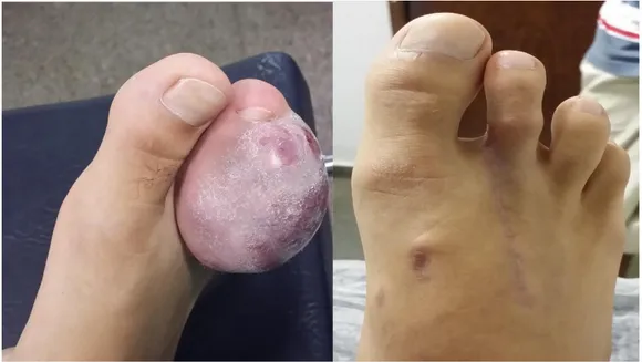
In the interval between the completion of an incisional biopsy and the result of the anatomopathological and immunohistochemical examination, the tumor evolved rapidly with large dimensions. Thus, it was opted for
immediate surgery without neoadjuvant chemotherapy. The surgical procedure consisted of the amputation of the affected radius, i.e., amputation of the second ray of the foot.
REVISTA UNIMONTES CIENTÍFICA
Montes Claros, II Congresso Nacional deOncologia da Associação Presente.
Figure 4: Before and after the surgical procedure.
It was proposed the realization of adjuvant chemotherapy in the schema of vincristine, doxorubicin, cyclophosphamide and actinomycin, considering the significant systemic involvement. After the initiation of
treatment there was improvement of the Karnofsky performance status , passing from 80% to 90%, despite the persistence of residual metastatic lesions.
DISCUSSION
The presence of an expansive lesion, painful in metadiaphyseal bone topography whose clinical sign is parallel to the appearance of nonspecific symptoms, as well as important weight loss and fever of intermittent characteristic should alert us to a possible bone neoplasm, in particular the Ewing's Sarcoma. 4.5
This tumor, also known as primary Neuroectodermal Tumor, consists of a primary bone neoplasm of small round cells and high malignancy, presenting commonly advanced degree of undifferentiation. It represents approximately 6% of the total number of primary malignant bone tumors, affecting commonly individuals from 5 to 15 years of age, with predominance in Caucasian individuals.5
The key locations are, in descending order: diaphysis of the
GOMES,S.I.S.;FILHO,S.T.C.;VELOSO,V.T.F;MEND ES,V.F.;SILVEIRA,T.M.R.;NETO,J.D;VELOSO,D.M. F.;CARVALHO,L.
femur, proximal region of the femur, diaphysis of the fibula, ulna and bones of the pelvis. The most common symptoms are pain and existence of a palpable mass, which occur in a progressive way. Pathological fracture, weight loss, weakness, anorexia and low-grade fever may also be present.5
The diagnostic and classification evaluation involve the performance of imaging examinations, namely, radiography, computed tomography, magnetic resonance imaging; in addition to inflammatory and histopathological markers. The radiographs in a general way showed permeative heterogeneous lesion, resembling "moth-eaten", and subperiosteal reaction, characterized by multiple layers (appearance of "onion skin").5.6
RNM is fundamental to demonstrate the e intramedullary and extramedullary extension and involvement of soft parts. NT has a high risk of extra compartmentation and metastasis in the distance. For that reason, image examinations should be requested of the chest and abdomen.5.7
This group of tumors has as one of its origins the chromosomal
abnormalities t(11;22)(q24;q12), which are found in more than 85% of the cases, causing a fusion of genes due to the translocations. Due to the similarity among the neoplastic cells of this tumor and those found in lymphoma, neuroblastoma and rhabdomyosarcoma, it is a difficult diagnosis, depending on molecular and immunohistochemical tests. In this sense, the detection of antigens such as CD99, a glycoprotein with high expression in these tumors, can be useful in the differential diagnosis.7
In the patient herein, after confirmed the suspicion, the examinations were carried out with the purpose of performing the staging according to the TNM system of the American Joint Committee on Cancer (AJCC). The presence of distant metastases allowed to classify it as stage IVB.
The treatment of advanced stages of Ewing's Sarcoma should include neoadjuvant or adjuvant chemotherapy, or both, for the treatment of metastases. Before the use of chemotherapy with multiple agents, the long-term survival was less than 10%. Currently, the majority of centers reported rates of long-term survival from 60 to 75%.
REVISTA UNIMONTES CIENTÍFICA
Montes Claros, II Congresso Nacional deOncologia da Associação Presente.
The most used drugs are those of the Rosen Scheme (T11), composed basically of adriacine (doxorubicin), vincristine, cyclophosphamide and actinomycin D.9.10
The local treatment of the lesion is still controversial. The Ewing's sarcoma
is radiosensitive, but some authors reported a decrease in the rate of local recurrence (<10%) and an increase in the rate of overall survival with wide resection of the primary tumor.3
FINAL CONSIDERATIONS
Considering the importance of broadening knowledge about the clinical suspicion and the treatment of advanced stages of Ewing's sarcoma, this report aims to disseminate
information about this neoplasm, as well as reporting the conduct of a clinical case of atypical presentation. Despite the high aggressiveness of this tumor, the clinical and surgical treatments can promote improvement of the quality of life in selected patients.
REFERENCES
1. HOROWITZ, M. E. et al. Ewing's sarcoma. CA Cancer J Clin, v. 42, n. 5, p. 300-320, 1992 2. YOUNG, S. C. et al. Incidence of malignance tumors in US. Children Pediatric, v. 86, p. 254-258, 1975 3. CANALE, S. T., BEATY, J. H. Ortopedic Surgery – Campbell. 12. ed. Tennessee: Elsevier Editora Ltd. 4. BATISTÃO, G. et al. Sarcoma de Ewing/Pnet primário pré-sacral: relato de caso. Medicina (Ribeirao Preto. Online). v. 48, n. 5, p. 518-522, 2015. 5. GONZÁLEZ, E. H. H. et al. Sarcoma de Ewing. Rev Archivo Médico de Camagüey. v. 17, n. 5, p; 623-640, 2013.
6. SASSI, L. M. et al. Sarcoma de Ewing. Rev Bras Cir
GOMES,S.I.S.;FILHO,S.T.C.;VELOSO,V.T.F;MEND ES,V.F.;SILVEIRA,T.M.R.;NETO,J.D;VELOSO,D.M. F.;CARVALHO,L. Cabeça Pescoço, v. 37, n. 1, p. 56-57, 2008. 7. LEONG, A. S. et al. Manual of Diagnostic Antibodies for Immunohistology. 1. ed. London: Greenwich Medical Media Ltd, 1999.
8. ABUL K. A.,
NELSON, F., VINAY, K. Robbins & Cotran. Patologia – Bases Patológicas das Doenças. 8. ed. Rio de Janeiro: Elsevier Editora Ltd, p. 1361, 2010.
9. CATALAN, J. et al. Sarcoma de Ewing: aspectos clínicos e radiográficos de 226 casos. Radiol Bras, v. 38, n.5, p. 333-336, 2005.
10. DELATTRE, O. et al. The Ewing family of tumors – a subgroup of small-round-cell tumors defined by specific chimeric transcripts. N Engl J Med, v. 331, n.5, p.294-299, 1994.
11. MIRRA, J. M. Bone tumors: diagnosis and treatment. 2. ed. Philadelphia: JB Lippincott, 1980.
12. BADER, J. L. et al. Intensive combined modality therapy of small round cell and undifferentiated sarcomas in children and young adults: local control and patterns of failure. Radiother Oncol, v. 16, n. 3, p. 189-201, 1989.