Hanna Fokt
Outubro de 2010
Universidade do Minho
Escola de Ciências
Actividade moduladora de extracto de
Ginkgo biloba no ciclo celular de
Saccharomyces cerevisiae e capacidade
de reparação de danos de DNA em células
sob stresse replicativo.
Dissertação de Mestrado
Mestrado em Biotecnologia e Bio-empreendedorismo em
Plantas Aromáticas e Medicinais
Hanna Fokt
Outubro de 2010
Universidade do Minho
Escola de Ciências
Actividade moduladora de extracto de
Ginkgo biloba no ciclo celular de
Saccharomyces cerevisiae e capacidade
de reparação de danos de DNA em células
sob stresse replicativo.
Trabalho efectuado sob a orientação do
iii
Acknowledgments/ Agradecimentos
First of all, I am heartily thankful to my supervisor, Prof. Rui Oliveira, whose knowledge; encouragement, patience and kindness have been invaluable to me during the development of the present work.
I am grateful to Prof. Manuela Côrte-Real for her aid and support in the realization of the present work.
Also, I want to express my thanks to all my laboratory partners, namely, Filipe Marques, Flávia Viana, Andreia Pacheco, Yolanda Delgado and Flávio Azevedo, since my work was easier with their help and, principally, their friendship.
I would like to show my gratitude to all technical and auxiliary staff of the Department of Biology for all assistance in the realization of this work, especially Cristina Ribeiro.
Finally, I am thankful to my parents for the constant source of support – emotional, moral and of course financial, and my friends for all love and support, because this thesis would certainly not have existed without them.
iv
Actividade moduladora de extracto de Ginkgo biloba no ciclo celular de
Saccharomyces cerevisiae e capacidade de reparação de danos de DNA em células
sob stresse replicativo Resumo
Ginkgo biloba L. possui uma ampla gama de actividades biológicas e o seu
extracto padronizado comercial, EGb 761, tem sido um dos produtos medicinais mais vendidos em todo o mundo, devido à sua actividade antioxidante. Efeitos benéficos sobre o sistema nervoso central, incluindo efeitos nos pacientes da doença de Alzheimer, e regulação da expressão genética relacionam-se com os dois principais constituintes do extracto de G. biloba: ginkgolidos e bilobalida. Entre os genes modulados pelo EGb 761, existem vários envolvidos na regulação do ciclo celular, como o gene XPC de mamíferos. A regulação do ciclo celular pode ser crucial para a sobrevivência do organismo, uma vez que, danos de DNA não reparados antes do processo de replicação, podem provocar mutações prejudiciais para as células filhas.
O objectivo do presente trabalho foi a investigação dos efeitos de extracto vegetal obtido a partir das folhas de G. biloba, no ciclo celular. Os ensaios envolveram incubações de células de Saccharomyces cerevisiae com o extracto de G. biloba antes da exposição ao choque oxidativo, provocado pelo peróxido de hidrogénio. A estimativa das proporções celulares em cada fase do ciclo celular foi efectuada através da contagem das células ao longo do tempo e da determinação dos índices de gemulação. Foram efectuadas ainda as medições de conteúdo de DNA nas células, através de citometria de fluxo. A capacidade de reparação de danos de DNA nas células com o ciclo celular parado e expostas ao choque oxidativo, também foi investigado. Os ensaios envolveram a incubação das células na presença da hidroxiurea, seguida de exposição a
H2O2. Os danos e a reparação de DNA foram determinados através do ensaio cometa.
Os resultados demonstraram o efeito protector do extracto de G. biloba contra os
danos de DNA provocados pelo H2O2 nas células de levedura, promovendo o
progressão do ciclo celular após o choque oxidativo. Além disso, as células sob stress replicativo são menos susceptíveis a danos oxidativos de DNA. No entanto, a hidroxyurea não provoca alterações na dinâmica de reparação de DNA.
v
Modulatory activity of Ginkgo biloba extract on Saccharomyces cerevisiae cell cycle and DNA damage repair ability in cells under replicative stress
Abstract
Ginkgo biloba L. is a plant with a wide range of biological activities and its leaf
extract (EGb 761) has been one of the best-selling medicinal products worldwide due to its antioxidant activity. Beneficial effects on the central nervous system, including in patients of Alzheimer's disease, and regulation of gene expression have been related to two main compounds of G. biloba extract: ginkgolides and bilobalide. Among EGb 761 modulated genes, are several involved in cell cycle regulation, like mammalian XPC gene. DNA lesions, which are not repaired before replication, may propagate harmful mutations to the daughter cells, thus, cell cycle control and regulation is crucial to the organism’s survival. DNA damage checkpoints temporarily block the cell cycle progression in G1 or G2 phases in response to genotoxic stress, that allow cells to repair damaged DNA.
The aim of the present work was the investigation of G. biloba leaf extract effects on cell cycle. The experiments involved incubation of Saccharomyces cerevisiae cells with G. biloba leaf extract, before the oxidative shock by hydrogen peroxide. Subsequent estimation of cell proportions in each phase of cell cycle by the budding index approach was performed, as well as measurement of cells DNA content by flow cytometry.
DNA damage repair ability in yeast cells under replicative stress, exposed to the oxidative shock, was also investigated. Typical experiments included incubation of cells in the presence of hydroxyurea, followed by exposure to hydrogen peroxide. Determination of DNA damage and repair was performed using the comet assay.
The results indicate that G. biloba extract protects yeast cells against DNA damage imposed by hydrogen peroxide, promoting progression of the cell cycle after oxidative shock. This protective effect is probably related to a lower degree of DNA damage and higher repair ability. In addition, cells under replicative stress are less susceptible to oxidative DNA damage, however, hydroxyurea does not provoke changes in the dynamics of DNA damage repair ability.
vi Index 1. Introduction ... 1 1.1. DNA damage ... 1 1.2. DNA repair ... 2 1.3. Cell cycle ... 3
1.4. Phytochemicals and biological activity of G. biloba ... 5
1.5. Methods for DNA damage/repair assessment ... 7
1.6. Biological problem and objectives of the present work... 8
2. Materials and Methods ... 10
2.1. Yeast strains, media and culture conditions ... 10
2.2. Plant material and extract preparation ... 10
2.3. Yeast cultures preparation ... 10
2.4. Comet assay ... 11
2.4.1. DNA repair ... 12
2.4.2. Pre-treatments with GBE ... 12
2.4.3. Hydroxyurea treatment ... 12
2.4.4. Image analysis ... 12
2.5. Budding index ... 13
2.5.1. Treatment with HU ... 13
2.5.2. Treatment with GBE ... 13
2.5.3. Oxidative shock and treatments with GBE... 13
2.6. Flow cytometry ... 14
2.6.1. Treatment of BY4741 with GBE ... 14
2.6.2. rad4 and rad23 treatment with GBE ... 15
2.7. Viability assay ... 15
2.8. Statistical analysis ... 15
3. Results ... 16
3.1. The effect of GBE on yeast cell cycle progression ... 16
3.2. Effect of GBE on cell cycle progression of rad4 and rad23 mutants ... 18
3.3. GBE protection against UVC radiation ... 19
3.4. Effect of oxidative shock on DNA of yeast cells under replicative stress. ... 21
vii
4. Discussion ... 26 5. References ... 29
viii
Abbreviations list:
8-oxoG, 8-oxo-7,8-dihydroguanine; AP, apurinic/apyrimidinic;
BER, base excision repair; CDK, cyclin-dependent kinase; DSBs, double-strand breaks;
EGb 761, standard Ginkgo biloba L. leaf extract; GBE, Ginkgo biloba L. leaf extract;
GGR, global genome repair; HR, homologous recombination; HU, hydroxyurea;
LMA, low melting agarose; MMR, mismatch repair;
N7-meG, N7-methylguanine;
NER, nucleotide excision repair; NHEJ, non-homologous end-joining; NMA, normal melting agarose; OD, optical density;
PCR, polymerase chain reaction; RNR, ribonucleotide reductase; ROS, reactive oxygen species;
SCGE, single cell gel electrophoresis; SSBs, single-strand breaks;
ix
TCR, transcription-coupled repair; Tg, 5,6-dihydroxy-5,6-dihydrothymine; TLS, translesion synthesis;
1
1. Introduction
1.1. DNA damage
The integrity of the genomic DNA of cells is crucial for survival and normal function of living organisms; however it is continuously faced by endogenous and exogenous DNA damaging agents [1]. Alkylating genotoxic agents and free radical damaging species from intracellular origin, cause DNA lesions such as modification, loss of DNA bases or DNA strand breaks. These events can alter coding specificity leading to mutations, a significant cause of human disease [2, 3]. Also, damaged bases may be cytotoxic or/and miscoding the latter is thought to be a major mechanism by which DNA-reactive agents cause diseases such as cancer [4]. Different types of DNA damage emerge as a result of genotoxic effects: DNA base modifications, abasic sites, deoxyribose damage, single and double strand breaks [5].
The most frequent types of DNA damage are apurinic/apyrimidinic sites (AP sites) produced by spontaneous and induced base loss [6]. These AP sites are formed as a result of depurination or DNA glycosylase-mediated hydrolytic cleavage at the N-glycosyl bond between the base and deoxyribose [7]. Studies of the depurination rate in calf thymus DNA demonstrated that, under physiological conditions, corresponds to approximately 9000 AP sites per cell per day [8]. These AP sites have cytotoxic and mutagenic potencial and can block DNA replication and transcription [9]. Also, it was previously demonstrated that a fraction of AP sites that escape repair, seems to contribute to mutations, transcription errors and chromosome aberrations associated with degenerative disorders and cancer.
Damaged DNA bases can occur by methylation, oxidation and deamination of normal bases and lead to a variety of lesions such as N7-methylguanine (N7-meG), 5,6-dihydroxy-5,6-dihydrothymine (Tg), uracil or 8-oxo-7,8-dihydroguanine (8-oxoG) [10, 11]. The latter is a major product of DNA oxidative damage and can produce GC to TA transversions, pairing with adenine. Thus, the cellular level and occurrence of 8-oxoG is important in terms of mutagenesis and carcinogenesis [12]. Moreover, inappropriate bases such as uracil can be incorporated into DNA during replication and repair [13].
2
Existence of a large variety of DNA lesions and crucial importance of DNA integrity for organisms’ survival led to the evolution of multiple repair and damage tolerance mechanisms in cells.
1.2. DNA repair
DNA repair is closely interrelated with cell cycle regulation, transcription and replication processes. Changes in genetic material may cause a variety of inheritable diseases, premature aging and cancer. All cancer cells are genetically unstable and contain mutations in genes involved in growth control [14].
Depending on the type of the DNA lesion, cells have evolved different repair pathways that allow the maintenance of the genome integrity. Removal or correction of endogenous/exogenous DNA damage in yeasts and mammals are possible due to the activity of a large variety of repair mechanisms, such as base excision repair (BER), nucleotide excision repair (NER), transcription-coupled repair (TCR), global genome repair (GGR), mismatch repair (MMR), translesion synthesis (TLS), homologous recombination (HR) and non-homologous end-joining (NHEJ) pathways [14, 15, 16].
Saccharomyces cerevisiae has been an informative model for investigating the
molecular biology and biochemistry of DNA repair in eukaryotes. The usefulness of yeast for this type of analysis is due to its simplicity; ease of handling and because of it being considered to have well-conserved DNA repair processes among eukaryotes [12]. Base excision repair seems to be the responsible for repair of a majority of endogenous DNA damages in yeast and mammalian cells [15]. This repair mechanism usually removes DNA lesions (8-oxoG, uracil, thymine glycols and hydrates, etc.) in two steps and involves the action of specific DNA glycosylases. In higher eukaryotes, these enzymes catalyze excision of the base by cleavage of the glycosylic bond, leaving noncoding AP sites in DNA [17, 18]. Subsequently, the resulting AP site is cleaved by an AP endonuclease and the one-nucleotide gap is filled by action of DNA polymerase β [6]. BER mechanism is similar in yeast, however, there are some important differences when compared to BER process of mammals. AP sites cleavage is also commenced by AP endonuclease in S. cerevisiae continuing with a participation of Rad27, Pol2 in the DNA synthesis and Cdc9 in the ligation step [12, 15].
3
In the absence of BER, the NER pathway repairs of a subset of cytotoxic DNA lesions, [13] mainly, the wide class of single-strand lesions that destabilize the double helix, including damages induced by ultraviolet light and DNA intrastrand and interstrand crosslinks [16]. This repair pathway is not essential for viability, however defects in genes involved in NER may result in sun-sensitive, cancer-prone genetic disorders [19]. In this repair process, the phosphodiester bonds on both sides of the lesion are hydrolyzed and damaged DNA (oligonucleotide of about 30 bases in length) is removed. Subsequently, two mechanisms could complete the removal: endonuclease-exonuclease mechanism and excision nuclease (excinuclease) mechanism. In the first case, an endonuclease makes an incision at a phosphodiester bond either 5’ or 3‘ to the lesion, and then an exonuclease digests the damaged strand past the lesion. The second mechanism involves an enzyme system that incises phosphodiester bonds on either side of and at some distance from the lesion, resulting in excised fragment of a relatively precise length [17]. This is followed by repair synthesis and ligation steps with the participation of DNA polymerase and DNA ligase, respectively [19].
Indeed, NER is a process that requires over 20 proteins and most of them are indispensable for DNA repair, such as Rad4. This protein is important for DNA damage recognition. In contrast, the yeast Rad23 represents a class of accessory NER proteins [20]. This evolutionarily conserved protein contains ubiquitin-like domain [21] and forms a high affinity interaction with Rad4, acting as the dimer in NER process. At the absence of Rad23, NER activity is reduced but not eliminated. Rad23 seems to have two roles in NER: direct participation through the stimulation of Rad4 binding to damaged DNA, and stabilization of Rad4 protein [20].
1.3. Cell cycle
DNA lesions, which are not repaired before replication, may propagate harmful mutations to the daughter cells. Thus, DNA damage checkpoints temporarily block the cell cycle progression in G1, S or G2 phases in response to genotoxic stress, that allow cells to repair damaged DNA [22]. Like other eukaryotes, budding yeast S. cerevisiae possesses DNA damage checkpoint response, leading to cell cycle arrest. Once damaged DNA is repaired, cells resume cell cycle progression. Though, cells with irreparable DNA lesions may escape the cell cycle arrest and reenter the cell cycle in the presence of DNA damage, resulting in DNA damage adaptation [23].
4
The cell cycle of S. cerevisiae commences with an unbudded cell in G1 phase (Fig. 1). Spindle plaque duplication, the initiation of DNA synthesis and the emergence of the bud characterize the start of S phase. In G2 phase of cell cycle occurs the formation of the complete spindle and the bud grows in size. M phase is marked by the nuclear division followed by cytokinesis results with the production of two unbudded cells [24]. Existing correspondence between the morphology and the phase of S.
cerevisiae cell cycle (Fig. 1) make it possible to use the size of yeast bud as a criterion
for identification of cell cycle progression.
Figure 1. Saccharomyces cerevisiae morphology variation throughout the different phases of the cell
cycle [adapted from 24]. Abbreviations: PD, plaque duplication; BE, bud emergence; iDS, initiation of DNA synthesis; DS, DNA synthesis; PS, plaque separation; NM, nuclear migration; mND, medial nuclear division; SE, spindle elongation; IND, late nuclear division; CK, cytokinesis: CS, cell separation.
Checkpoint control pathways are complex genetic mechanisms that consist of stimuli (different types of DNA damage), transduction machinery (secondary messenger systems that can transmit the signal) and targets (which are presumably required for cell cycle progression) [25]. A large variety of genes required for the cell cycle arrest was identified in S. cerevisiae, depending on the type of the damage [26].
Passage through Start point (in mid G1 phase) is promoted through the activation of the G1-phase cyclin-dependent kinase (CDK). Most of the cells preferentially arrest in G1, inhibiting CDK activity in response to DNA damage [27]. This arrest allows repair of damaged DNA before the duplication in subsequent S phase.
5
During the exit from G1, activation of yeast Clb5,6-Cdc28 CDK complex, spindle pole body duplication and beginning of DNA synthesis take place. Succeeding bud growth continues through S phase and mitosis (Fig. 1). Finally, return to G1 is accompanied by cytokinesis and bud separation [24, 28]. In budding yeast, RAD24 and a complex composed of MEC3, RAD17 and DDC1 are required for most DNA damage checkpoints [28]. RAD9 is shown to be a negative regulator that inhibits progression from G2 so that cell viability is preserved and fidelity of chromosome transmission is maintained [29]. However, even required for the G2/M checkpoint, RAD9 has not been demonstrated to function in the DNA replication checkpoint [28].
It is possible to arrest yeast cell cycle in the specific phases, enabling studies of all the events related with cell cycle in synchronous cultures. Using treatment with some chemicals, mating factors, radiation or submitting yeast cultures to stress conditions, like nutrient starvation, it is possible to obtain cells arrested in a specific phase of cell cycle. For example, hydroxyurea is a compound capable to block DNA replication, arresting the cell cycle in S phase. This drug blocks an enzyme ribonucleotide reductase (RNR) which catalyzes deoxyribonucleotides production for DNA synthesis by reducing the corresponding ribonucleotides [30].
1.4. Phytochemicals and biological activity of G. biloba
Plants have been used as an important source of drugs from an ancient time until today. Plants contain a large variety of metabolites which provide nutraceuticals and pharmaceuticals for the treatment of different diseases [31]. A wide range of plants and their biological activities: cytoprotective, immunomodulatory, chemotherapeutic and anti-mutagenic, among others, was previously reviewed [32].
Nowadays, significant attention has been paid to phytochemicals with antimutagenic and anticarcinogenic properties. Natural compounds with antigenotoxic activity and inhibitors of carcinogenesis are of particular importance [33].
Polyphenolic compounds are natural antioxidants, which able to protect against many chronic diseases associated with oxidative stress. Supplying the diet with such ingredients it is possible to destroy some reactive oxidative species (ROS) that damage DNA, reducing the risk of cancer [34]. Antigenotoxic activity in most of the cases is
6
related to the presence of antioxidant properties, but additional mechanisms cannot be excluded.
Anticancer chemotherapy relies basically on DNA-damaging and microtubule-targeting drugs, with considerable side effects. One of the hallmarks of cancer cells proliferation is deregulation of the cell cycle, allowing cells to continuously proliferate despite the state of integrity of the genome. Therefore, cell cycle regulating proteins are potential targets for efficient anticancer drugs as they could allow efficient cell cycle arrest, so that DNA damage could be repaired or apoptosis triggered [35, 36]. In addition, the common development of chemotherapy resistance mediated by cell cycle could be avoided by combination therapies of cell cycle-targeting drugs and the chemotherapeutic agents used in clinical practice [37]. Cell cycle-modulating drugs include the recently identified compounds flavopiridol and silibinin that inhibit cyclin-dependent kinases (CDK) positive regulators, which allow cell cycle arrest [38].
Ginkgo biloba L. is a plant with a broad spectrum of important biological
effects. Leaf extract from this tree was standardized and commercialized under the name of EGb 761. The extract contains 24% ginkgo-flavone glycosides, 6% terpene lactones (ginkgolides and bilobalide), and approximately 7% proanthocyanides, and other uncharacterized compounds [39]. The main compounds of the G. biloba extracts, which believed to be responsible for most pharmacological properties, are the terpene trilactones: bilobalide and ginkgolides [40, 41].
Bilobalide contains three lactones and the characteristic tert-butyl group, as well as a secondary and a tertiary hydroxyl group, but only one carbocycle [42]. The ginkgolides are characterized by a presence of a cage skeleton consisting of six five-membered rings: a spiro (4, 4) - nonane carbocyclic ring, three lactones, and a tetrahydrofuran ring. They also contain an unprecedented tert-butyl group and vary only in the number and positions of their hydroxy groups [40, 42]. Ginkgolides are stable to light, humidity and easily dissolved in dilute alkali and recovered quantitatively on subsequent acidification [40].
Flavonoids have high affinity to react with hydroxyl radicals to form an additional product, due to the presence of aromatic rings with double bonds and phenolic hydroxyl groups. This reactivity is thought to be related to scavenging the
7
hydroxyl radicals directly. The phenolic hydroxyl groups possibly chelate the ferrous
cation and decrease the formation of hydroxyl radicals generated from H2O2.
Among a variety of properties, EGb 761 is an excellent antioxidant and seems to be a potential drug for neuronal diseases associated with the excessive production of reactive oxygen species [41, 43, 44, 45]. Non-standardized extract obtained from G.
biloba leaves (GBE), was shown to have strong antioxidant and antigenotoxic activity
similar to EGb 761 effect [46]. G. biloba extract has a number of beneficial effects on central nervous system, including to patients of Alzheimer's disease [42]. More recently, EGb 761 was reported to be able to up- and down-regulate a variety of genes involved in cell cycle regulation, cellular growth, nervous system development and function [47].
1.5. Methods for DNA damage/repair assessment
Characterization of structural integrity of DNA is of great importance in a large variety of biological applications. Assessing DNA damage and repair is possible using specific methods of DNA lesions measurement, independent of biological responses to such lesions [48]. Currently, there are some methods of repairable and nonrepairable DNA damage/repair assessment, carried out by measuring DNA fragmentation. Some of them are single cell gel electrophoresis (SCGE) or comet assay and other approaches based on electrophoresis on agarose gel, micronuclei detection assay, DNA alkaline unwinding method, polymerase chain reaction (PCR), SDS-DNA PAGE, TUNEL assay, flow cytometry, FISH, HPLC-electrospray tandem mass spectrometry and GC-MS. Each of the referred methods encloses advantages/disadvantages and used depending on the type of the DNA lesion.
Comparative to other methods, comet assay and its variations (neutral, alkaline and use of lesion-specific enzymes) have some important advantages, like a possibility to evaluate DNA damage at single cell level, using a wide spectrum of parameters for its measurement that can be chosen to each particular situation. The simplicity, sensitivity, versatility, speed and economy of the comet assay enable its wide use in a large number of laboratories worldwide. This method can be used in eukaryotic and prokaryotic cells [49], detect low levels of DNA damage (one break per 1010 Da of DNA) [51] and requires small number of cells (approximately 10,000) per sample. Generated data allow application of robust types of statistical analysis. On the other hand, comet assay has some limitations. The method provides no information on DNA fragment size since
8
fragments are not separated during electrophoresis and requires viable single-cell suspension. If the suspension consists predominately of necrotic or apoptotic cells, true information on the presence of specific lesions (strand brakes or base damage) cannot be obtained. Variability of the samples (between cells and cultures), image analysis systems or visual scoring and interpretations are the other disadvantages of comet assay that contribute to inter-laboratory variations in results [51, 52, 53].
Nevertheless, the advantages of the method far outnumber the disadvantages, and therefore, it has been widely used as a tool for DNA damage and repair studies [50, 54, 55], genotoxicity testing [54, 56], human and environmental biomonitoring [54, 57].
1.6. Biological problem and objectives of the present work
Nowadays, the pollution of ecosystems increases every year, when new chemicals are added to the existing amount of toxic substances in the environment. Intensive agricultural and industrial activities harmfully affect biodiversity of ecosystems, constituting a high risk to the survival of the species and disease risks to humans [50]. Attending to this problem, the interest to obtain biologically active compounds from natural sources for use in treatment of human diseases has renewed. Complete biodegradability of such compounds, low or absent toxicity and availability from renewable sources constitute important advantages if compared with those of compounds obtained by total chemical synthesis [31].
A considerable number of plant extracts and phytochemicals have been reported to have antigenotoxic and anticancer effects. Like the flavonoids flavopiridol and silibinin, many other compounds are expected to exist in plants, especially those with high content of compounds of this chemical family. In fact, Ginkgo biloba leaf extract has been demonstrated to have an antigenotoxic effect concomitant with higher DNA repair capacity in yeast cells [46]. However, in a very few cases, antimutagenic and anticancer activities were caused by a mechanisms involving cell cycle modulation.
Saccharomyces cerevisiae has been used successfully as model organism in
studies of DNA damage, DNA repair and cell cycle due to the similitude of these processes with higher eukaryotes. This system has the advantages of simplicity of culture, fast growth, genetic tractability and the possibility to create conditional mutants in essential genes, which allows the study of their phenotype by loss of function.
9
Another advantage in the case of cell cycle is the possibility to visually identify the phase of the cell cycle of a given cell simply by the observation of the presence and size of the bud (Fig. 1). Therefore, comet assay constitutes a valuable tool for the evaluation of DNA damage and antigenotoxic effects of plant extracts or phytochemicals. To the best of our knowledge, there are few reports for comet assay successful application on yeast cells. Moreover, the protocol optimized for S. cerevisiae was developed previously in our laboratory and successfully used for DNA damage and repair assessment [55].
The present work aims to contribute to knowledge of therapeutic potential of G.
biloba leaf extract and its effects on yeast cell cycle. Experiments of DNA damage and
repair measurement were performed, involving pre-incubation of S. cerevisiae cells with
GBE, before oxidative stress induced by H2O2, which produces base oxidation and
single-strand breaks, mediated by the highly reactive hydroxyl radicals [58]. Also, DNA damage repair ability in yeast cells with arrested cell cycle and exposed to the oxidative shock, was also investigated. The replicative stress was provoked by chemical compound hydroxyurea, in order to arrest yeast cell cycle in S phase. Alternatively, the budding index and flow cytometry of S. cerevisiae BY4741 cells and respective mutants’ rad4 and rad23 pre-treated with GBE and submitted to oxidative shock, were achieved.
10
2. Materials and Methods
2.1. Yeast strains, media and culture conditions
In the present work, the haploid Saccharomyces cerevisiae strain BY4741 (MATa his3Δ1 leu2Δ0 met15Δ0 ura3Δ0) [59] was used for all experiments and derived mutants rad4 (BY4741 MATa his3Δ1 leu2Δ0 met15Δ0 ura3Δ0 YEL037c::kanMX4) and
rad23 (BY4741 MATa his3Δ1 leu2Δ0 met15Δ0 ura3Δ0 YER162c::kanMX4) were used
for the analysis by flow cytometry. Stock cultures were maintained on solid (1% w/v yeast extract, 2% w/v peptone, 2% w/v glucose and 2% w/v agar) and liquid (lacking agar) YPD medium at 4ºC. For the experiments, cell cultures were grown on liquid YPD medium, Difco™ Yeast Nitrogen Base without amino acids and ammonium sulfate medium (YNB-N) and Difco™ Yeast Nitrogen Base (YNB), at 0,17% and 0,67% w/v, respectively, using an orbital shaker at 30ºC and 200 rpm. Yeast growth
was monitored by optical density at 600 nm (OD600).
2.2. Plant material and extract preparation
The plant material used in the present study were Ginkgo Biloba L. leaves collected at spring (May) from a specimen located in an urban area of Braga, Portugal. The leaves were washed, air-dried at room temperature and subsequently, pulverized with a pestle into a fine powder. Ginkgo biloba L. leaves extract (GBE) was prepared as described by Ding and co-workers [60]. Five grams of leaf powder were transferred to a 200 ml polypropylene centrifuge tube and 30 ml of boiling deionized water were added. Subsequently, the mixture was heated to 100ºC for 5 min. After centrifugation at 2000 g for 15 min, the supernatant was obtained. This operation was repeated twice and the supernatants were joined together, pH was adjusted to 6.5 with NaOH and stored at -20 ºC.
2.3. Yeast cultures preparation
Saccharomyces cerevisiae cells from solid stock culture were removed with an
inoculation loop, then suspended in 5 ml of liquid YPD medium (pre-inoculum) and incubated overnight at 30ºC, 200 rpm. An appropriate volume of the pre-inoculum was
diluted in 10 ml of liquid YPD medium to obtain a culture with an OD600 0.1 and
incubated under the same conditions until OD600 0.4-0.8. In order to obtain yeast cells
11
an appropriate volume was suspended in liquid YPD medium and incubated overnight
at 30ºC, 200 rpm to obtain a culture with an OD600 0.2 next day. Subsequently,
incubation during two generations under the same conditions was effectuated. 2.4. Comet assay
Saccharomyces cerevisiae cultures were prepared as described in 2.3. Cells were
harvested by centrifugation at 5000 rpm, 4ºC for 2 min and washed twice with the same volume of cold deionized water. The pellet was suspended in a double volume of cold S
buffer (1 M sorbitol, 25 mM KH2PO4, pH 6.5). Subsequently, 1 ml of cell suspension
was centrifuged and the pellet was suspended in lyticase buffer (200 μl lyticase (87
U/ml), 500 μl S buffer 2x, 300 μl deionized H2O and β-mercaptoethanol to 50 mM final
concentration) and, incubated at 200 rpm and 30ºC for 30 min in order to obtain spheroplasts. Subsequently, 80 μl of spheroplasts suspension were distributed by 1 ml aliquots and spheroplasts were collected by centrifugation at 15300 rpm, 4ºC for 2 min. Each pellet was suspended in 80 μl of low melting agarose (LMA) 1.5% (w/v in S buffer) at 35ºC and was applied onto glass slides coated with 0.5% normal melting agarose (NMA) (w/v) and covered with cover slips. All subsequent steps were performed at 4ºC.
The cover slips were removed, 250 μl of the oxidant solution (10 mM H2O2) was
applied on each gel and 20 min incubation was performed. After the incubation, the slides were washed with S buffer for 5 min and submerged in the lysing buffer (30 mM NaOH, 1 M NaCl, 0.05 % (w/v) lauroylsarcosine, 50 mM EDTA, 10 mM Tris-HCl, pH 10) for 20 min. After cell lysis, the slides were washed with the electrophoresis buffer (30 mM NaOH, 10 mM EDTA, 10 mM Tris-HCl, pH 10) for 20 min and electrophoresis was performed with the same buffer for 10 min and 0.7 V/cm. Afterwards, gels were neutralized with 10 mM Tris-HCl buffer, pH 7.4, for 10 min and samples were fixed, first with ethanol 76% v/v and subsequently with 96% v/v, both for 10 min. Subsequently, the slides were dried in a laminar flow chamber or at room temperature (RT) and stored at 4ºC until visualization. Immediately before the analysis in fluorescence microscope, gels were stained with 3 fold concentrated Gel Red™ prepared from the stock 10,000 fold concentrated solution.
12 2.4.1. DNA repair
In these experiments, the slides treated with 10 mM H2O2 for 20 min, were
washed with S buffer for 5 min and incubated at 37ºC for 5, 10, 15 and 20 min. A control with deionized water and a control without incubation at 37ºC were included in the experiment. The described procedure of comet assay was followed afterwards.
2.4.2. Pre-treatments with GBE
Before the incorporation in LMA in the comet assay procedure, spheroplasts were suspended in 80 μL of GBE diluted in S buffer for the maintenance of osmotic protection and were incubated at 30ºC for 20 min. After incubation, spheroplasts were collected by centrifugation at 15,300 rpm, 4 ºC for 2 min, washed with 80 μL S buffer and subsequently, suspended in LMA. The described procedure of the comet assay was followed afterwards.
2.4.3. Hydroxyurea treatment
Saccharomyces cerevisiae cell cultures were prepared from liquid stock culture
as described in 2.3. Afterwards, 1 ml of cells was collected by centrifugation at 5000 rpm, 4 ºC for 2 min and suspended in 1 ml of YPD medium containing 200 mM hydroxyurea (HU). The control sample was suspended in 1 ml of YPD medium lacking HU. Subsequently, incubation during two generations under the same conditions was effectuated, and the procedure of comet assay as described above was followed afterwards.
2.4.4. Image analysis
The images were obtained by fluorescence microscopy (Leica DMB 5000, black and white camera) with 400X magnification. Tail length (in µm) was used as a parameter for DNA damage quantification. Fifty comets per each treatment were analyzed with the CometScore software. GraphPad Prism software was used for data processing.
13 2.5. Budding index
Saccharomyces cerevisiae cells from the liquid and solid stocks were used for
cultures preparation as described in 2.3. Afterwards, 1 ml cell cultures were submitted
to treatments with HU, H2O2 and GBE as described below and samples 50 µl each, were
taken at different time points. All the samples were stored on ice and at the end of the experience were observed under a light microscope. For each time point 100 cells were counted and the fraction of cells with no bud, a small bud (smaller than one half of the yeast cell) or a large bud (equal or larger than one half of the yeast cell) was recorded.
2.5.1. Treatment with HU
According to the procedure described above, S. cerevisiae cell cultures from the
solid stock grown to OD600 0.1 and cultures from the liquid stock grown to an OD600 0.2
were harvested by centrifugation at 5000 rpm, 4ºC for 2 minutes and suspended in 1 ml of liquid YPD medium containing 200 mM hydroxyurea. Next, cultures were incubated under the same conditions and samples were taken for microscope observation at 0, 1, 3 and 4 hours after HU addition. Cells incubated in 1 ml of YPD medium lacking HU were used as a control.
2.5.2. Treatment with GBE
Yeast cell cultures from the solid and liquid stock grown to OD600 0.1 and 0.2,
respectively and harvested by centrifugation as described for HU treatment, were suspended in YPD medium containing GBE 2 fold diluted (500 µl of GBE were added to 500 µl of YPD medium 2 fold concentrated). Cultures were incubated at 30ºC, 200 rpm and samples for microscope observation were taken at 0, 1, 3 and 4 hours after GBE addition. Cells suspended in 1 ml of YPD medium without GBE were used as a control.
2.5.3. Oxidative shock and treatments with GBE
According to the procedure of budding index, 8 ml S. cerevisiae cell cultures
from liquid stock, grown to OD600 0.2 were harvested by the centrifugation at 5000 rpm,
4 ºC for 2 min and suspended in YPD medium containing 10 mM H2O2. The first and
the second samples were taken immediately before H2O2 addition and after 20 minutes
14
water for H2O2 elimination, suspended in YPD medium containing GBE 2 fold diluted
(4 ml of GBE were added to 4 ml of YPD medium 2 fold concentrated) and incubated at 30ºC, 200 rpm. Samples were taken at 1, 3, 4 and 6 hours after GBE addition. Yeast cells suspended in 8 ml of YPD without extract were used as a control.
2.6. Flow cytometry
Saccharomyces cerevisiae cell cultures from liquid stock were grown as
described in 2.3 and submitted to oxidative shock by H2O2. Samples were taken at
different time points and stored on ice until the end of the experience. Then, 1x10⁶
cells/ml of each sample were harvested by centrifugation at 15300 rpm, 4ºC for 2 min and fixed in 70% v/v ethanol, by adding 700 µL 100% ethanol to 300 µL of deionized
H2O. Fixed samples were stored at 4ºC for 2-4 days before the continuation of protocol.
Afterwards, the cells were harvested by centrifugation at 15300 rpm, 4ºC for 2 min and suspended in 1 ml of 50 mM sodium citrate, pH 7. To avoid cell aggregation, each sample was sonicated by three 1 second pulses at 35 % of duty cycle, harvested by centrifugation at 15300 rpm, 4ºC for 2 min and suspended in 1ml of the same solution. Cell suspensions were incubated overnight at 37ºC in water bath with 5 mg/ml RNase A and 20 mg/ml Proteinase K in order to degrade RNA and proteins. Next day, cells were harvested at 15300 rpm, 4ºC for 2 min, then suspended in 50 mM sodium citrate solution containing 1µM Sytox Green and incubated in the dark at RT for 2-4 hours. Twenty thousand cells of each sample were analyzed with Epics® XLTM cytometer (Beckman Coulter) equipped with an argon-ion laser 16 emitting a 488 nm beam at 15 mW. Data were analyzed and histograms were obtained with the WinMDI 2.8 software.
2.6.1. Treatment of BY4741 with GBE
Immediately after 20 minutes of 10 mM H2O2 exposure, yeast cells were washed
once with deionized water, suspended in YPD medium containing GBE 2 fold diluted and incubated at 30ºC, 200 rpm. Samples for the cytometry analysis were taken at the same time points as for budding index (oxidative shock and treatment with GBE). Cells suspended only in YPD after oxidative shock exposure, were used as a control.
15 2.6.2. rad4 and rad23 treatment with GBE
Mutant cells from the liquid stock cultures were grown to OD600 0.2 and
submitted to the same procedure as for BY4741 cells until the point of GBE addition to the medium. The first and the second samples for the cytometry analysis were taken immediately before and after 20 min of oxidative shock, respectively. Then, samples were taken at 30 min, 1 and 3 hours after GBE addition. Mutant cells suspended only in YPD after oxidative shock exposure, were used as a control.
2.7. Viability assay
Saccharomyces cerevisiae cells (BY4741, rad4 and rad23) from solid stock
cultures were prepared as described in 2.3. Subsequently, 250 µl of cell suspension were incubated with 250 µl of GBE at 30ºC, 200 rpm for 20 min or with 250 µl of deionized sterilized water as a control. After the incubation, cells were washed once with S buffer
and diluted to OD600 0.1. Then, serial dilutions to 10-3 in S buffer were performed, 5
µl-drops of each suspension were placed on solid YPD medium and allowed to dry at RT.
Afterwards, Petri dishes were irradiated with UVC 25, 50 and 100 J/m2, incubated
overnight in the dark, at 30ºC and compared to the control situation (without irradiation). Images were taken using ChemiDoc™XRS.
2.8. Statistical analysis
The experiments were done in duplicate or triplicate and results are presented as a mean ± standard deviation (SD). One-way analysis of variance (ANOVA) was used for comparison of more than two means and Tukey´s test to multiple comparisons. All asterisks indicate differences considered statistically significant: * means p < 0.05, ** means p < 0.01, and *** means p < 0.001, when compared to the respective control.
16
3. Results
3.1. The effect of GBE on yeast cell cycle progression
Bidon and co-workers [47] in their study referred a range of genes modulated by EGb 761 in rats, some of them involved in cell cycle progression. Based on these results, we decided to investigate the effect of GBE in yeast cell cycle progression. Firstly we decided to do the analysis by the budding index. Yeast cell cultures grown for approximately 18 hours were suspended on fresh YPD medium containing 2-fold diluted GBE. Cultures incubated on YPD medium lacking GBE were used as controls. Budding index was measured along time up to 4 hours by observation under a light microscope, and the proportion of cells with no bud, a small bud (smaller than one half of the yeast cell) and a large bud (equal or larger than one half of the yeast cell) was calculated. A decrease of the fraction of small-budded cells in yeast culture without GBE from the first hour of incubation was observed (Fig. 2A). When GBE was added to the medium, after 3 h a small increase of the amount of small-budded cells (an early stage of S phase) was observed (Fig. 2B). However, taking in consideration that all values are not significantly different in all the remaining time-points, we considered that GBE does not affect cell cycle progression until 4 h incubation.
Figure 2. Effect of GBE on S. cerevisiae cell cycle progression. Budding index of S. cerevisiae
cell culture, grown on YPD medium for approximately 18 h. A: Start corresponds to the culture immediately before addition of fresh medium. B: Start corresponds to the time point of GBE addition. Budding index was measured along time (1, 3 and 4 h) and a fraction of non-budded (circles), small-budded (squares) and large-small-budded (triangles) cells was plotted. Mean ± SD values are from three independent experiments.
17
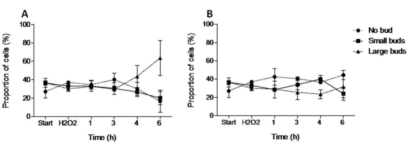
The effect of GBE on S. cerevisiae cell cycle progression after the perturbation by oxidative shock was also tested. The assays were performed under the same
conditions as mentioned for the previous tests. Exposure to 10 mM H2O2 was included
to cause oxidative shock to the yeast cell cultures. Afterwards, the toxic was washed out and the cells were suspended on YPD medium containing 2-fold diluted GBE. Cultures incubated on YPD medium lacking GBE were used as controls. Budding index was measured along time up to 6 h, as fractions of non-budded, small-budded and large
budded cells. In cell cultures incubated without GBE, H2O2 caused an increase in the
proportion of large-budded cells (G2/M) (Fig. 3A), suggesting that cells arrest cell cycle, allowing efficient DNA damage repair. However, in a similar experiment in the presence of GBE, G2/M cells did not increase in proportion as in its absence, suggesting that GBE improves cell cycle progression upon oxidative shock (Fig. 3B). This revocation of cell cycle arrest is probably related to a lower degree of DNA damage and higher repair ability, so that DNA of cells does not get as damaged to trigger cell cycle arrest.
Figure 3. GBE effect on cell cycle progression of S. cerevisiae cells after exposure to H2O2.
Budding index of S. cerevisiae culture, grown on YPD medium for approximately 18 h. A: Start corresponds to the culture immediately before addition of fresh medium. B: Start corresponds to the culture immediately before addition of fresh medium containing GBE 2-fold diluted. H2O2 corresponds to
the budding index of the culture after 20 min 10 mM H2O2 incubation. Afterwards, budding index was
measured along time (1h, 2h, 3h, 4h and 6h after H2O2 incubation) and a fraction of non-budded (circles;
dashed line), small-budded (squares; full line) and large-budded (triangles; dashed line) cells was plotted. Mean ± SD values are from three independent experiments.
18
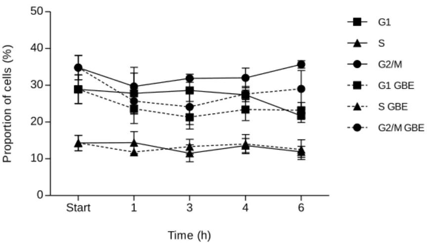
Results depicted in Fig. 3 suggest that GBE appears to protect cells from H2O2,
promoting the progression of the cell cycle after oxidative shock. To confirm this, we decided to discriminate fractions of yeast cells in each phase of cell cycle, measuring DNA content of cells by flow cytometry. The assays were performed under the same conditions as mentioned for the budding index. Oxidative stress was provoked by 10
mM H2O2, followed by incubation of the cells with GBE 2-fold diluted in medium, up
to 6 h. Yeast cell cultures incubated on YPD lacking GBE were used as controls. In cell cultures incubated without GBE and in cell cultures incubated in the presence of the extract, the proportion of G2/M cells increased, comparing to G1 cells fraction (Fig 4). However in cultures with GBE the proportion of G2/M cells compared with G1 is not as higher as in its absence. This is in accordance with results obtained by the budding index (Fig. 3). Start 1 3 4 6 0 10 20 30 40 50 G1 S G2/M G1 GBE S GBE G2/M GBE Time (h) P ro p o rt io n o f c e ll s ( % )
Figure 4. GBE effect on cell cycle progression of S. cerevisiae cells after exposure to H2O2.
Yeast cultures grown for approximately 18 h were exposed to 10 mM H2O2 for 20 min and incubated on
YPD containing 2-fold diluted GBE (dashed lines) or lacking GBE (full lines). The samples were taken along time after H2O2 treatment (1, 2, 3, 4 and 6 h). 0 h corresponds to the sample taken immediately
before the oxidative shock. Cells were stained with Sytox Green and DNA content were analyzed with Epics® XLTM cytometer (Beckman Coulter). Mean ± SD values are from three independent experiments.
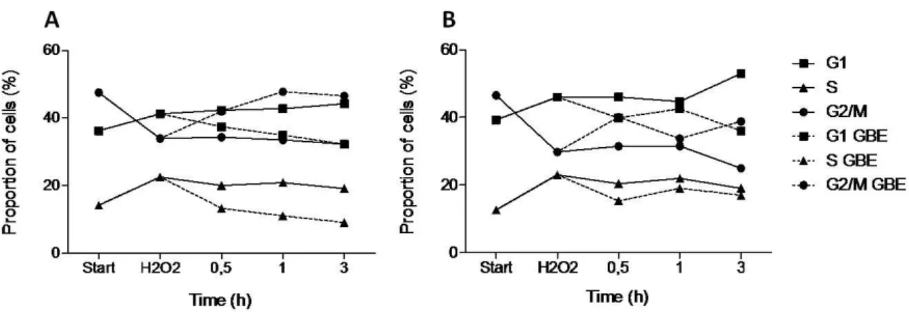
3.2. Effect of GBE on cell cycle progression of rad4 and rad23 mutants
XPC, a rat gene involved in cell cycle regulation, was reported to have an increased expression when rats were treated with EGb 761 [47]. Taking into account these results, we decided to test GBE effect on S. cerevisiae mutant lacking RAD4 gene, the orthologue of mammalian XPC. Moreover, yeast mutant lacking RAD23 gene was
19
used in the experiment, since RAD4 and RAD23 proteins act as a dimmer. Cultures of mutants were grown for approximately 18 h, then submitted to oxidative shock by 10
mM H2O2 and incubated on YPD containing 2-fold diluted GBE up to 3 h. Cell cultures
incubated in medium lacking GBE were used as control situations. The profile of cells from rad4 culture after 3 h was different from the parental strain (Fig. 4), since it was detected a larger fraction of G1 than G2/M cells, when incubated on YPD medium lacking GBE (Fig. 5A; full lines). However, when the cells were incubated with GBE, the profile was reversed to the one observed with the parental strain BY4741, with a major fraction of G2/M cells in culture. Results of rad23 cell culture, when incubated with GBE were similar to the ones of rad4 culture (Fig. 5B) except that both proportions were similar after 3 h incubation. Therefore, the influence of GBE on cell cycle progression is different regarding the existence of functional RAD4 or RAD23 genes. These results suggest a direct activity of component(s) of GBE on cell cycle regulation.
Figure 5. GBE effect on cell cycle progression of S. cerevisiae rad4 and rad23 mutants after
exposure to H2O2. rad4 (A) and rad23 cultures (B) grown for approximately 18 h were exposed to 10 mM
H2O2 for 20 min and incubated on YPD containing 2-fold diluted GBE (dashed lines) or lacking GBE
(full lines). Start corresponds to the culture immediately before addition of 10 mM H2O2; H2O2
corresponds to the culture after 20 min 10 mM H2O2 incubation. The samples were taken along time (0,5;
1 and 3 h after H2O2 incubation). Cells were stained with Sytox Green and DNA content were analyzed
with Epics® XLTM cytometer (Beckman Coulter). Fractions of yeast cells in each phase of the cell cycle were calculated. Mean values are from two independent experiments.
3.3. GBE protection against UVC radiation
Among a variety of biological activities, G. biloba extract was reported to possess radioprotective effect [61]. Most of the studies reported protective activity of
20
G.biloba extract against ionizing radiation. In addition, XPC gene encodes a protein that
recognizes DNA damage caused by UV radiation, besides the participation in cell cycle regulation. In the present experiments, protective effect against UVC radiation in S.
cerevisiae cells was tested. For this, yeast cell cultures of BY4741 and the UV-sensitive
mutants, rad4 and rad23, were pre-incubated with 2-fold diluted GBE for 20 min,
followed by serial dilutions to 10-3. Drops of the cellular suspension were placed on
YPD medium and irradiated with different doses of UVC. The results suggest the absence of GBE protective effect against UVC radiation (Fig. 6). As expected, BY4741 was the most UVC-resistant strain, followed by rad23. The first one has the NER mechanism entirely functional and rad23 mutant possesses Rad4 activity, indispensable for NER. On the other hand, rad4 was the most UVC-sensitive strain, since its NER mechanism was non-functional as this strain lacks functional Rad4. These results, together with data from cell cycle analyses, suggest that the GBE interaction with Rad4 could be via cell cycle regulation rather than by modulation of NER.
Figure 6. GBE effect on UVC-irradiated S. cerevisiae cells. BY4741, rad4 and rad23 cell
cultures were pre-incubated with 2-fold diluted GBE (GBE + BY4741; GBE + rad4; GBE + rad23) for 20 min. BY4741, rad4 and rad23 pre-incubated with deionized water were used as a control (BY4741;
rad4; rad23). Serial dilutions 10-1 , 10-2 and 10-3 were performed (0 corresponds to non-diluted culture). 5 µl-drops of each suspension were placed on solid YPD medium and irradiated with UVC 0, 25, 50 and 100 J/m2. Afterwards, Petri dishes were incubated overnight in the dark, at 30ºC and the images were taken using ChemiDoc™XRS.
21
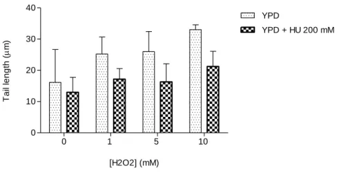
3.4. Effect of oxidative shock on DNA of yeast cells under replicative stress. We started to evaluate the effect of oxidative shock provoked by different
concentrations of H2O2 on yeast cell culture arrested by HU in S phase (by replicative
stress), and on non-arrested cell cultures grown on YPD medium. As reported before, HU, a specific inhibitor of ribonucleotide reductase, inhibits DNA replication in a wide variety of cells, including S. cerevisiae [62]. This drug arrests yeast cells that have not completed DNA replication in S phase, preventing cell division. Cells with completed S phase divide in the presence of hydroxyurea and become arrested in the subsequent S phase of the cell cycle [63]. Cell cultures were grown overnight, and suspended in fresh YPD medium containing or lacking 200 mM HU. Yeast spheroplasts obtained from
these cultures were exposed to 1, 5, and 10 mM H2O2 for 20 min and analyzed for DNA
damage. Additionally, incubation with deionized H2O as negative control of the
oxidative agent H2O2 was included (Fig. 7).
0 1 5 10 0 10 20 30 40 YPD YPD + HU 200 mM [H2O2] (mM) T a il le n g th ( m)
Figure 7. Toxicity and DNA damage induced by the exposure of S. cerevisiae cells to H2O2.
Yeast cell cultures were incubated on YPD medium containing (YPD + HU 200 mM) or lacking 200 mM HU (YPD), for two generations, and obtained spheroplasts were exposed to 1, 5 and 10 mM H2O2 for 20
min. The control experiment (0 mM H2O2) reflects the amount of DNA damage in cells without exposure
to H2O2. DNA damage was analyzed by the comet assay method (see Material and Methods). Mean ± SD
values are from three independent experiments.
Cells grown in medium containing HU are less susceptible to oxidative stress
provoked by different concentrations of H2O2 (Figs. 7 and 8). The difference of the
amount of DNA damage between the control situation and the rest of the treatments for each of the cultures was not statistically significant. However, a clear tendency of
22
cases. In the case of yeast culture grown on YPD medium lacking HU, 10 mM H2O2
increases twice the comet tail length, when compared to the control situation. These results suggest that HU protects DNA from oxidative DNA damage.
Figure 8. Images of the yeast comet assay after DNA labelling with Gel Red. A, B, C and D
correspond to S. cerevisiae cell cultures grown on YPD medium. A: control experiment with deionized water for cell treatment; B, C, D: cells treated with 1, 5 and 10 mM H2O2, respectively. E, F, G and H
correspond to yeast cell cultures grown on YPD medium containing 200 mM HU. E: control experiment with deionized water for cell treatment; F, G, H: cells treated with 1, 5 and 10 mM H2O2, respectively. All
images were obtained at 400X magnification.
3.5. Effect of cell cycle arrest on DNA repair ability of yeast cells.
To investigate the effect of cell cycle arrest in DNA repair ability in S. cerevisiae cells, a modification of the comet assay optimized for DNA damage repair in yeast cells was used (see Materials and Methods). The experiments consisted in provoking DNA damage in yeast spheroplasts from HU-lacking and HU-containing cultures with 10 mM
H2O2 for 20 min to induce damage in DNA, and then incubating at 37 ºC up to 20 min
to allow DNA repair by the spheroplasts. Additionally, two controls were included in
this experiment: incubation with deionized H2O as negative control of the oxidative
agent H2O2 and incubation with H2O2 alone as control of this reagent. As expected, a
statistically significant decrease in comet tail length during the 20 min incubation was observed in both, arrested and non-arrested yeast cell cultures, when compared to respective positive controls (Fig. 9).
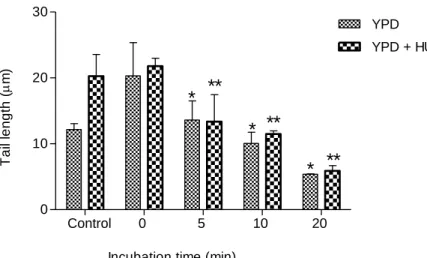
23 Control 0 5 10 20 0 10 20 30 YPD YPD + HU 200 mM
Incubation time (min) *** *** *** *** * * * T a il le n g th ( m)
Figure 9. The effect of cell cycle arrest on DNA repair ability of S. cerevisiae cells grown
overnight. Yeast spheroplasts from asynchronous (YPD) and HU-containing (YPD + HU 200 mM) cell cultures were exposed to 10 mM H2O2 for 20 min, and subsequently, incubated at 37 ºC for different
incubation times (0, 5, 10 and 20 min). The control experiment reflects the amount of DNA damage that cells have without exposure to H2O2. DNA damage was analyzed by the comet assay method (see
Material and Methods). Mean ± SD values are from three independent experiments (*** represents p < 0.001 and * represents p < 0.05).
Yeast cell cultures treated with HU had a lower basal DNA damage (Fig. 9; control), although not statistically different. As can be seen in fig. 9 (0 min repair
incubation), cells treated with 10 mM H2O2 displayed different sensitivities whether
they were HU-treated as shown in Fig. 7; these cells were less susceptible for DNA damage upon oxidative shock. These results suggest that cells with arrested cell cycle are less susceptible to oxidative stress. However, HU did not provoke changes in the repair dynamics, since the repair rates for HU-containing and HU lacking cultures were approximately equal (Table 1).
Table 1. Repair rates of S. cerevisiae cultures.
Cultures Repair rates
(µm of comet tail/min)
YPD -0,08
24
Yeast cell cultures used in these experiments were grown overnight (for approximately 18 h), collected and suspended in fresh medium, following by incubation for two generations. This way, a considerable proportion of the cells in the culture are aged cells when we performed experiments. Therefore, the presence of considerable amount of DNA damage accumulated in cells in the control (Fig. 9) was not surprising. In an attempt to verify the extent the yeast culture growth affects the basal levels of DNA damage in cells, we decided to perform the DNA repair assay described above, using a small volume of cell cultures grown overnight, diluted with fresh medium and incubated for two generations.
For the following experiments, S. cerevisiae cell cultures were grown to exponential phase followed by the addition of fresh YPD medium containing or lacking 200 mM HU. This way, the proportion of aged cells used in the experiment was considerably low. DNA damage in yeast spheroplasts was provoked by exposure to 10
mM H2O2 for 20 min, and incubation at 37 ºC up to 20 min was performed to activate
DNA damage repair mechanisms of cells. Negative and positive (incubation with
deionized H2O and incubation with H2O2 alone, respectively) controls were included in
the experiment. Interestingly, tail length values of the HU-containing culture negative control were higher, when compared to the negative control of the non-arrested culture (Fig. 10). The results of this experiment indicate that HU-treated cells are equally tolerant to oxidative stress (measured as DNA damage). In the previous experiment
(Fig. 9; 0 mM H2O2), HU-treated cells were more resistant; therefore oxidative stress
has different impacts on DNA, depending on the activity of DNA replication.
A statistically significant decrease in comet tail length during the 20 min incubation was observed in both, HU-containing and non-arrested yeast cell cultures, when compared to respective 0 min incubation controls (Fig. 10). This is in accordance with DNA repair activity measured in the first experiment with higher proportion of aged cells, suggesting that DNA repair ability is not considerably affected with aging.
25 Control 0 5 10 20 0 10 20 30 YPD YPD + HU 200 mM
Incubation time (min)
T a il le n g th ( m) * * * ** ** **
Figure 10. Effect of cell cycle arrest on DNA repair ability of S. cerevisiae cells. Yeast
spheroplasts from asynchronous (YPD) and HU-containing (YPD + HU 200 mM) cell cultures were exposed to 10 mM H2O2 for 20 min, and subsequently, incubated at 37 ºC for different incubation times
(0, 5, 10 and 20 min). The control experiment reflects the amount of DNA damage that cells have without exposure to H2O2. DNA damage was analyzed by the comet assay method (see Material and Methods).
Mean ± SD values are from three independent experiments (** represents p < 0.01 and * represents p < 0.05).
26
4. Discussion
We were particularly interested in G. biloba tree species because standard
Ginkgo biloba L. leaf extract (EGb 761) has been one of the best-selling medicinal
products worldwide due to its antioxidant activity. Memory improvement, decrease of cerebral insufficiency, increase of cerebral blood flow and circulation, and beneficial effects in patients of Alzheimer's disease are some of the effects of GBE, which can be explained by its antioxidant activity [39, 41, 42, 43, 44, 64]. In addition, in vivo and in
vitro experiments have demonstrated the efficacy of EGb 761 in protecting against
age-related processes such as increase of oxidative stress, brain mitochondrial dysfunction [65] and chronic age-dependent neurological disorders [66, 67]. Moreover, its antioxidant and antigenotoxic effects in S. cerevisiae cells were previously investigated and confirmed in our laboratory [46]. The analyses of DNA (microarrays) made possible the discrimination of genes regulated by G. biloba L. leaf extract [68, 69], however, the mechanisms of its action are still unclear. Recently, a set of genes, modulated by EGb761 extract have been identified [47], some of them are involved cell cycle regulation. Thus to investigate such activity, the budding index approach used to identify yeast cells with phases of the cell cycle was applied with an extract from the
leaves of G. biloba tree species. Results suggest that GBE protects cells from H2O2,
promoting progression of the cell cycle after oxidative shock (Fig. 3B). This protective effect correlates with the antioxidant properties of GBE, which have been reported in previous studies performed in our laboratory [46].
Experimental conditions used for the budding index approach were applied to flow cytometry assay of BY4741 strain and two mutant strains, rad4 and rad23. Yeast mutant affecting in the RAD4 gene, a homolog of mammalian XPC, up-regulated by EGb 761 [47], was used in the present study. The results for the parental (BY4741) strain (Fig. 4) were in accordance with those obtained by the budding index (Fig. 3A). Moreover, BY4741 and rad4 cultures had inverted profiles in the proportion of G1 and G2/M cells after exposure to oxidative shock (Fig. 4; Fig. 5A, full lines). Application of GBE caused the reversal of rad4 profile, regarding the proportion of cells in G1 and G2/M, to the one similar to BY4741 (Fig. 5A; dashed lines). Thus, GBE activity in modulation of cell cycle is different when RAD4 is affected. This suggests that RAD4 and/or factors involved in its expression can be a target of GBE biological activity.
27
Ginkgo biloba leaf extract has been reported to possess a radioprotective activity
against ionizing radiation, but, to our knowledge, no reports concerning its protective activity against UVC radiation have been published. The extract attenuates irradiation-induced oxidative organ injury in rats [70], protects against radiation-irradiation-induced lethality, lipid peroxidation and DNA damage in humans [71], however, mechanisms of its actions are steel unclear. Among a variety of UV-induced DNA damage repair mechanisms (recombinational repair, dimer bypass), the most important and frequent is NER, besides photoreactivation with the participation of enzyme photolyase that repairs pyrimidine dimmers [19]. To assess G. biloba extract activity against UVC, the most energetic of the UV subtypes of radiation, a viability assay including pre-treatment with GBE, was performed. The results indicated no protective effect of GBE against UVC radiation for each S. cerevisiae strain (Fig. 6), suggesting no interference of GBE with yeast DNA repair mechanisms, particularly with NER because cells were incubated in the dark to prevent photoreactivation of photolyase.
Another objective of the present work was an investigation of the effect of H2O2
on yeast cell culture with arrested cell cycle and its effect on DNA repair ability. Results suggested that cells under replicative stress are less susceptible to oxidative stress, regarding DNA damage assessed by the comet assay (Fig. 7 and Fig.8). Hydroxyurea, a classical inhibitor for deoxyribonucleotide synthesis is a radical scavenger and can react with the iron/radical site of ribonucleotide reductase [46], which may confer a protective effect against oxidative stress. Moreover, the results depicted in Fig. 9 suggest that HU do not provoke changes in DNA damage repair ability dynamics.
Growth conditions of S. cerevisiae cell cultures used in previous experiments contained a considerable proportion of aged cells. We changed these conditions in order to obtain a culture with essentially non-aged cells. These cells were apparently more susceptive to HU effect (Fig. 10), presumably because they had more replication forks functioning. In addition, under the alkaline conditions of the comet assay, replication forks possibly have a behavior as single-strand breaks in electrophoresis increasing migration of S-phase DNA in the gel [72], resulting in a larger length of the comet tails.
The pharmacological properties of G. biloba extracts are attributed mostly to terpene trilactones [40, 41]. Thus, separation and characterization of GBE compounds by chromatography techniques can contribute to the identification of biologically active
28
compounds. Budding index or cytometric approaches could be applied to the isolated compounds and could help to identify the ones with cell cycle modulating activity. Moreover, a construction of reporter systems using genes involved in cell cycle regulation upon DNA damage (MEC1, CHK1, RAD53 and cyclin-dependent kinases) could be performed for fast and simple screening of GBE compounds with cell cycle modulating activities.
Also, a characterization of the interference of different DNA-damaging agents
(MMS, H2O2 or UV radiation, among others) on S. cerevisiae cell cycle may be
assessed. Using as a method the budding index or flow cytometry, it is possible to estimate the percentage of the cell population in each phase of cell cycle, upon treatment with genotoxic agents. Subsequently, results can be compared with those of cells treated with drugs, which arrest cell cycle in specific phases. The knowledge of the effects of these DNA-damaging agents on cell cycle will allow assessing correctly the effect of GBE or its constituents on cell cycle.
![Figure 1. Saccharomyces cerevisiae morphology variation throughout the different phases of the cell cycle [adapted from 24]](https://thumb-eu.123doks.com/thumbv2/123dok_br/17944539.853307/15.892.283.613.400.712/figure-saccharomyces-cerevisiae-morphology-variation-different-phases-adapted.webp)