Pho to dynam ic D NA dam age induce d
by phyco cyanin and its re pair in
Saccharom yces cerevisiae
1Laboratoire de Radiobiologie de l’ADN, Commissariat à l’Energie Atomique, Département de Radiobiologie et Radiopathologie, UMR217 Centre National de la Recherche Scientifique, BP6, Fontenay-aux-Roses, France
2Departamento de Fármacos, Faculdade de Farmácia,
Universidade Federal do Rio de Janeiro, Rio de Janeiro, RJ, Brasil M. Pádula1,2 and
S. Boiteux1
Abstract
In the present study, we analyzed DNA damage induced by phycocya-nin (PHY) in the presence of visible light (VL) using a set of repair endonucleases purified from Escherichia coli. We demonstrated that the profile of DNA damage induced by PHY is clearly different from that induced by molecules that exert deleterious effects on DNA involving solely singlet oxygen as reactive species. Most of PHY-induced lesions are single strand breaks and, to a lesser extent, base oxidized sites, which are recognized by Nth, Nfo and Fpg enzymes. High pressure liquid chromatography coupled to electrochemical detection revealed that PHY photosensitization did not induce 8-oxo-7,8-dihydro-2'-deoxyguanosine (8-oxodGuo) at detectable levels. DNA repair after PHY photosensitization was also investigated. Plasmid DNA damaged by PHY photosensitization was used to transform a series of Saccharomyces cerevisiae DNA repair mutants. The results revealed that plasmid survival was greatly reduced in rad14 mutants, while the ogg1 mutation did not modify the plasmid survival when compared to that in the wild type. Furthermore, plasmid survival in the ogg1 rad14 double mutant was not different from that in the rad14 single mutant. The results reported here indicate that lethal lesions induced by PHY plus VL are repaired differently by prokaryotic and eukaryotic cells. Morever, nucleotide excision repair seems to play a major role in the recognition and repair of these lesions in Saccharo-myces cerevisiae.
Co rre spo nde nce M. Pádula
Departamento de Fármacos
Faculdade de Farmácia, UFRJ CCS, Bloco B sub-solo (S 219) 21941-590 Rio de Janeiro, RJ Brasil
E-mail: depadula@ openlink.com.br
Research supported by the
Commissariat à l’Energie Atomique (CEA), the Centre National de la Recherche Scientifique (CNRS) and CNPq.
Received January 20, 1999 Accepted June 22, 1999
Ke y wo rds
·Phycocyanin
·Saccharom yces cerevisiae ·O GG1
·Nucleotide excision repair
Intro ductio n
Photosensitizers have been extensively used in DNA repair research due to their ability to induce oxidative damage (1-4). Recently, the relationship among oxidative stress, aging and some degenerative diseases has been demonstrated in humans (5-7). Therefore, photodynamic compounds may constitute an important tool that can be used
(15,16). Its structure is based on an open-chain tetrapyrrolic chromophore covalently bound to an apoprotein, which possesses two chromophores attached to the alpha sub-unit and one to the beta subsub-unit (17,18). Indeed, the photosensitizing properties of PHY are linked to light activation, which seems to be related to the tetrapyrrolic chro-mophore light absorption, rather than to light absorption by the apoprotein (8,10,19). On the other hand, the anti-inflammatory and scavenging effects of PHY are not related to light activation and may be due, in these cases, to an apoprotein protective action (11,13). If PHY is considered to be a photo-sensitizer, it may exert its biological effect by two different pathways (type I and type II reactions), with the prevalence of one over the other being dependent on the chemical nature of the sensitizer and on the molecular oxygen content in the reaction (20). In the type I reaction, there is a direct electron transfer from the photosensitizer to the DNA, while in the type II reaction there is an energy transfer or electron transfer from the photosensitizer to molecular oxygen (20). Recently, we demonstrated that DNA dam-age induced by PHY plus visible light (VL) could be repaired in vitro by Fpg protein and that it depended on the uvrA gene of E. coli in vivo. Furthermore, the photodynamic ac-tion of PHY could be inhibited by the addi-tion of atoxic concentraaddi-tions of sodium azide, indicating the production of singlet oxygen (10). The Fpg, as well as Nth, Nfo and Xth proteins belong to the base excision repair (BER) and the UvrABC complex belongs to the nucleotide excision repair (NER), the most effective pathways that bacterial cells possess to counteract oxidative damage in DNA (21,22). BER and NER are also present in yeast and in human cells (22).
Saccharomyces cerevisiae possesses a func-tional homolog of the bacterial fpg gene, the OGG1 gene, which, like fpg, is respon-sible for the recognition and repair of 8-oxo-7,8-dihydro-2'-deoxyguanosine
(8-oxodGuo) and 2,6-diamino-4-hydroxy-5-N-methylformamidopyrimidine (FapyGua) (23).
In the present study we used purified DNA repair endonucleases to characterize the chemical profile of PHY-induced lesions in the presence of VL. The identification of DNA damage was based on the substrate specificity of the following DNA repair en-zymes from E. coli: the Fpg and Nth proteins for photo-oxidized purines and pyrimidines, respectively, and the Nfo protein for regular and modified abasic sites or AP sites (3,4, 21,24). The production of 8-oxodGuo was monitored in PHY and methylene blue (MB)-photodamaged DNA using HPLC coupled to an electrochemical detector (HPLC-ECD). We have also investigated the repair of DNA lesions induced by the PHY and MB photo-sensitization using the YEplac181 plasmid DNA, which was treated with both photo-sensitizers in a cell-free system and then used to transform a series of S. cerevisiae
DNA repair mutants. The yeast cells were also directly submitted to the photodynamic action of PHY and MB.
The results reported here demonstrate that PHY induces photooxidative damages in DNA that represent mostly single strand breaks followed, to a lesser extent, by base-oxidized sites. Moreover, only the NER system of Saccharomyces cerevisiae, but not the OGG1 gene, was found to be in-volved in the repair of lethal lesions induced by PHY plus VL. Thus, oxidative damage induced by PHY is repaired in different man-ners in prokaryotic and eukaryotic organ-isms.
Mate rial and Me tho ds
Che micals
purified from Spirulina platensis as previ-ously described (10). Salmon sperm DNA was purchased from Sigma, rehydrated in PBS and kept at -80oC. Proteinase K was
purchased from Sigma.
Ye ast and bacte rial strains
Escherichia coli JM105 [supE endA
sbcB15 hsdR4 rpsL thi D (lac-proAB)/F (traD36 lacIqD (lacZ) M15 proA+B+)] was
from our laboratory stock in France. All
Saccharomyces cerevisiae strains used in the present study and derived from FF18733 [MATa, his7, leu2, lys1, ura3, trp1] were from Dr. Francis Fabre, Institut Curie, Paris. CD138 [ogg1::TRP1], BP10 [rad14::URA3] and BP20 [ogg1::TRP1 rad14::URA3] were from our stock laboratory in France (25,26).
Ce ll gro wth
Escherichia coli JM105 was grown in LB broth at 37oC with shaking. The medium
was supplemented with ampicillin (100 µg/ ml) when cells were hosting plasmid YEplac181. Yeast strains were grown in YPD medium (1% yeast extract, 1% bacto peptone and 2% glucose) or YNBD minimal medium (0.7% yeast nitrogen base without amino acids and containing 2% glucose) at 30oC with shaking (27). YNBD medium was
supplemented with histidine (100 µg/ml), leucine (100 µg/ml), lysine (40 µg/ml), uracil (20 µg/ml) or tryptophan (20 µg/ml) accord-ing to the auxotrophic requirements of each yeast strain. Solid medium was prepared by the addition of 2% agar to yeast medium and of 1.5% to the bacterial medium.
Plasmid and D NA re pair e ndo nucle ase s
Plasmid YEplac181 [LEU2-2µ, AmpR]
(28) was prepared from transformed JM105 using the Qiagen midi-prep kit (Qiagen, Paris, France) and stored at -20oC in TE buffer.
The DNA repair endonucleases Fpg, Nth
and Nfo were prepared from overproducing
E. coli strains and purified to apparent ho-mogeneity according to previously described methods and were from our laboratory stock (29-31).
Pho to se nsitizatio n o f ye ast and plasmid D NA
Yeast cells were grown in YPD medium and incubated for 24 h at 30oC with shaking.
An aliquot was then taken and diluted in fresh YPD medium to obtain a 0.2 A650 nm.
This culture was incubated at 30oC with
shaking to reach a density of 107 cells/ml.
The cells (10 ml) were washed twice with PBS and resuspended in PBS containing PHY (500 µg/ml). The cell suspension was incubated in the dark for 1 h at 30oC with
shaking. Irradiation with VL was performed at room temperature in Petri dishes (3.0 x 1.0 cm) containing 2.0 ml of cell suspension as previously described (10). Plasmid YEplac181 (25 µg/ml) was diluted in PBS buffer and incubated in the presence of PHY (500 µg/ml) or MB (2 µg/ml). Each mixture (200 µl) was placed in a 96-well microtiter plate and irradiated at 0oC. Irradiation with
VL was performed using a GE PAR 38 Cool beam 220 V/150 W lamp as previously described (10). Fluence was measured with a YSI-Kettering model 65A radiometer (Yel-low Spring Instruments, Yel(Yel-low Spring, OH, USA). After PDA treatment, the cell suspen-sion was diluted and plated onto YPD and scored after 48-h incubation at 30oC.
MB-treated DNA was ethanol precipitated, washed with 70% ethanol and resuspended in 50 µl of BE16 buffer (26).
Phycocyanin-treated DNA was incubated with proteinase K (5 µg/ml in BE16 buffer) for 30 min at 37oC
and extracted with phenol/chloroform be-fore ethanol precipitation.
Q uantificatio n o f D NA strand bre aks and
e ndo nucle ase -se nsitive site s
fi-nal volume) contained 0.15 µg of plasmid YEplac181 DNA, either untreated or treated with PHY or MB PDA, and 10 ng of Fpg, Nth or Nfo proteins (4). The reactions were carried out at 37oC for 15 min and stopped
by the addition of 3 µl 10% SDS. The reac-tion mixtures were then submitted to 0.8% gel agarose electrophoresis. The fraction of supercoiled and open circular DNA was de-termined after ethidium bromide staining and analysis using the Image Store System V.5 and quantified with the NIH Image Pro-gram. The average number of strand breaks per circle was calculated assuming a Poisson distribution of the lesions (24).
Pre paratio n o f co m pe te nt ce lls and transfo
r-m atio n
Transformation of the Saccharomyces ce-revisiae strains with YEplac181 plasmid DNA was performed by the lithium acetate method (28). Yeast transformants were plated onto YNBD-agar without leucine and scored after 72-h incubation at 30oC.
D e te ctio n o f 8-o xo dGuo in D NA tre ate d with
PHY o r MB plus visible light
The presence of 8-oxodGuo was deter-mined by HPLC-ECD. The analyses were performed using a Waters chromatographic system coupled to an electrochemical detec-tor (Waters, model 460). A C18 µBondapack column equilibrated with methanol/water (5:95, v/v) containing 50 mM sodium cit-rate, pH 5.0 (BSC solution), was used. The electrochemical detector was calibrated at 700 mV with 100 µl of an 8-oxodGuo stan-dard solution (0.1 µM in BSC). All the analy-ses were performed under isochratic condi-tions at 1 ml/min. Salmon sperm DNA (500 µg/ml) was treated with PHY (1 mg/ml) or MB (5 µg/ml) plus VL as described for plasmid DNA in Material and Methods. The samples were ethanol precipitated and re-suspended in BE15 (10). A 5-µl aliquot of
each sample was used to react with Fpg protein (50 µg/ml) for 1 h at 37oC in BE
15
(final volume 10 µl). The reactions were stopped by the addition of 90 µl ice cold BSC. The samples were then injected into the HPLC-ECD apparatus. The number of 8-oxodGuo/105 DNA bases was calculated by
the method of Karahalil et al. (32).
Re sults
Enzymatic re co gnitio n o f D NA le sio ns
induce d by PHY
D e te ctio n o f 8-o xo dGuo in D NA tre ate d with
PHY and MB plus VL using HPLC-ECD
In early studies, it was reported that PHY photosensitization may involve the produc-tion of reactive oxygen species. The addiproduc-tion of sodium azide protected bacteria against the lethal effects of PHY plus VL (10). Fur-thermore, the Fpg protein, which recognizes 8-oxodGuo, was able to nick DNA in vitro
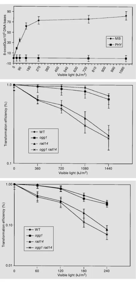
after PHY plus VL treatment (10). More-over, 8-oxodGuo is a coding lesion that in-creases the incidence of GC®TA transver-sions in bacterial and yeast mutants defec-tive in Fpg and Ogg1 functions, respecdefec-tively (25,33). These results led us to monitor the formation of 8-oxodGuo in DNA after PHY photosensitization. Using HPLC-ECD, the production of 8-oxodGuo was measured in DNA treated with MB or PHY in the pres-ence of increasing VL doses (Figure 2). In the presence of MB, increasing VL induced 8-oxodGuo in DNA, reaching a plateau at 720 kJ/m2 (1-h light exposure), which may
be due to an equilibrium in 8-oxodGuo deg-radation/formation in DNA (34). On the other hand, after PHY photosensitization, no sig-nificant amount of 8-oxodGuo was detected in DNA, indicating that 8-oxodGuo is not produced in DNA or is being produced at a very low rate (Figure 2).
Survival o f Saccharom yces cerevisiae ce lls and o f YEplac181 plasm id D NA tre ate d with
PHY plus VL
None of the yeast strains tested was sen-sitive to phycocyanin PDA. The FF18733 (wild type), CD138 (ogg1) BP10 (rad14) and BP20 (ogg1rad14) strains were submit-ted to PHY plus VL treatment under differ-ent conditions: increasing dye concdiffer-entra- concentra-tion, enhanced incubation temperature, in-creased incubation periods in the dark (over 2 h) and increasing visible light doses. How-ever, even in these conditions, survival of PHY plus VL-treated cells remained the same
as that of the control cells, without PHY (data not shown). Therefore, we decided to sensitize plasmid DNA and measure its sur-vival after transformation in the S. cerevi-siae strains. Figure 3 shows the survival of PHY photodamaged plasmid YEplac181 af-ter transformation into different S. cerevi-siae DNA repair mutants. Survival of the PHY photodamaged YEplac181 in ogg1
single mutant was not significantly different from that found in the wild type strain. On the other hand, survival of YEplac181 after PHY photosensitization was greatly reduced in rad14 mutants. The survival of PHY photodamaged YEplac181 in the ogg1rad14
double mutant was not different from that in the rad14 single mutant. Taken together, these results indicate that the ogg1 mutation does not modify plasmid survival. There-fore, the rad14 mutation, which inactivates NER in S.cerevisiae, seems to play a major role in the elimination of lethal lesions in-duced by phycocyanin PDA.
Survival o f Saccharom yces cerevisiae ce lls and o f YEplac181 plasm id D NA tre ate d with
m e thyle ne blue plus visible light
Survival of Saccharomyces cerevisiae
was measured after sensitization with MB. Although all strains proved to be sensitive to MB PDA, there were no significant differ-ences in lethality between the wild type strain (FF18733) and the DNA repair mutants (data not shown). Therefore, YEplac181 plasmid
S
e
n
s
it
iv
e
s
it
e
s
/c
ir
c
le
1.0
0.6 0.8
0.4
0.2
0
PHY M B
SSB Nfo Nth Fpg
Figure 2 - Detection of 8-oxodGuo in DNA treated w ith phycocyanin (PHY) or methylene blue (M B) plus visible light. Salmon sperm DNA w as first treated w ith PHY or M B plus visible light and then w ith Fpg protein (50 µg/ ml) for 1 h at 37oC as described in M aterial and M eth-ods. Each final mixture (100 µl) w as injected into the HPLC-ECD system. The number of 8-oxodGuo/105 DNA bases w as calculated and the limit of sensitivity w as six 8-oxodGuo per 105 DNA bases (32). Total expo-sure time w as 1.5 h (1,080 kJ/m2).
Figure 3 - Survival of YEplac181 plasmid DNA treated w ith PHY plus visible light w hen transformed into w ild type (WT) and excision repair mutants of Saccharomy-ces cerevisiae. YEplac181 DNA w as incubated w ith 500 µg/ml PHY in PBS buffer and irradiated w ith differ-ent doses of visible light. Untreated and treated DNA w ere used to transform the follow ing competent yeast hosts: FF18733 (w ild type), CD138 (ogg1), BP10 (rad14) and BP20 (ogg1rad14). Total exposure time w as 2.0 h (1,440 kJ/m2).
Figure 4 - Survival of YEplac181 treated w ith M B plus visible light w hen transformed into w ild type (WT) and excision repair mutants of Saccharomyces cerevisiae. YEplac181 DNA w as incubated w ith 2 µg/ml M B in PBS buffer and irradiated w ith visible light. Untreated and treated DNA w as used to transform the follow ing com-petent yeast hosts: FF18733 (w ild type), CD138 (ogg1) BP10 (rad14) and BP20 (ogg1rad14). Total exposure time w as 16 min (240 kJ/m2).
T
ra
n
s
fo
rm
a
ti
o
n
e
ff
ic
ie
n
c
y
(
%
)
1.00
Visible light (kJ/m2) WT
ogg1
0.01
0 60 120 180 240
rad14
ogg1 rad14
0.10
8
-o
x
o
d
G
u
o
/1
0
5 D
N
A
b
a
s
e
s
90
70
50
30
10
-10
M B
PHY
90 180 270 360 450 540 630 720 810 900 990 1080
Visible light (kJ/m2) 0
T
ra
n
s
fo
rm
a
ti
o
n
e
ff
ic
ie
n
c
y
(
%
)
1.0
Visible light (kJ/m2) 0.1
0 360 720 1080 1440
WT
DNA was sensitized to determine the role of BER and NER in the repair of MB plus VL-induced lethal lesions in yeast. Figure 4 shows survival of YEplac181 plasmid DNA treated with MB plus VL after transformation in S. cerevisiae wild type and DNA repair mu-tants. Similarly to YEplac181 survival with PHY plus VL, when plasmid DNA was sen-sitized with MB and transformed into S.
cerevisiae, its survival was reduced only in the rad14 mutants. The ogg1 mutation had no effect in reducing plasmid survival com-pared to the wild type strain.
D iscussio n
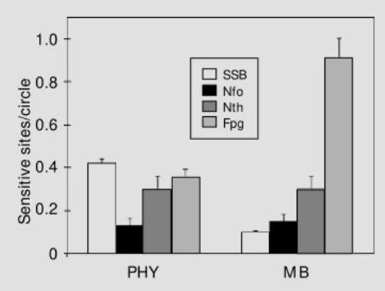
In the present study, we analyzed the damage profile and repair of DNA lesions induced by the photodynamic action of phy-cocyanin in vitro and in a cellular system. The damage profile of DNA exposed to PHY plus VL was determined using a set of DNA repair endonucleases purified from E. coli. The DNA damage profile of PHY-induced lesions was different from that of MB-in-duced lesions. While MB plus VL induces a large excess of Fpg-sensitive sites, PHY pho-tosensitization mostly induced single strand breaks (Figure 1). The major difference be-tween these two profiles is in the number of Fpg-sensitive sites generated by the treat-ments. The excess of Fpg-sensitive sites gen-erated by MB photosensitization can be ex-plained by the production of 8-oxodGuo, which was not detected after PHY plus VL treatment. In fact, the profile of DNA dam-age induced by phycocyanin was quite simi-lar to that induced by ionizing radiation or by hydrogen peroxide in the presence of transi-tion metals (3,35), but was different from that induced by molecules which involve solely singlet oxygen as the major species responsible for the DNA modifications, such as heat decomposition of 3,3'-(1,4 naphthyl-idene)-dipropionate endoperoxide, NDPO2
(3). Since PHY photosensitization did not induce 8-oxodGuo in DNA (Figure 2), we
conclude that singlet oxygen is not being produced or is immediately quenched by the apoprotein itself. This reasoning is valid when PHY anti-inflammatory properties are asso-ciated with oxygen free radical scavenging (12). Since the tetrapyrrolic chromophores are found in the apoprotein core (19), the production and diffusion of singlet oxygen may be hindered by the shielding and quench-ing effects of the apoprotein. Therefore, PHY photosensitization may occur via a type I reaction. The Fpg-sensitive sites found in DNA after PHY photosensitization may be due to the induction of open imidazole ring purines, such as 2,6-diamino-4-hydroxy-5-N-methylformamidopyrimidine (Me-FapyGua) or 4,6-diamino-5 formamidopyri-midine (FapyAde), which can be found in DNA after reaction with ·OH radical (34). In
addition, PHY-photosensitized DNA also presents Nth- and Nfo-sensitive sites, which may constitute oxidized pyrimidine-derived lesions and AP sites, respectively.
Phycocyanin was incapable of sensitiz-ing Saccharomyces cerevisiae probably due to a cell wall barrier that prevents phycocya-nin from penetrating and exerting its effects. We have shown that phycocyanin could in-teract with positive, but not Gram-negative, bacteria, which could justify its effectiveness in sensitizing Staphylococcus epidermidis, but its failure in sensitizing Es-cherichia coli (10). This same problem arose when MB was used to sensitize DNA repair mutants of E. coli, a fact that led researchers to sensitize plasmid DNA in cell-free sys-tems to study DNA repair (36). For the rea-sons considered above, YEplac181 plasmid DNA was photosensitized in vitro and used to transform a series of S.cerevisiae DNA repair mutants. Considering the two binomes
fpg/uvrA double mutant of E. coli, while its survival in the single mutants was similar to that in the wild type strain (10). Similar results were also obtained when plasmid DNA was treated with MB plus VL and used to transform these same E. coli repair mu-tants (36). On the other hand, in S. cerevi-siae, plasmid survival after PHY photosensi-tization was greatly reduced only in rad14
mutants (Figure 3). Plasmid survival in the double mutant ogg1rad14 was the same as that in the single mutant rad14, indicating that an ogg1 mutation against a rad14 back-ground had no effect on plasmid survival. Moreover, plasmid survival in the ogg1 single mutant was similar to that in the wild type strain, showing that the OGG1 gene itself does not play a role in the repair of the lethal lesions induced by PHY plus VL. The same results were obtained for plasmid DNA treated with MB plus VL and used to trans-form the S. cerevisiae strains (Figure 4). In
E. coli, the Fpg protein and NER system are able to recognize PHY- or MB-induced le-sions independently and to perform DNA repair. Plasmid survival is only diminished in the absence of both activities, indicating that Fpg and NER recognize the same DNA lesions. In S. cerevisiae, only the NER sys-tem is able to recognize PHY- or MB-in-duced lesions since plasmid survival is not altered by inactivation of the OGG1 gene. Apparently, the difference between bacte-rial and yeast repair of PHY- and MB-in-duced lesions may reside in the Fpg/Ogg1 substrate specificity, in a possible BER backup conferred by the Ntg1 protein and, finally, in the NER backup of S. cerevisiae
(32,37). If PHY photosensitization does not induce 8-oxodGuo in DNA, the
Fpg-sensi-tive sites found after DNA treatment may be due to Me-FapyGua or FapyAde lesions. Both are lethal lesions that can be well rec-ognized by Fpg protein, but only Me-FapyGua is recognized by Ogg1 (21,32). In addition, the Ogg1 protein has a marked preference for AP sites opposite cytosine, while the Fpg protein cleaves AP sites opposite any one of the four DNA bases (32). Although MB produces high levels of 8-oxodGuo, this may not be important in terms of plasmid lethality since it is a pre-mutagenic rather than a lethal lesion (25,38). Besides, if Me-FapyGua, FapyAde, and even 8-oxodGuo lesions were not repaired by the Ogg1 pro-tein, they may be recognized by the Ntg1 protein in S. cerevisiae (37,39). Since E. coli
does not possess a backup system for oxi-dized purine lesions, the simultaneous inac-tivation of the fpg/uvrA will result in plasmid lethality. In S. cerevisiae, since Ntg1 may serve as a backup for Ogg1, plasmid lethality is only observed in the rad14 mutants. Con-sequently, one may suppose that the NER system of S. cerevisiae differs from that of E.
coli in terms of lesion recognition and repair, since the former is responsible for the repair of PHY and MB lethal lesions that are not recognized by Ogg1 or Ntg1.
In conclusion, the results reported here demonstrate that PHY photosensitization induces mainly single strand breaks in DNA and, to a lesser extent, base oxidized sites. Furthermore, oxidative damage induced by PHY plus VL is repaired differently in Es-cherichia coli and in Saccharomyces cerevi-siae, a fact that may be due to a BER backup conferred by the Ntg1 protein and to a NER backup of Saccharomyces cerevisiae.
Re fe re nce s
1. Epe B, Hegler J & Wild D (1989). Singlet oxygen as ultimately reactive species in
Salmonella typhimurium damage induced by methylene blue/visible light. Carcino-genesis, 10: 2019-2024.
2. M enezes S, Capella M AM & Caldas LR (1990). Photodynamic action of methyl-ene blue: repair and mutation in Escheri-chia coli. Journal of Photochemistry and Photobiology. B, Biology, 5: 505-517.
3. Epe B, Pflaum M & Boiteux S (1993). DNA damage induced by photosensitizers in a cellular and cell-free systems. M utation Research, 299: 135-145.
Enzymatic recognition and biological ef-fects of DNA dam age induced by 3-carbethoxypsoralen plus UVA. M utation Research, 294: 43-50.
5. Breimer LH (1990). M olecular mecha-nisms of oxygen radical carcinogenesis and mutagenesis, the role of base dam-age. M olecular Carcinogenesis, 3: 188-197.
6. Feig DI, Reid TM & Loeb LA (1994). Reac-tive oxygen species in tumorigenesis.
Cancer Research, 54 (Suppl): 1890-1894. 7. Demple B & Harrison L (1994). Repair of oxidative damage to DNA: enzymology and biology. Annual Review of Biochem-istry, 63: 915-948.
8. M orcos NC, Berns M & Henry WL (1988). Phycocyanin laser activation, cytotoxic ef-fects, uptake and human aterioesclerotic plaque. Lasers in Surgery and M edicine, 8: 10-17.
9. M orcos NC, Zaldivar F, Lo Hsueh M & Henry WL (1991). Bovine coronary artery endothelium: Culture, characterization, angiogenesis and sensitivity to laser pho-todynamic treatment modalities. Journal of Clinical and Laboratory Immunology, 34: 99-106.
10. Pádula M , Boiteux S, Felzenszw alb I & M enezes S (1996). Photodynamic action of phycocyanin: damage and repair. Jour-nal of Photochemistry and Photobiology. B, Biology, 32: 19-26.
11. Rom ay C, Arm est o J, Rem ierz D, Gonzales R, Ledon N & Garcia I (1998). Antioxidant and anti-inflammatory proper-ties of C-phycocyanin from blue-green al-gae. Inflammation Research, 47: 36-41. 12. Romay C, Ledon N & Gonzales R (1998).
Further studies on anti-inflammatory ac-tivity of phycocyanin in some animal mod-els of inflammation. Inflammation Re-search, 47: 334-348.
13. Vadiraja BB, Gaikw ad NW & M adyastha KM (1998). Hepatoprotective effect of C-phycocyanin: protection for tetrachloride and R-(+)-pulegone-mediated hepatotox-icity in rats. Biochemical and Biophysical Research Communications, 249: 428-431. 14. Araoz R, Lebert M & Hader DP (1998). Electrophoretic applications of phycobili-proteins. Electrophoresis, 19: 215-219. 15. Yoshida A & Takagaki Y (1995). Detection
of food additives by enzyme immunoas-say. Nippon Rinsho, 53: 2316-2321. 16. Yoshida A, Takagaki Y & Nishimune T
(1996). Enzyme immunoassay for phyco-cyanin as the main component of spirulina color in foods. Bioscience, Biotechnology andBiochemistry, 60: 57-60.
17. Killilea SD, O’Carra P & M urphy RF (1980).
Structures and apoprotein linkages of phycoerythrobilin and phycocianobilin.
Biochemical Journal, 187: 311-320. 18. Brow n B & Holroyd JA (1984).
Biosynthe-sis of the chromophore of phycobilipro-teins. Biochemical Journal, 217: 265-272. 19. Berns DS & M acColl R (1989). Phycocya-nin in physical-chemical studies. Chemi-cal Review s, 89: 807-825.
20. Foote CS (1991). Definition of type I and type II photosensitized oxidation. Photo-chemistry and Photobiology, 54: 659. 21. Boiteux S (1993). Properties and
biologi-cal functions of the Nth and Fpg proteins of Escherichia coli: tw o DNA glycosylases that repair oxidative damage in DNA. Jour-nal of Photochemistry and Photobiology. B, Biology, 19: 87-96.
22. Friedberg E, Walker GC & Siede W (1995).
DNA Repair and M ut agenesis. ASM Press, Washington, DC.
23. Auffret van der Kem p P, Thom as D, Barbey R, de Oliveira R & Boiteux S (1996). Cloning and expression in Escheri-chia coli of the OGG1 gene of Saccharo-myces cerevisiae, w hich codes for a DNA glycosylase that excises 7,8-dihydro-8
-oxoguanine and
2,6-diamino-4-hydroxy-5-N-methylformamidopyrimidine. Proceed-ings of the NationalAcademy of Sciences, USA, 93: 5197-5202.
24. Epe B, Herzl H, Adam W & Saha-M öller CR (1993). Endonuclease sensitive DNA modifications induced by acetone and ac-etophenone as photosensitizers. Nucleic Acids Research, 21: 863-869.
25. Thomas D, Scott AD, Barbey R, Pádula M & Boiteux S (1997). Inactivation of OGG1
increases t he incidence of GC®TA transversions in Saccharomyces cerevi-siae: evidence of endogenous oxidative damage to DNA in eukaryotic cells. M o-lecular and General Genetics, 254: 171-178.
26. Pádula M , Averbeck S, Boit eux S & Averbeck D (1997). Enzymatic recognition and biological effects of photodynamic damage induced in DNA by 1.6-dioxapy-rene plus UVA. Journal of Photochemis-try and Photobiology. B, Biology, 41: 60-66.
27. Cherest H & Surdin-Kerjan Y (1992). Ge-netic analysis of new mutations confer-ring cystein auxotrophy in Saccharomy-ces cerevisiae: updating of the sulphur metabolism pathw ay. Genetics, 130: 51-58.
28. Gietz RD & Sugino A (1988). New
yeast-Escherichia coli shut t le vect ors con-structed w ith in vitro mutagenized yeast genes lacking six-base pair restriction
sites. Gene, 74: 527-534.
29. Cunningham RP, Saporito SM , Spitzer SG & Weiss B (1986). Endonuclease IV (nfo) mutant of Escherichia coli. Journal of Bac-teriology, 168: 1120-1127.
30. Asahara H, Wistort PM , Bank JF, Bakerian RH & Cunningham RP (1989). Purification and characterization of Escherichia coli
endonuclease III from the cloned nth
gene. Biochemistry, 28: 4444-4449. 31. Boit eux S, O’ Connor TR, Lederer F,
Gouyette A & Laval J (1990). Homoge-neous Escherichia coli Fpg protein. A DNA glycosylase w hich excises imidazole ring-opened purines and nicks DNA at apurinic/ apirimidinic sites. Journal of Biological Chemistry, 265: 3916-3922.
32. Karahalil B, Girard PM , Boit eux S & Dizdaroglu M (1998). Substrate specificity of the Ogg1 protein of Saccharomyces cerevisiae: excision of guanine lesions produced in DNA by ionizing radiation- or hydrogen peroxide/metal ion-generated free radicals. Nucleic Acids Research, 26: 1228-1233.
33. Boiteux S & Huisman O (1989). Isolation of a formamidopyrimidine DNA glycosyl-ase (fpg) mutant of Escherichia coli K12.
M olecular and General Genetics, 215: 300-305.
34. Cadet J, Berger M , Douki T & Ravanat JL (1997). Oxidative damage to DNA: forma-tion, measurement and biological signifi-cance. Review s of Physiology, Biochem-istry and Pharmacology, 131: 3-87. 35. Epe B (1995). DNA damage profiles
in-duced by oxidizing agents. Review s of Physiology, Biochemistry and Pharmacol-ogy, 127: 223-249.
36. Czeczot H, Tudek B, Lambert B, Laval J & Boiteux S (1991). Escherichia coli Fpg pro-tein and UvrABC endonuclease repair DNA damages induced by methylene blue plus visible light in vitro and in vivo. Jour-nal of Bacteriology, 173: 3419-3424. 37. Sentürker S, Auffret van der Kemp P, You
HJ, Doetsch PW, Dizdaroglu M & Boiteux S (1998). Substrate specificities of the Ntg1 and Ntg2 protein of Saccharomyces cerevisiae for oxidized DNA bases are not identical. Nucleic Acids Research, 26: 5270-5276.
38. Grollman AP & M oriya M (1993). M u-tagenesis by 8-oxoguanine: an enemy w ithin. Trends in Genetics, 9: 246-249. 39. Bruner SD, Nash HM , Lane WS & Verdine