UNIVERSIDADE DA BEIRA INTERIOR
Ciências da Saúde
Desenvolvimento de nanomateriais para a terapia
fototermal do cancro
Cleide Isabel Pais Silva
Dissertação para obtenção do Grau de Mestre em
Ciências Biomédicas
(2º ciclo de estudos)
Orientador: Professor Doutor Ilídio Joaquim Sobreira Correia
Co-orientador: Mestre Duarte Miguel de Melo Diogo
iii
“Do you wish to rise? Begin by descending. You plan a tower that will pierce the clouds? Lay first the foundation of humility.”
v
vii
Acknowledgements
Just for today, be grateful.
First of all, I would like to acknowledge my supervisor, Professor Ilídio Correia, for the possibility to integrate his group and to develop my master thesis with them. His orientation, expertise and wise advices throughout this year were crucial to accomplish this work. He made me improve and surpass myself, either at the professional and personal level, and I am likewise grateful to him for that. I am also deeply thankful to Duarte Diogo, my co-supervisor, for his guidance, for believing in me and in my work and for always keeping me motivated. The constant discussion of ideas and the know-how he transmitted to me contributed to my growth as a researcher. All his support made the development of this work easier.
I also thank Diana Dias, Elisabete Costa and Marco Carvalho, not only for their accompaniment and help during the long lab protocols, but mainly for their friendship, talks and laughs. To my lab partners, I thank their fellowship and moments of conviviality. To all, I wish the best. To my family, there is no word that expresses how grateful I am. Foremost, I profoundly thank my mom, my all-time guide, for the endless encouragement, belief, prudent advices and love. To my father, I acknowledge his attention and trust, which I assume his silence hides. I thank my brother for his affection, concern and for always having faith in its “little princess”. To my lovely and brave grandmother, the cornerstone of the family, I address an enormous thank for the encouragement and for being an inspiration to me. To all of you, thank you, thank you from the bottom of my heart.
To Telmo, my longtime friend, I thank for our daily talks, support and for never giving up on our friendship. “Out of the sight, close to the heart”.
Last, but not least, words cannot express how extremely grateful I am to my boyfriend, Davide, for his continuous support, comprehension and endless love throughout these years. Most of all, I have to recognize the unconditional patience over this last year, that made every single day seems easier. Just for today… I am grateful for having you by my side.
Thank. A little word with so much meaning. Thank you all.
ix
Resumo
O cancro da mama é um dos mais prevalentes e uma das principais causas de morte das mulheres em todo o mundo. No caso específico da população Portuguesa, as taxas de incidência e mortalidade associadas ao cancro da mama têm vindo a aumentar nos últimos anos. Este cenário deve-se maioritariamente à ineficácia dos tratamentos atualmente disponíveis (cirurgia, radioterapia e terapias baseadas em pequenas moléculas), cuja eficácia terapêutica é reduzida devido a problemas de segurança, toxicidade não específica e ainda devido aos mecanismos de resistência a fármacos que as células cancerígenas apresentam. Estes factos evidenciam a necessidade de desenvolver novas abordagens terapêuticas.
Nas últimas décadas, as fototerapias têm sido investigadas como estratégias alternativas para combater as células cancerígenas. Estas modalidades terapêuticas incluem a terapia fotodinâmica e a fototérmica. Ambos os tratamentos dependem da irradiação da região tumoral com radiação com um comprimento de onda próximo do infravermelho (near-infrared (NIR)) que ativa moléculas fotoresponsivas, as quais induzem a produção de espécies reativas de oxigénio (terapia fotodinâmica) ou um aumento de temperatura (terapia fototérmica), que têm um efeito citotóxico nas células cancerígenas. Porém, estes tratamentos ainda têm que ser otimizados para incrementar a sua eficácia e seletividade para o tumor.
A nanotecnologia tem permitido ultrapassar algumas das principais limitações da aplicação das fototerapias no tratamento do cancro, através da encapsulação de agentes fotoresponsivos em nanopartículas. A encapsulação de moléculas fotoresponsivas à luz NIR em nanoveículos é crucial para reverter a baixa solubilidade destas moléculas em fluidos biológicos. Para além disso, estes nanotransportadores acumulam-se preferencialmente no tumor devido às suas propriedades físico-químicas, o que permite aumentar a biodisponibilidade das moléculas fotoresponsivas encapsuladas nestes, melhorando assim a eficácia e seletividade destas abordagens terapêuticas.
Na presente tese é descrito o desenvolvimento de um nanoveículo com capacidade de encapsular um agente fotoresponsivo à luz NIR, para aplicação na fototerapia do cancro da mama. Para tal, foram utilizados dois derivados de vitamina E, succinato de D-α-tocoferil polietilenoglicol 1000 (TPGS) e succinato de D-α-tocoferil (TOS), para formular micelas anfifílicas com uma estrutura tipo “núcleo-concha”. O TPGS e o TOS foram selecionados por apresentarem atividade anticancerígena intrínseca (por exemplo, através da produção de espécies reativas de oxigénio) e pela sua capacidade para encapsular moléculas pouco solúveis em água. O iodeto de 2-[2-[2-cloro-3-[(1,3-dihidro-3,3-dimetil-1-propil-2H-indol-2-ilideno) etilideno]-1-ciclohexeno-1-il]etenil]-3,3-dimetil-1-propilindólio (IR780) foi escolhido devido à sua versatilidade como molécula fotoresponsiva, uma vez que possui características que
permitem a sua aplicação como agente fototerapêutico (terapia fotodinâmica e fototermal) e de diagnóstico. No presente estudo foram obtidas micelas de TPGS-TOS contendo IR780 (IR780-TTM) com características físico-químicas adequadas, utilizando proporções de TPGS e TOS específicas aquando da sua formulação. As micelas produzidas permitiram a encapsulação do IR780 com elevada eficiência. Nos ensaios in vitro realizados verificou-se que as IR780-TTM medeiam um efeito citotóxico nas células cancerígenas após irradiação com luz NIR, através da produção de espécies reativas de oxigénio (terapia fotodinâmica). Esta eficiente ablação das células cancerígenas foi alcançada usando uma concentração de IR780 mais baixa do que a que foi descrita até agora na literatura. Aliado a isto, constatou-se que as IR780-TTM demonstram potencial para serem utilizadas como agentes de fototermia e imagiologia.
Em suma, os nanoveículos desenvolvidos na presente tese apresentam um enorme potencial para a terapia fotodinâmica do cancro da mama. Por outro lado, as propriedades fototérmicas e imagiológicas que as IR780-TTM apresentam aumentam a aplicabilidade destas micelas no tratamento e diagnóstico do cancro.
Palavras-chave
Resumo alargado
Na atualidade, o cancro é um dos principais problemas de saúde pública que afeta a população a nível mundial. Em particular, o cancro da mama é uma das neoplasias mais comuns e com maior taxa de mortalidade entre o sexo feminino. Este cenário deve-se essencialmente à ineficiência das terapias anticancerígenas convencionais, uma vez que as moléculas terapêuticas apresentam problemas intrínsecos, como sejam a baixa solubilidade, fraca disponibilidade no local do tumor e acumulação não específica em tecidos saudáveis. Por outro lado, o desenvolvimento de mecanismos de resistência a fármacos por parte das células cancerígenas causa também efeitos adversos para o paciente. Todos estes factos evidenciam a necessidade urgente de desenvolver novas terapias anticancerígenas.
Neste contexto, as terapias fotodinâmica e fototérmica têm-se revelado como alternativas promissoras para o tratamento do cancro. Estas abordagens terapêuticas exploram a aplicação de radiação com comprimento de onda próximo do infravermelho (near-infrared (NIR); 750-1000 nm) na região tumoral para ativar moléculas fotoresponsivas, o que se traduz na formação de espécies reativas de oxigénio (terapia fotodinâmica) ou num aumento de temperatura (terapia fototérmica), que causam citotoxicidade às células cancerígenas. Porém, este tipo de terapias necessitam de melhorias no que diz respeito à sua seletividade para as células cancerígenas. Para além disto, estas moléculas são geralmente lipofílicas, o que limita a dose que pode ser administrada.
Na última década, os investigadores têm explorado a encapsulação de moléculas fotoresponsivas em nanotransportadores. Esta estratégia visa aumentar a solubilidade destas moléculas, bem como protegê-las da degradação. Para além disto, estes nanotransportadores, devido às suas dimensões, podem permitir uma acumulação preferencial no tecido tumoral. Desta forma, é possível submeter o tumor a espécies reativas de oxigénio e calor após irradiação dos nanotransportadores com radiação NIR.
Na presente tese desenvolveu-se um nanotransportador micelar para mediar a entrega de um composto fotoresponsivo à luz NIR a células cancerígenas do cancro da mama. Para tal, foram utilizados dois derivados de vitamina E, succinato de D-α-tocoferil polietilenoglicol 1000 (TPGS) e succinato de D-α-tocoferil (TOS), uma vez que apresentam atividade anticancerígena intrínseca (nomeadamente através da produção de espécies reativas de oxigénio) e a sua combinação em meio aquoso leva à formação de micelas com carácter anfifílico, o que permite a encapsulação de compostos lipossolúveis responsivos à luz NIR. O iodeto de 2-[2-[2-cloro-3- [(1,3-dihidro-3,3-dimetil-1-propil-2H-indol-2-ilideno)etilideno]-1-ciclohexeno-1-il]etenil]-3,3-dimetil-1-propilindólio (IR780) foi selecionado como agente fotoresponsivo a encapsular nas micelas de TPGS-TOS devido à absorção que apresenta a 780 nm, que possibilita a sua interação
xiii
com a luz NIR. Esta propriedade é de extrema importância, uma vez que a radiação NIR penetra mais profundamente nos tecidos e apresenta interações mínimas com os componentes biológicos. Além disto, o IR780 é um composto fotoresponsivo versátil, uma vez que é capaz de produzir espécies reativas de oxigénio e/ou variações de temperatura, quando irradiado com luz NIR. Por outro lado, a capacidade que o IR780 tem de emitir fluorescência, permite ainda a sua aplicação na deteção de tumores, isto é, imagiologia de tumores.
Durante a otimização do processo de formulação das micelas de TPGS-TOS contendo IR780 (IR780-TTM) foram testadas várias proporções de TPGS e TOS, e verificou-se que a utilização de uma percentagem de 40, 33 ou 25 de TPGS permite produzir micelas com propriedades físico-químicas adequadas para encapsular e entregar IR780 a células cancerígenas. Para além disto, verificou-se que as IR780-TTM possuem excelentes propriedades como agentes fototerapêuticos devido à sua alta absorção de radiação NIR. Aliado a este facto, observou-se que as IR780-TTM são capazes de produzir, quando irradiadas com radiação NIR, uma variação de temperatura de até 18 °C, o que demonstra o potencial destas micelas para serem usados em fototermia.
Nos ensaios in vitro constatou-se que as IR780-TTM são internalizadas pelas células de cancro da mama, o que é fundamental para aumentar a concentração de IR780 no interior das células, visto que o composto na sua forma livre tem uma baixa translocação para o espaço intracelular. Estes estudos permitiram ainda confirmar o potencial das IR780-TTM para serem usadas em aplicações imagiológicas. Os estudos de viabilidade celular permitiram verificar que a utilização combinada de radiação NIR e das IR780-TTM, encapsulando apenas 1.5 µg mL-1 de IR780, reduziu a viabilidade celular das células cancerígenas para 19 %. Nestas mesmas condições, as células tratadas com IR780-TTM, mas que não foram irradiadas, permaneceram viáveis, o que demonstra que a fototerapia mediada pelas IR780-TTM é extremamente eficaz. Em estudos subsequentes verificou-se que a fototoxidade mediada pelas IR780-TTM é induzida pela produção de espécies reativas de oxigénio (terapia fotodinâmica). Aliado a isto, a capacidade intrínseca do TPGS e do TOS para produzirem espécies reativas de oxigénio parece dar um contributo importante para esta eficiente ablação celular. Por fim, é importante salientar que o efeito fototerapêutico mediado pelas IR780-TTM foi alcançado usando a menor concentração de IR780 descrita até ao momento na literatura, o que atesta a eficácia e o potencial das micelas desenvolvidas.
Em suma, o sistema micelar desenvolvido apresenta um enorme potencial para ser utilizado na terapia fotodinâmica do cancro da mama. Além disso, a natureza versátil das IR780-TTM é fundamental para a sua futura aplicação como agentes fototérmicos e de diagnóstico.
xv
Abstract
Breast cancer is one of the most prevalent and one of the leading causes of cancer-related deaths among women worldwide. In the specific case of the Portuguese population, breast cancer incidence and mortality rates increased in the past years. This scenario is mostly owed to the ineffectiveness of the currently available treatments (surgery, radiotherapy and small molecule-based therapies), whose therapeutic success is hindered by safety issues, non-specific toxicity and resistance mechanisms displayed by cancer cells to drugs. Such emphasizes the demand for a plethora of novel therapeutic approaches.
In the past decades, light-induced therapies have started to be investigated as alternative strategies to combat cancer. These treatment modalities comprise photodynamic therapy (PDT) and photothermal therapy (PTT). Both therapeutic approaches depend on the external irradiation of the tumor region with near-infrared (NIR) light for activating photosensitizers or photothermal agents, that lead to the generation of reactive oxygen species (ROS; in PDT) or to a temperature increase (in PTT), which have a cytotoxic effect on cancer cells. Yet, such treatments still need to be further improved in what concerns their efficacy and selectivity towards the tumor region.
Recently, the entrapment of NIR photoabsorbers in nanoparticles surpassed problems like the poor solubility of these molecules in biological fluids. Moreover, nanoparticles due to their physicochemical properties can display a preferential tumor accumulation, which is crucial for increasing the loaded NIR photoabsorbers bioavailability and ultimately the efficacy and selectivity of this therapeutic approach.
Herein, a novel nanovehicle loaded with a NIR dye aimed for breast cancer phototherapy was developed. To accomplish that, two vitamin E derivatives, D-α-tocopheryl polyethylene glycol 1000 succinate (TPGS) and D-α-tocopheryl succinate (TOS), were used to assemble amphiphilic micelles with a core-shell structure. TPGS and TOS were selected due to their intrinsic anticancer activity (e.g. through the generation of ROS) and ability to encapsulate poorly water-soluble molecules. 2-[2-[2-Chloro-3-[(1,3-dihydro-3,3-dimethyl-1-propyl-2H-indol-2-ylidene)ethylidene]-1-cyclohexen-1-yl]ethenyl]-3,3-dimethyl-1-propylindolium iodide (IR780) was chosen due to its versatile nature as a NIR light-responsive compound (photothermal agent, photosensitizer and NIR imaging dye). IR780-loaded TPGS-TOS micelles (IR780-TTM) with suitable sizes were obtained by using specific TPGS and TOS weight feed ratios during micelles formulation and these were able to encapsulate IR780 with high efficiency. In in vitro assays, the IR780-TTM induced a cytotoxic effect in cancer cells upon exposure to NIR irradiation through the generation of reactive oxygen species (PDT). This effective ablation of cancer cells
was achieved using an ultra-low IR780 concentration. Moreover, IR780-TTM also demonstrated the ability to act as photothermal and imaging agents.
Overall, the novel micellar nanoplatforms developed in this study possess a huge potential for breast cancer PDT. Moreover, IR780-TTM also demonstrate promising results to act as photothermal and imaging agents, which widens their applicability for the treatment and diagnosis of cancer.
Keywords
List of Publications
Articles submitted for publication in international peer reviewed
journals:
Pais-Silva, C., de Melo-Diogo, D., and Correia, I. J. IR780-loaded TPGS-TOS micelles for breast cancer photodynamic therapy. European Journal of Pharmaceutics and Biopharmaceutics (3.975), under review.
de Melo-Diogo, D., Pais-Silva, C., Costa, E. C., Louro, R. O., and Correia, I. J. TPGS functionalized nanographene oxide for cancer phototherapy. Carbon (6.198), submitted.
Poster communications:
Pais-Silva, C., de Melo-Diogo, D., and Correia, I. J. Vitamin E-based micelles for cancer cell imaging, V Encontro Nacional de Estudantes de Materiais (ENEM), 29th of September, Universidade da Beira Interior, Covilhã, Portugal.
Index
Chapter 1 ... 1
1. Introduction ... 2
1.1. Cancer ... 2
1.1.1. Cancer: an evolutionary pathology with specific hallmarks ... 2
1.1.2. Breast cancer ... 5
1.1.3. Phototherapies ... 7
1.2. Nanomaterials for application in cancer phototherapy ... 10
1.2.1. The potential of unifying the best of both worlds ... 10
1.2.2. NPs-mediated phototherapy: the design drives the journey ... 11
1.2.3. Nanovehicles classes: different characteristics for multiple applications ... 14
1.2.4. Polymeric micelles ... 15
1.2.4.1. Polymeric micelles application in cancer phototherapy ... 15
1.2.4.2. Vitamin E-based micelles aimed for cancer phototherapy ... 16
Aims ... 19 Chapter 2 ... 20 2. Experimental Section ... 21 2.1. Materials ... 21 2.2. Methods ... 21 2.2.1. Production of IR780-TTM ... 21
2.2.2. Physicochemical characterization of IR780-TTM ... 22
2.2.3. IR780-TTM encapsulation efficiency and loading content ... 22
2.2.4. Phototherapeutic capacity of IR780-TTM ... 22
2.2.5. Cell culture ... 23
2.2.6. IC50 determination ... 23
2.2.7. Evaluation of IR780-TTM biocompatibility ... 23
2.2.8. Assessment of IR780-TTM internalization in cancer cells ... 23
2.2.9. Evaluation of phototherapy mediated by IR780-TTM ... 24
2.2.10. In vitro detection of ROS generation by IR780-TTM ... 24
2.2.11. Statistical analysis ... 24
Chapter 3 ... 25
xxi
3.1. Characterization of IR780-TTM ... 26
3.2. Phototherapeutic ability of IR780-TTM ... 29
3.3. IC50 determination and evaluation of IR780-TTM biocompatibility ... 30
3.4. Cellular internalization of IR780-TTM ... 32
3.5. Phototherapy mediated by IR780-TTM ... 33
Chapter 4 ... 37
4. Conclusions and Future Perspectives ... 38
Chapter 5 ... 39
xxiii
Figures Index
Figure 1. Representation of the cellular and non-cellular components found in tumor microenvironment and that have an active role in the process of carcinogenesis ... 2 Figure 2. Description of the major types and sub-types of stromal cells that populate the tumor microenvironment and particular functions that contribute to the development and regulation of the carcinogenic process ... 3 Figure 3. Hallmarks displayed by cancer cells ... 4 Figure 4. Most commonly diagnosed types of cancer in women worldwide in 2012, according to the estimates of the International Agency for Research on Cancer ... 5 Figure 5. Schematic representation of the breast cancer carcinogenesis ... 6 Figure 6. Representation of the biological transparency window and the ultraviolet-visible-near-infrared absorption spectra of the main biological components of the human tissues ... 8 Figure 7. Representation of the mechanism of action of nanoparticles-mediated photodynamic therapy and photothermal therapy ... 10 Figure 8. Illustration of the different stages of nanoparticles-mediated phototherapy ... 11 Figure 9. Examples of the different types of nanoparticles that can be used to encapsulate near-infrared photoabsorbers ... 14 Figure 10. Representation of the polymeric micelle assembling process and of the core-shell organization ... 15 Figure 11. Schematic illustration of the protocol used to produce IR780-loaded TPGS-TOS micelles ... 18 Figure 12. Determination of the size and polydispersity index of IR780-TTM ... 26 Figure 13. Morphological characterization of IR780-TTM ... 28 Figure 14. Characterization of IR780 encapsulation efficiency and loading content in TTM .. 28 Figure 15. Evaluation of IR780-TTM NIR absorption capacity ... 29 Figure 16. Determination of temperature variations induced by IR780-TTM at different concentrations over 5 min of NIR irradiation ... 30 Figure 17. Determination of the IC50 value of TPGS, TOS and IR780 in MCF-7 cells ... 31 Figure 18. Evaluation of the biocompatibility of IR780-40TTM, IR780-33TTM and IR780-25TTM, using MCF-7 as model cell line ... 32
Figure 19. Representative CLSM images of free-IR780 and IR780-TTM internalization in MCF-7 cells ... 33 Figure 20. Breast cancer phototherapy mediated by IR780-TTM ... 34 Figure 21. Photodynamic effect mediated by IR780-TTM ... 35
Tables Index
Table 1. Formulation conditions used for preparing IR780-TTM ... 21 Table 2. Size and PDI values of blank TTM ... 27 Table 3. Summary of the different IR780-loaded NPs produced for cancer phototherapy ... 36
List of Abbreviations
ADR adriamycin-resistant
ANOVA analysis of variance
Bcl-2 B-cell lymphoma 2
BxPC-3 human primary pancreatic adenocarcinoma cell line CD4 T cluster of differentiation 4-positive lymphocyte CLSM confocal laser scanning microscopy
CT-26 murine colon adenocarcinoma cell line
CTL cytotoxic T lymphocyte
DMEM-F12 Dulbecco’s modified Eagle’s medium F-12
DNA deoxyribonucleic acid
Dox doxorubicin
ECM extracellular matrix
EE encapsulation efficiency
EPR enhanced permeability and retention
FBS fetal bovine serum
FDA food and drug administration
H2DCF-DA 2',7'-dichlorofluorescein diacetate
Hb deoxyhemoglobin
HbO2 oxyhemoglobin
HCT-116 human colon carcinoma cell line HepG2 human hepatocyte carcinoma cell line
HF heparin-folic acid
HSA human serum albumin
IARC international agency for research on cancer IC50 half maximal inhibitory concentration
ICG indocyanine green
IFP interstitial fluid pressure IR780
2-[2-[2-Chloro-3-[(1,3-dihydro-3,3-dimethyl-1-propyl-2H-indol-2- ylidene)ethylidene]-1-cyclohexen-1-yl]ethenyl]-3,3-dimethyl-1-propylindolium iodide
IR780-TTM IR780-loaded TPGS-TOS micelles
LC loading content
MCF-7 Michigan cancer foundation-7 cell line MDSC myeloid-derived suppressor cell
MetOH methanol
mPEG methoxy poly(ethylene glycol)
MSC mesenchymal stem cell
xxix
NK/T natural killer lymphocyte/natural killer T lymphocyte
NP nanoparticle
PBMA poly(n-butylmethacrylate)
PBS phosphate buffered saline
PCL poly(caprolactone)
PDI polydispersity index
PDT photodynamic therapy
PEG poly(ethylene glycol)
PLGA poly(lactic-co-glycolic acid)
PMDPC poly(12-methacryloyloxy dodecyl phosphorylcholine) PMPC poly(2-methacryloyloxyethyl phosphorylcholine)
PTT photothermal therapy
RES reticuloendothelial system
ROS reactive oxygen species
RT room temperature
SD standard deviation
TEM transmission electron microscopy
Tf transferrin
Th2 helper type 2 lymphocyte
TME tumor microenvironment
TOS D-α-tocopheryl succinate
TPGS D-α-tocopheryl polyethylene glycol 1000 succinate
Treg regulatory T lymphocyte
TTM TPGS-TOS micelles
UV-vis-NIR ultraviolet-visible-near-infrared VEGF vascular endothelial growth factor
1
Chapter 1
1. Introduction
1.1. Cancer
1.1.1. Cancer: an evolutionary pathology with specific hallmarks
Cancer is a challenging global health problem with a growing incidence that has high morbidity and mortality rates associated (Fitzmaurice et al., 2015). The International Agency for Research on Cancer (IARC) of World Health Organization estimates that there were more than eight million of cancer-related deaths worldwide in 2012 (Ferlay et al., 2015). In 2016, more than one and a half million of new cases are expected to be diagnosed just in the United States of America and almost six hundred thousand cancer-related deaths will occur within the same period (Siegel et al., 2016). In Portugal, the Direção-Geral da Saúde predicts that in 2035 more than sixty thousand people will be diagnosed with this disease (Miranda et al., 2015).
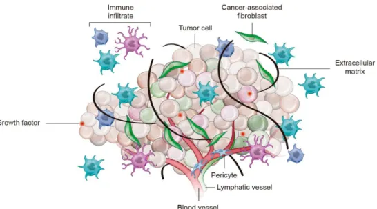
Cancer is triggered by an uncontrolled proliferation process – termed carcinogenesis - where healthy cells go through profound genetic and epigenetic transformations that lead to the formation of malignant cells (Gillies et al., 2012). The causes for the development of this malignant phenotype are essentially related to: (i) genetic susceptibility, (ii) environmental factors (radiation, pollution and infections), and (iii) lifestyle factors (diet, physical inactivity, tobacco and alcohol consumption) (Gillies et al., 2012; Vineis and Wild, 2014). Nowadays, cancer is viewed as a complex and heterogeneous tissue formed by a mass of cancer cells in continuous proliferation that are in a dynamic interplay with surrounding/recruited stromal cells and non-cellular elements that constitute the tumor microenvironment (TME) (Figure 1) (Hanahan and Weinberg, 2000).
Figure 1. Representation of the cellular and non-cellular components found in tumor microenvironment
and that have an active role in the process of carcinogenesis. The pro-survival, proliferation and invasion pathways of cancer cells are supported by the cross-talk between the malignant cells and the stromal cells, extracellular matrix and various signaling molecules (Adapted from (Junttila and de Sauvage, 2013)).
3
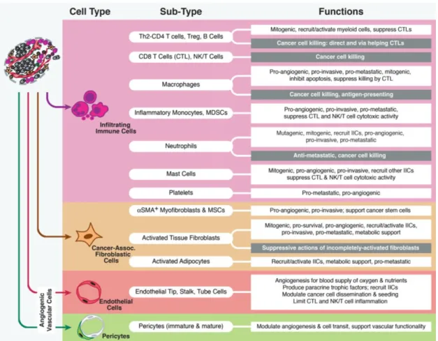
The tumor-associated stroma that populates the TME encompasses endothelial cells, pericytes, immune inflammatory cells and cancer-associated fibroblasts (Barcellos-Hoff et al., 2013; Hanahan and Coussens, 2012), whose particular functions are summarized in Figure 2.
Figure 2. Description of the major types and sub-types of stromal cells that populate the tumor
microenvironment and particular functions that contribute to the development and regulation of the carcinogenic process. Helper type 2 lymphocyte (Th2), cluster of differentiation 4-positive lymphocyte (CD4 T), regulatory T lymphocyte (Treg), cytotoxic T lymphocyte (CTL), natural killer lymphocyte/natural killer T lymphocyte (NK/T), myeloid-derived suppressor cell (MDSC), alpha smooth muscle actin (αSMA), mesenchymal stem cell (MSC) (Adapted from (Hanahan and Coussens, 2012)).
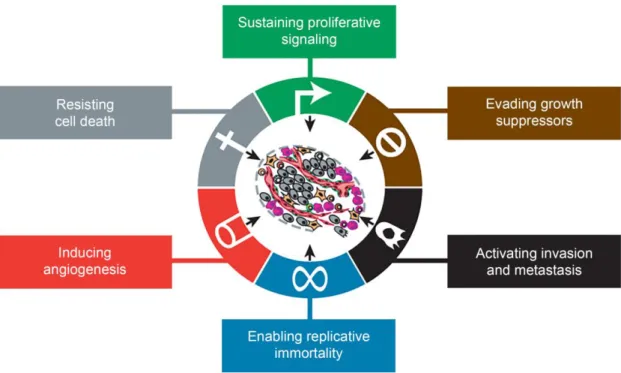
Apart from the cellular components, extracellular matrix (ECM), various signaling molecules and soluble mediators are also found in the TME (Pickup et al., 2014; Witsch et al., 2010; Xu et al., 2014). The synergistic interaction between the TME elements contribute to the development and the maintenance of unique features displayed by cancer cells that Hanahan and Weinberg termed as “hallmarks of cancer” (Hanahan and Weinberg, 2000). Cancer hallmarks include the capacity to: (i) sustain proliferative signaling, (ii) evade growth suppressors, (iii) avoid programmed cell death, (iv) enable replicative immortality, (v) induce angiogenesis, and (vi) activate invasion and metastasis (Figure 3).
Figure 3. Hallmarks displayed by cancer cells. These hallmarks are responsible for the development and
maintenance of their malignant characteristics (Adapted from (Hanahan and Weinberg, 2011)).
Cancer cells are able to proliferate by deregulating growth signaling pathways. In this process, cells can acquire autonomy in the synthesis of their own mitogenic growth factors (e.g. platelet-derived growth factor) or overexpress receptors (such as those containing intracellular tyrosine kinase domains) that are involved in growth signaling pathways (Hanahan and Weinberg, 2000; Pietras and Östman, 2010). The continuous proliferation of cancer cells is also prompted by their capacity to evade growth suppressors, like tumor suppressor protein 53 and retinoblastoma protein (Di Fiore et al., 2013; Giampazolias and Tait, 2015). Moreover, cancer cells can also resist to death mechanisms by favoring the expression of anti-apoptotic proteins (like those from the B-cell lymphoma 2 (Bcl-2) family) and by circumventing pro-apoptotic signals commonly present in healthy cells (Caino et al., 2009; Giampazolias and Tait, 2015; Kelly and Strasser, 2011). Furthermore, these cells also acquire replicative immortality through the overexpression of telomerase, an enzyme that adds repeated segments of hexanucleotides to the ends of telomeric deoxyribonucleic acid (DNA). The expression of high levels of telomerase by cancer cells renders them the capacity to surpass senescence, by preventing DNA damage and cell death associated with end-to-end chromosomal fusion (Caino et al., 2009; Hanahan and Weinberg, 2000).
Additionally, cancer cells induce angiogenesis and metastasis, in order to have access to nutrients, oxygen and also to remove metabolic waste and carbon dioxide generated by their metabolism. The angiogenic capacity of cancer cells is induced by the switch of the quiescent state of vasculature to a permanently activated state during tumor progression, resulting from an increased expression of angiogenic inducers (e.g. vascular endothelial growth factor (VEGF) and fibroblast growth factor) (Saharinen et al., 2011; Turner and Grose, 2010). Furthermore,
5
cancer cells may attain certain features that allow them to extravasate, invade and colonize other tissues throughout the body, resulting in the formation of metastasis (Hanahan and Weinberg, 2000). The complex and rich vascular network generated during the tumor-associated angiogenesis supports cancer cells metastization (Hanahan and Coussens, 2012). Along with this, variations in the shape and tethering of cancer cells (specifically the lack of expression of cell-cell adhesion molecules or integrins) also contribute to the mestastization process (Canel et al., 2013; Chaffer and Weinberg, 2011; Hanahan and Weinberg, 2011; Yilmaz and Christofori, 2009).
More recently, owing to the remarkable progress in the field of cancer research, Hanahan and Weinberg added two additional hallmarks to the ones previously described: the ability of cancer cells to reprogram their metabolism, namely through adjustments in the glycolytic pathways, and their capacity to evade the immune system (Hanahan and Weinberg, 2011).
1.1.2. Breast cancer
Breast cancer is the most frequently diagnosed type of cancer among women (Figure 4). It is expected that around 249 thousand new cases will occur in 2016 in the United States of America, accounting for 29 % of all female cancer cases (Siegel et al., 2016). Furthermore, according to the latest IARC report, in 2012 more than half a million women died from breast cancer, placing it as the leading cause of female cancer-related deaths worldwide (Torre et al., 2016).
Figure 4. Most commonly diagnosed types of cancer in women worldwide in 2012, according to the
estimates of the International Agency for Research on Cancer (Adapted from (Torre et al., 2016)).
Breast cancer is intimately associated with gender, since females have a higher propensity for its development (Colditz et al., 2014; Trichopoulos et al., 2008). Moreover, the risk of developing this disease also increases with aging and it is influenced by the family history, inherited mutations (e.g. in breast cancer susceptibility genes 1 and 2), and reproductive factors (Rudolph et al., 2016; Singletary, 2003; Trichopoulos et al., 2008). Furthermore, environmental (e.g. exposure to radiation) and lifestyle factors (e.g. obesity, diet or physical
inactivity) can also influence the emergence of this disease (Colditz et al., 2014; Singletary, 2003; Trichopoulos et al., 2008).
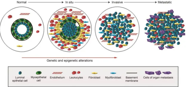
Breast cancer carcinogenesis comprises several stages that are governed by genetic and epigenetic alterations (Figure 5) (Polyak, 2007). In normal breast ducts, a layer of luminal epithelial and myoepithelial cells is found in the basement membrane, which is surrounded by the cells that compose the stroma (fibroblasts, myofibroblasts, leukocytes and endothelial cells) (Polyak, 2007). When myoepithelial cells decrease in number (presumably due to degradation of the basement membrane) and undergo through epigenetic and phenotypic alterations, together with an increment of the stromal cells, an in situ carcinoma is developed (Polyak, 2007). The subsequent total loss of myoepithelial cells and basement membrane results in the establishment of an invasive carcinoma (Polyak, 2007). Ultimately, tumor cells may invade the surrounding tissues and colonize distant sites in the body, leading to the appearance of breast cancer metastasis (Polyak, 2007).
Figure 5. Schematic representation of the breast cancer carcinogenesis. Breast cancer emerges as a
consequence of genetic and epigenetic alterations that occur in myoepithelial cells, together with a reduction in their number. At the same time, the basement membrane starts to degrade and the population of stromal cells (lymphocytes, fibroblasts, myofibroblasts and endothelial cells) increases, resulting in the formation of an in situ carcinoma. The subsequent total loss of myoepithelial cells and basement membrane results in the development of invasive carcinomas, which can spread to distant sites of the body, leading to the formation of breast cancer metastasis (Adapted from (Polyak, 2007)).
Breast cancer can have a different answer to treatments due to its heterogeneity, which is characterized by several clinical, morphological and molecular features (Polyak, 2011; Zardavas et al., 2015). Currently, the standard treatment options for breast cancer include surgery, radiation therapy and small molecule-based therapies (chemo, hormone or targeted therapies) (DeSantis et al., 2014). When cancer is diagnosed at an early stage, the surgical resection of the tumor is usually performed to remove the majority of the tumor mass (Wyld et al., 2015). This treatment may also be complemented with radiotherapy and/or small molecule-based therapies (Wyld et al., 2015). Late-stage breast cancer is treated with small molecule-based
7
therapies due to the existence of metastasis throughout the body (Tryfonidis et al., 2015; Twelves et al., 2016).
However, the currently available therapies are not selective since they damage both cancer and healthy cells, leading to the occurrence of adverse side effects that may span from fatigue, nausea or hair loss to organ failure (Runowicz et al., 2016). Moreover, cancer cells often develop resistance mechanisms to radio and small molecule-based therapies, which further decreases the effectiveness of the available treatments (Blanco and Ferrari, 2014; Tang et al., 2016). Due to these drawbacks, researchers and clinicians are currently investigating new therapeutic approaches to increase the efficacy and safety of breast cancer treatments.
1.1.3. Phototherapies
Within the panoply of cancer therapies under investigation, light-induced treatments have been demonstrating promising results (Huggett et al., 2014; Lucky et al., 2015). After the administration of light-responsive molecules, the tumor zone is irradiated with light and thus the compounds accumulated within this area will produce the desired therapeutic effect (Cheng et al., 2014). The compounds that may have accumulated in other sites will not produce any cytotoxic effects since those areas are not exposed to light. Exceptionally, compounds accumulated within organs that are in the light path (e.g. skin) can induce some degree of toxicity. Therefore, this therapeutic modality offers some tumor selectivity when compared to conventional therapies (Cheng et al., 2014).
The most promising phototherapeutic agents under investigation are those that strongly absorb in the near-infrared (NIR) region (750 – 1000 nm) due to their capacity to interact with NIR light. NIR radiation is within the so-called biological transparency window, meaning that its interaction with biological components (e.g. hemoglobin, myoglobin, melanin or water) is minimal (Figure 6) (Cheng et al., 2014; Rwei et al., 2015; Thakor and Gambhir, 2013). Moreover, NIR light also ensures deep tissue penetration, which is fundamental for the radiation to reach the photoabsorbers accumulated in the tumor zone.
Figure 6. Representation of the biological transparency window (750 – 1000 nm) and the
ultraviolet-visible-near-infrared (UV-vis-NIR) absorption spectra of the main biological components of the human tissues. Within 750 - 1000 nm the biological components display a minimal absorbance, conferring NIR light deep tissue penetration and low off-target interactions. Oxyhemoglobin (HbO2), deoxyhemoglobin
(Hb) (Adapted from (Rwei et al., 2015)).
There are two classes of NIR photoabsorbers: the photosensitizers that are used in photodynamic therapy (PDT) and the photothermal agents that are employed in photothermal therapy (PTT) (Cheng et al., 2014). In PDT, the administrated photosensitizers are activated after their exposition to light, that leads to an energy transfer between the photosensitizers and the molecular oxygen that culminates in the generation of reactive oxygen species (ROS). ROS cause tumor ablation through three main mechanisms: (i) by killing the tumor cells, (ii) damaging the normal and the tumor-associated vasculature, and (iii) activating inflammatory and immune responses against tumor cells (Dolmans et al., 2003; Lucky et al., 2015; Tong and Kohane, 2012). The extent of the PDT-induced cytotoxicity is affected by the optical properties and the concentration of the photosensitizers used and by its localization (intra- or extracellular space), the time span between the photosensitizers administration and the light exposure, and the oxygen availability in the tumor area (Dolmans et al., 2003; Lucky et al., 2015). Photofrin® is a Food and Drug Administration (FDA)-approved photosensitizer used for cancer PDT (Lucky et al., 2015; Tong and Kohane, 2012). However, this photosensitizer is usually irradiated with 630 nm light, due to its intrinsic properties, which reduces the penetration depth attained by this therapy. Moreover, Photofrin® also displays a low molar extinction coefficient, which demands the administration of high concentrations of this molecule and the application of intense irradiation to achieve the desired therapeutic effect (Dolmans et al., 2003; Lucky et al., 2015).
Similarly, PTT starts with the administration of photothermal agents to the patient, followed by the irradiation of the tumor zone. However, the mechanism of action of PTT relies on the thermal ablation of cancer cells, through the ability of the photothermal agents to convert the absorbed light into heat (Cheng et al., 2014; Tong and Kohane, 2012). If temperature increases to above 50 °C, cell death through coagulation necrosis process can occur. When lower
9
temperatures (41-45 °C) are reached, cellular functions can be compromised (e.g. DNA repair or mitochondrial activity), resulting in damage to cancer cells (Chu and Dupuy, 2014). Moreover, this hyperthermic effect can also sensitize cancer cells to radio and chemotherapies (Jaque et al., 2014). In this way, the optical properties and photothermal conversion efficiency of the photothermal agents are crucial for the therapeutic outcome (Song et al., 2015). Furthermore, the availability of the photothermal agents within the TME, their cellular distribution and the onset of the irradiation also affect therapeutic effectiveness (Tong and Kohane, 2012). NIR dye indocyanine green (ICG) is approved by FDA for clinical applications and has been widely investigated for cancer PTT. Nevertheless, the poor stability of ICG in aqueous media and concentration-dependent aggregation limit its applications in medicine (Cheng et al., 2014; Song et al., 2015).
More recently, 2-[2-[2-Chloro-3-[(1,3-dihydro-3,3-dimethyl-1-propyl-2H-indol-2-ylidene) ethylidene]-1-cyclohexen-1-yl]ethenyl]-3,3-dimethyl-1-propylindolium iodide (IR780), a hydrophobic heptamethine indocyanine dye, started to be explored as a light-responsive agent for cancer therapy (Peng et al., 2011; Wang et al., 2016). The characteristic absorption peak of IR780, at 780 nm, enables its effective interaction with NIR light (Han et al., 2015). Additionally, IR780 presents a good quantum yield and reduced photobleaching (Zhang et al., 2010). In fact, as a NIR dye, IR780 presents a higher molar extinction coefficient than Photofrin® and it is also able to reach deeper diseased tissues (Bazylińska et al., 2014; Dolmans et al., 2003). Moreover, comparatively to ICG, IR780 has higher photostability and fluorescence intensity, endowing IR780-based therapies an even higher potential for cancer phototherapy (Yuan et al., 2015). Currently, IR780 has been reported to produce ROS, temperature increase or both effects upon NIR irradiation, that along with its good NIR fluorescence enables its use as a phototherapeutic (PDT and PTT) and imaging agent (Peng et al., 2011; Wilk et al., 2012; Zhang et al., 2010). However, the clinical application of this versatile photoresponsive molecule is strongly impaired by its low solubility in pharmaceutically acceptable solvents (Jiang et al., 2015). To surpass such handicap, IR780 encapsulation in nanoparticles (NPs) has been proposed for improving its therapeutic efficacy.
1.2. Nanomaterials for application in cancer phototherapy
1.2.1. The potential of unifying the best of both worlds
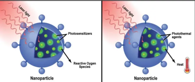
For improving the effectiveness and selectivity of PDT and PTT towards cancer cells, researchers have been encapsulating NIR light-responsive molecules in NPs (Figure 7) (Dolmans et al., 2003; Tong and Kohane, 2012; Wust et al., 2002).
NPs can be composed by organic or inorganic materials and display a size that spans from 1 to 1000 nm (Ferrari, 2005; Jain and Stylianopoulos, 2010; Petros and DeSimone, 2010; Schroeder et al., 2012). These nanostructures can be used to encapsulate NIR photosensitizer and photothermal agents, which is fundamental to overcome their low water solubility, degradation and photobleaching issues (Lucky et al., 2015; Thakor and Gambhir, 2013; Tong and Kohane, 2012). Moreover, NPs can also display a preferential accumulation in tumors, which in turn improves the availability of the loaded NIR photoabsorbers at the tumor zone. Thereby, this approach also reduces the non-specific toxicity of phototherapies towards healthy cells/tissues and decreases the light exposure (duration and intensity) required to achieve a suitable therapeutic effect (Farokhzad and Langer, 2009; Lucky et al., 2015; Rwei et al., 2015; Thakor and Gambhir, 2013; Tong and Kohane, 2012).
Figure 7. Representation of the mechanism of action of nanoparticles-mediated photodynamic therapy
(left) and photothermal therapy (right). In this type of therapeutic approach, nanoparticles are used to load photoabsorbers and deliver them to tumor cells. Within the cells, the entrapped photoabsorbers are stimulated by near-infrared light to generate reactive oxygen species (photodynamic therapy) or heat (photothermal therapy) that induce cellular damage (Adapted from (Thakor and Gambhir, 2013)).
For a successful NP-mediated phototherapy, these nanosized agents must be carefully designed in order for them to display a specific size, morphology, charge and corona composition. These parameters influence the pharmacokinetic profile of the NPs and will, ultimately, determine the dose of photosensitizers/photothermal agents delivered into the tumor by the NPs and thus the therapeutic outcome (Ernsting et al., 2013; Kaur et al., 2016; Lucky et al., 2015).
11
1.2.2. NPs-mediated phototherapy: the design drives the journey
NPs loading NIR photoabsorbers can be administered by intratumoral or intravenous injections (Etheridge et al., 2013; Petros and DeSimone, 2010). Since the majority of the FDA-approved NPs for cancer therapy are administered intravenously, this section will be focused on the presentation of the design requirements that NPs loaded with NIR photoabsorbers should fulfill considering this administration route (Figure 8) (Ernsting et al., 2013; Ferrari, 2005).
Figure 8. Illustration of the different stages of nanoparticles-mediated phototherapy. Factors that
influence nanoparticles blood circulation (e.g. size), extravasation from the blood flow to the tumor zone (e.g. nanoparticles features) and penetration in the tumor mass (nanoparticles features and tumor-related factors) are represented. Reticuloendothelial system (RES), near-infrared (NIR) (Adapted from (Luo et al., 2015)).
Once intravenously injected, NPs must sustain their structural integrity and protect the loaded photoabsorbers (Figure 8). Such is fundamental to avoid the leakage of the loaded compounds, which in turn could lead to its degradation and clearance from blood circulation or even result in undesirable pharmacokinetics (accumulation in off-target organs) (Blanco et al., 2015; Ernsting et al., 2013). NPs must be able to avoid renal filtration and opsonization by blood proteins (serum albumin, complement components, apolipoproteins and immunoglobulins). These opsonins adsorb on the surface of NPs enabling their recognition and phagocytosis by the reticuloendothelial system (RES) cells (Aggarwal et al., 2009; Blanco et al., 2015). NPs size, charge and corona composition have a key role in this phase of the phototherapeutic procedure (Ernsting et al., 2013).
NPs must not be smaller than 5 nm or otherwise they will be removed from circulation through renal filtration (Blanco et al., 2015). Moreover, nanostructures with a size lower than 50 nm tend to become accumulated in the liver since these can extravasate through the liver
fenestrations, which have a size that is comprehended between 50-100 nm (Blanco et al., 2015; Ernsting et al., 2013). On the other hand, the splenic filtration governs the upper size limit. Splenic interendothelial slits present a size range from 200 to 500 nm and impose 200 nm as the maximal NP size that is able to evade retention in the spleen (Blanco et al., 2015; Petros and DeSimone, 2010). Furthermore, NPs with a size higher than 200 nm are also more likely to be phagocytized by liver Kupffer cells and by splenic macrophages (Blanco et al., 2015; Ernsting et al., 2013; Luo et al., 2015).
NPs surface charge also determines their biological fate during blood circulation. Generally, negatively charged (zeta potential < 10 mV) nanocarriers tend to have a high uptake by RES macrophages, while those with a positive surface charge (zeta potential > 10 mV) display a high propensity for protein adsorption on their surface (Ernsting et al., 2013). Hence, NPs should have a superficial charge of ± 10 mV, the so-called neutral charge range, in order to exhibit low protein adsorption and low accumulation in RES organs (Blanco et al., 2015; Ernsting et al., 2013; Stylianopoulos and Jain, 2015).
The surface composition of NPs is another important parameter that mediates their interaction with serum proteins and RES components. To avoid such undesirable interactions, the surface of NPs is commonly modified with poly(ethylene glycol) (PEG). This type of functionalization stabilizes the NPs and also prevents their opsonization and phagocytosis by RES cells (Blanco et al., 2015; Ernsting et al., 2013; Jain and Stylianopoulos, 2010; Stylianopoulos and Jain, 2015). Still, the benefits of PEGylation are not always attained since these depend on of multiple factors such as the molecular weight and density of the PEG chains (Aggarwal et al., 2009; Ernsting et al., 2013). Currently, other polymers are also being investigated as alternatives to PEG, such as poly(aminoacid)s (e.g. poly(glutamic acid)) and poly(oxazolines) (Barz et al., 2011; Knop et al., 2010).
Besides avoiding its clearance by kidneys and RES, NPs must be able to extravasate from the blood flow to the tumor site (Figure 8). Tumor vasculature presents fenestrations that vary from 400 to 600 nm, which are permeable to NPs. In this way, NPs may benefit from this leaky vasculature to extravasate into the tumor interstitial space (Danhier et al., 2010; Ernsting et al., 2013). Moreover, the impaired lymphatic drainage present in this tissue promotes NPs retention in the TME (Bertrand et al., 2014; Danhier et al., 2010). This phenomenon is known as the Enhanced Permeability and Retention (EPR) effect (Bertrand et al., 2014; Danhier et al., 2010; Ernsting et al., 2013). Taking these and the previous size impositions into consideration, NPs should have a size of 100-200 nm in order to be minimally cleared by off-target organs and display an enhanced tumor accumulation through the EPR effect (Blanco et al., 2015; Petros and DeSimone, 2010).
Once in the tumor interstitial space, NPs must penetrate through the tumor biomass in order to reach the cancer cells (Figure 8). Their efficient penetration depends on factors intrinsic to
13
the tumor physiology and NPs features (Ernsting et al., 2013). Tumor physiological factors comprise tumor vasculature, interstitial fluid pressure (IFP) and the presence of cells that populate the TME (Danhier et al., 2010; Ernsting et al., 2013).
As a consequence of the aberrant and heterogeneous tumor vasculature, the tumor periphery is highly perfused whereas its core is hypoxic (Blanco et al., 2015; Ernsting et al., 2013). In this way, most of the NPs become accumulated in tumor periphery leading to a heterogeneous NP distribution (Ernsting et al., 2013). Moreover, the tumor vasculature and the impaired lymphatic drainage are responsible for the presence of a high IFP in the tumor, which also hinders the penetration of NPs through the tumor biomass (Blanco et al., 2015; Jain and Stylianopoulos, 2010). Furthermore, the cells that populate the TME can also hamper NPs penetration (Blanco et al., 2015; Ernsting et al., 2013; Jain and Stylianopoulos, 2010).
On the other hand, NPs-related characteristics such as their size and charge also determine their penetration through the tumor tissue (Ernsting et al., 2013). Generally, smaller NPs have a better tumor penetration capacity than those with larger dimensions (Bertrand et al., 2014; Ernsting et al., 2013; Jain and Stylianopoulos, 2010; Stylianopoulos and Jain, 2015). Moreover, positively charged NPs have a higher propensity to interact with the hyaluronic acid present in tumor ECM than those with a negative charge, that are more prone to interact with collagen (Ernsting et al., 2013; Jain and Stylianopoulos, 2010). In this way, neutral charged NPs display a better penetration capacity since they establish a lower number of interactions with tumor ECM components (Ernsting et al., 2013).
Finally, the photoabsorber-loaded NPs must then be internalized by cancer cells (Figure 8). Such is fundamental to achieve an effective and selective therapeutic effect upon NIR laser irradiation. The main parameters affecting NPs cellular internalization are related to their size, charge and the presence of targeting ligands on their surface (Blanco et al., 2015; Ernsting et al., 2013).
NPs usually have an optimal size for cellular internalization, which varies for each type of NP (Hillaireau and Couvreur, 2009; Nel et al., 2009; Petros and DeSimone, 2010). In terms of charge, particles with a positive surface charge are more likely to be internalized by cells, since these are able to establish interactions with the negatively charged components of cells’ membrane (Stylianopoulos and Jain, 2015). However, the previous considerations about positively charged NPs should still be taken into consideration since these are more likely to adsorb proteins, which may result in their clearance during blood circulation (Bertrand et al., 2014; Ernsting et al., 2013). Additionally, NPs surface can be decorated with targeting ligands in order to promote their uptake by cancer cells overexpressing the receptor for the immobilized ligand (Nel et al., 2009).
Therefore, the efficacy of NPs-mediated phototherapies is strongly influenced by NPs physicochemical properties and also by the properties of the photoabsorbers and external light, as previously described.
1.2.3. Nanovehicles classes: different characteristics for multiple
applications
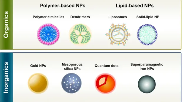
Currently, there are different types of NPs that have shown the capacity to encapsulate hydrophobic molecules, such as NIR photoabsorbers, and to deliver them to cancer cells (Cheng et al., 2014; Jaque et al., 2014; Kim et al., 2016; Lucky et al., 2015; Pekkanen et al., 2014; Rwei et al., 2015; Tong and Kohane, 2012). In a broad sense, these NPs can be divided into two main classes: organic and inorganic NPs (see Figure 9 for further details) (Wicki et al., 2015).
Figure 9. Examples of the different types of nanoparticles that can be used to encapsulate near-infrared
photoabsorbers. These types of nanoparticles have specific characteristics that allow their application in cancer therapy and/or diagnosis (Adapted from (McCarthy et al., 2015) and (Wicki et al., 2015)).
Organic NPs are produced with natural or synthetic compounds and comprise polymer-based nanocarriers (micelles and dendrimers) and lipid-based nanocarriers (liposomes and solid-lipid NPs) (Figure 9). Polymeric micelles are formulated with amphiphilic polymers and have a core-shell organization, that comprises a hydrophobic core capable of encapsulating hydrophobic molecules and a hydrophilic shell that provides stability to the nanostructure (Nazir et al., 2014). Dendrimers are hyperbranched nanostructures with a central core, that can be used to encapsulate molecules through covalent and non-covalent binding (Cheng and Xu, 2008). Liposomes are closed spherical vesicles containing an aqueous core surrounded by one or more concentric lipid bilayers that are able to entrap both hydrophobic and hydrophilic molecules (Al-Jamal and Kostarelos, 2011). Solid-lipid NPs are produced with solid lipids (at room and body temperature) that are stabilized by surfactants (Wissing et al., 2004). These type of NPs
15
possess a rigid core that can be used to encapsulate hydrophobic and hydrophilic molecules (Thakor and Gambhir, 2013; Wissing et al., 2004).
Gold NPs, quantum dots, mesoporous silica NPs and superparamagnetic iron oxides are examples of inorganic nanocarriers (Figure 9). Gold NPs and quantum dots are nanostructures that have been widely applied in cancer imaging and that can also be used for encapsulating therapeutic molecules (Jia et al., 2013; Sun et al., 2014). Mesoporous silica NPs due to their large pore volume and high superficial area have a high drug loading capacity (Rosenholm et al., 2010). Superparamagnetic iron oxides can be employed as imaging and magnetic field-responsive therapeutic agents (Nazir et al., 2014).
Each type of NP has specific characteristics that enable their utilization in cancer therapy. Polymeric micelles will be the focus of the next section since these are promising candidates for the delivery of NIR photoabsorbers to cancer cells.
1.2.4. Polymeric micelles
1.2.4.1. Polymeric micelles application in cancer phototherapy
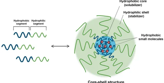
Polymeric micelles display a core-shell organization resulting from the self-assembly of amphiphilic molecules in aqueous environment. In this process, the hydrophobic blocks form the core which is responsible for entrapping the hydrophobic NIR photoabsorbers. The hydrophilic segments form the corona that is responsible for the stabilization of the nanostructure (Figure 10) (Jones and Leroux, 1999; Kedar et al., 2010; Owen et al., 2012).
Figure 10. Representation of the polymeric micelle assembling process and of the core-shell organization
Furthermore, the unique features presented by micelles make them very effective vehicles for the delivery of NIR photoabsorbers, such as: (i) straightforward formulation, (ii) high encapsulation efficiency (EE), (iii) great stability in circulation, (iii) suitable physicochemical characteristics to benefit from the EPR effect, (iv) biocompatibility, and (v) biodegradability (Gong et al., 2012; Kedar et al., 2010; Owen et al., 2012).
Micelles’ corona is typically composed by PEG since this hydrophilic polymer provides stealth properties to nanostructures, thereby preventing their opsonization and uptake by RES cells (Owen et al., 2012). Such is fundamental to enhance micelles’ blood circulation time, which in turn favors their accumulation in the tumor through the EPR effect (Knop et al., 2010; Owen et al., 2012). Regarding micelles’ core, this can be composed of hydrophobic polymers such as poly(caprolactone) (PCL) or poly(lactic-co-glycolic acid) (PLGA), or hydrophobic blocks such as cholesterol or deoxycholic acid (Kedar et al., 2010; Park et al., 2004; Yu et al., 2013) due to their good loading capacity. For instance, Master and co-workers prepared PEG-PCL micelles loaded with the photosensitizer silicon phtalocyanine 4, that produced a cytotoxic effect towards cancer cells under NIR laser irradiation (Master et al., 2012). In another work, Zheng and co-workers synthesized micellar carriers composed of PLGA-lecithin-PEG loading the photothermal agent ICG. Their results demonstrated that these nanovehicles are efficient agents for cancer PTT (Zheng et al., 2013). These observations attest the suitability of using polymeric micelles in cancer PDT and PTT.
Currently, other materials are being explored to form novel and improved micellar nanocarriers. Particularly, vitamin E derivatives arise as promising candidates to produce micelles that are able to deliver NIR photoabsorbers. Here, they were selected to produce the carriers.
1.2.4.2. Vitamin E-based micelles aimed for cancer phototherapy
Vitamin E family is constituted by eight forms of both tocopherols and tocotrienols naturally occurring as alpha (α), beta (β), gamma (δ) and delta (γ) isomers. Vitamin E derivatives have been applied in different areas of the biomedical field (Youk et al., 2005).
D-α-tocopheryl succinate (TOS) is a vitamin E derivative with anticancer properties (Duhem et al., 2014b; Youk et al., 2005). TOS exert its antitumoral activity through two main mechanisms: inhibition of tumor cells proliferation and induction of apoptosis. The antiproliferative activity of TOS is related to its capacity to: (i) inhibit DNA synthesis, (ii) induce cell cycle arrest, and (iii) interfere with the expression of cell cycle proteins (e.g. downregulating nuclear transcription factor-kappa B) (Constantinou et al., 2008; Duhem et al., 2014b). Furthermore, TOS induces apoptosis through activation of extrinsic and intrinsic apoptotic pathways (Constantinou et al., 2008; Duhem et al., 2014b; Youk et al., 2005). The former involves the upregulation of Fas/Fas receptor and restoration of transforming growth factor pathway in cancer cells, while the latter relies on the production of ROS that leads to mitochondrial
17
dysfunctions (Duhem et al., 2014b). In addition, TOS may also hinder angiogenesis and metastization by inhibiting VEGF and matrix metalloproteinases-9 functions, respectively (Duhem et al., 2014b). However, the biomedical use of TOS is limited by its low water solubility (Duhem et al., 2014b; Youk et al., 2005).
D-α-tocopheryl polyethylene glycol 1000 succinate (TPGS), a PEGylated vitamin E derivative produced through the esterification of TOS with a PEG chain of 1000 Da, has also been employed in cancer therapy (Zhang et al., 2012). In fact, TPGS has been described as being more potent in generating ROS, inducing apoptosis and inhibiting tumor cells growth than TOS (Youk et al., 2005). Moreover, the mechanisms of TPGS-mediated apoptosis seem to result from its capacity to inhibit protein kinase B phosphorylation that is involved in the downregulation of the anti-apoptotic proteins Survivin and Bcl-2, leading to cell death through the activation of mediated apoptotic pathways. It was also verified that TPGS induces the activation of caspase-independent apoptosis and cell cycle arrest (Neophytou et al., 2014). Additionally, TPGS has also demonstrated the capacity to inhibit P-glycoprotein, a drug efflux pump implied in cancer cells drug-resistance mechanisms (Collnot et al., 2007).
The amphiphilic character of TPGS together with its intrinsic anticancer activity provide several advantages for its application in micelles’ production (Zhang et al., 2012). Moreover, TPGS-based nanomedicines have an increased cellular uptake, which also accounts for their good therapeutic potential (Kulkarni and Feng, 2013). For instance, Kutty and Feng demonstrated that Docetaxel-loaded TPGS micelles produce an anticancer therapeutic effect that is superior to that of the free drug (Kutty and Feng, 2013). However, these micelles demonstrated a size of about 18 nm, which may have a negative impact on their biodistribution. More recently, Danhier et al. prepared micelles using a combination of TPGS and TOS, and they were used to encapsulate doxorubicin (Dox). In TPGS-TOS micelles (TTM), the tocopherol (hydrophobic) moieties of TOS and TPGS form the core (encapsulates the hydrophobic drug), while the PEG chains (hydrophilic) of TPGS originate the shell (stabilizes the micelle). However, the Dox-loaded TTM had a size of 350 nm, which is not suitable for passive tumor accumulation and emphasizes the need to optimize the physicochemical properties of this promising delivery system (Danhier et al., 2014).
TTM appear to be promising vehicles for the encapsulation and delivery of NIR photoabsorbers, since the final physicochemical properties of these micelles can be adjusted by controlling their TPGS and TOS content, and also due to the intrinsic anticancer activity of both vitamin E derivatives. Moreover, TPGS and TOS are already approved by FDA and European Medicines Agency as pharmaceutical solubilizers, which attests their biological safety (Danhier et al., 2014). In this context, the encapsulation of IR780 in TTM seems to be a promising strategy for cancer phototherapy since it aims to: (i) overcome IR780 poor water solubility, (ii) take advantage of the excellent properties of micelles in cancer drug delivery, (iii) benefit from vitamin E intrinsic anticancer activities, and (iv) produce an anticancer treatment responsive
to NIR light by exploring the photoversatility of IR780 (photosensitizer, photothermal agent and imaging agent). Herein, TPGS and TOS were used to produce micelles that were exploited to encapsulate the NIR agent IR780 (IR780-TTM; Figure 11) and their therapeutic efficacy under NIR irradiation was evaluated using breast cancer cells as models.
Figure 11. Schematic illustration of the protocol used to produce IR780-loaded TPGS-TOS micelles.
19
Aims
The main objective of this masters’ thesis was to develop a vitamin E-based micellar nanocarrier to deliver a NIR photoabsorber to breast cancer cells and subsequently evaluate the effect of NIR irradiation in these cells.
The specific aims of this thesis comprise the:
Preparation of IR780-loaded TPGS-TOS micelles;
Characterization and modulation of micelles’ physicochemical properties;
Investigation of micelles’ ability to absorb NIR light and photothermal efficiency under NIR laser irradiation;
Evaluation of IR780-loaded TPGS-TOS micelles biocompatibility;
Assessment of micelles capacity to be internalized in breast cancer cells;
Evaluation of IR780-loaded micelles therapeutic effect under NIR laser irradiation in breast cancer cells.
Chapter 2
21
2. Experimental Section
2.1. Materials
Dulbecco’s Modified Eagle’s Medium F-12 (DMEM-F12), IR780, resazurin, TOS, TPGS, trypsin and 2',7'-dichlorofluorescein diacetate (H2DCF-DA) were acquired from Sigma–Aldrich (Sintra, Portugal). Michigan Cancer Foundation-7 (MCF-7; ATCC® HTB-22TM) cell line was purchased from ATCC (Middlesex, United Kingdom). Fetal bovine serum (FBS) was obtained from Biochrom AG (Berlin, Germany). Cell culture plates and T-flasks were acquired from Orange Scientific (Braine-l’Alleud, Belgium) and cell imaging plates were purchased from Ibidi GmbH (Munich, Germany).
2.2. Methods
2.2.1. Production of IR780-TTM
IR780-TTM were produced by using the nanoprecipitation technique as previously described by Duhem et al., with slight modifications (Duhem et al., 2014a).First, a 1 mL acetone solution containing 250 μg of IR780 and 5 mg of different weight feed ratios of TPGS:TOS was prepared (Table 1). This solution was then added dropwise to 5 mL of double deionized water (0.22 μm filtered, 18.2 MΩ·cm) under constant magnetic stirring for 1 h at room temperature (RT). Afterward, the aqueous solution was dialyzed (1000 Da molecular weight cut-off) for 2 h at RT, yielding IR780-TTM. Samples from three different batches (n = 3) were analyzed and each sample was screened three times (three measurements per n). Blank micelles were prepared according to the above described procedure but without adding IR780.
Table 1. Formulation conditions used for preparing IR780-TTM.
Sample abbreviation TPGS feed (%) TPGS feed (mg) TOS feed (mg) IR780 feed (µg)
IR780-100TTM 100 5 0 250 IR780-67TTM 67 3.33 1.67 250 IR780-60TTM 60 3 2 250 IR780-50TTM 50 2.5 2.5 250 IR780-40TTM 40 2 3 250 IR780-33TTM 33 1.67 3.33 250 IR780-25TTM 25 1.25 3.75 250 IR780-0TTM 0 0 5 250