1
This is the pre-peer reviewed version of the following article: [Teixeira D, Carrilho M,Silva M, et al. MediterraneanEucalyptusplantationsaffect small mammal ectoparasites abundance but notindividual body condition.Ecol Res. 2019;34:415–427], which has been published in final form at [DOI: 10.1111/1440-1703.12003]. This article may be used for non-commercial purposes in accordance with Wiley Terms and Conditions for Use of Self-Archived Versions
A Membrane-Bound Cytochrome c3 – Type II Cytochrome c3 from
Desulfovibrio vulgaris Hildenborough
Filipa M. A. Valente[a], Lígia M. Saraiva[a], Jean LeGall[a,b], António V. Xavier[a], Miguel Teixeira[a] and Inês A. C. Pereira[a]*
[a] F. M. A. Valente, Dr. L. M. Saraiva, Prof. J. LeGall, Prof. A. V. Xavier, Prof. M. Teixeira, Prof. I.A.C. Pereira Instituto de Tecnologia Química e Biológica, Universidade Nova de Lisboa,
Apt. 127, 2780-156 OEIRAS, Portugal. Fax: 351-214428766
Email: [email protected]
[b] Prof. J. LeGall
Department of Biochemistry, The University of Georgia, Athens, Georgia 30602, USA.
Abbreviations: 16Hcc: 16-heme high molecular weight cytochrome c; 9Hcc: nine-heme cytochrome c; Da: Desulfovibrio africanus;
2
DvH: Desulfovibrio vulgaris Hildenborough; DvM: Desulfovibrio vulgaris Miyazaki; Dg: Desulfovibrio gigas; Dd27k: Desulfovibrio desulfuricans ATCC 27774; Dmn: Desulfomicrobium norvegicum; TpII-c3: Type II cytochrome c3; TpI-c3: Type I cytochrome c3; FeS: Iron-sulfur.
KEY WORDS: cytochrome c3; Desulfovibrio; Electron transfer; Hydrogenases; Membrane proteins.
Abstract
A new tetraheme cytochrome c3 was isolated from the membranes of Desulfovibrio vulgaris Hildenborough (DvH). This cytochrome has a molecular mass of 13.4 kDa, a pI of 5.5, and contains four hemes c with apparent reduction potentials of –170 mV, -235 mV, -260 mV and – 325 mV at pH 7.6. The complete sequence of the new cytochrome, retrieved from the preliminary data of the DvH genome, shows that this cytochrome is homologous to the “acidic” cytochrome c3 from Desulfovibrio africanus (Da). A model for the structure of the DvH cytochrome was built based on the structure of the Da cytochrome. Both cytochromes share structural features that distinguish them from other cytochromes c3, such as a solvent-exposed heme 1 surrounded by an acidic surface area, and a heme 4 which lacks most of the surface lysine patch proposed to be the site of hydrogenase interaction in other cytochromes c3. Furthermore, in contrast to previously discovered cytochromes c3, the genes
3
coding for these two cytochromes are adjacent to genes coding for two membrane associated FeS proteins, which indicates that they may be part of membrane-bound oxido-reductase complexes. Altogether these observations suggest that the DvH and Da cytochromes are a new type of cytochromes c3 (Type II: TpII-c3) with different redox partners and physiological function than the other cytochromes c3 (Type I: TpI-c3).
The DvH TpII-c3 is reduced with considerable rates by the two membrane-bound [NiFe] and [NiFeSe] hydrogenases, but catalytic amounts of TpI-c3 increase these rates two and four-fold, respectively. With the periplasmic [Fe] hydrogenase TpII-c3 is reduced much slower than TpI-c3, and no catalytic effect of TpI-c3 is observed.
Introduction
Sulfate-reducing bacteria are anaerobes that can grow by oxidation of hydrogen or organic compounds using sulfate as terminal electron acceptor. The reduction of sulfate is a true respiratory process, leading to oxidative phosphorylation via a still poorly understood electron transfer pathway. A characteristic feature of this electron transfer pathway is the involvement of multiheme cytochromes c of low redox potential.[1] Several elements of this family of electron transfer proteins have been identified in Desulfovibrio, the most studied genus of sulfate-reducing bacteria: The tetraheme
4
cytochrome c3 (Mr 13 000)[2] is the most abundant member, and is the only one present in all the species studied so far. In addition, it is also present in other genera of sulfate-reducing bacteria such as Desulfomicrobium,[3] Desulfobulbus[4] and Thermodesulfobacterium.[5] Other members are the Split-Soret cytochrome that is a dimer of a diheme subunit,[6] the octaheme cytochrome c3 (Mr 26 000), a dimer of a tetraheme subunit,[7] the monomeric nine-heme cytochrome c (9Hcc)[8] and the high molecular weight cytochrome c which is a monomer containing 16 hemes (16Hcc).[9] Desulfovibrio species contain different cytochrome compositions: for example, in Desulfovibrio gigas (Dg) the tetraheme cytochrome c3, the octaheme cytochrome c3 (Mr 26 000) and the 16Hcc were described, whereas in Desulfovibrio desulfuricans ATCC 27774 (Dd27k) the tetraheme cytochrome c3, the split-Soret cytochrome and the 9Hcc were reported. In Desulfovibrio vulgaris Hildenborough (DvH) the tetraheme cytochrome c3 and 16Hcc were isolated, but this bacterium contains also a monoheme cytochrome called cytochrome c553.[10]
The physiological function of some of these cytochromes is still unclear, as is the need for several similar cytochromes in the same bacterium. The ubiquitous tetraheme cytochrome c3 is generally considered to act as the physiological partner for hydrogenases, which are also always present in these bacteria, and has been proposed to play a central role in their metabolism by acting as an energy transducing device which converts high
5
energy electrons (low redox potential) and low energy protons (high pKa), resulting from the periplasmic oxidation of hydrogen, into lower energy electrons for the reduction of sulfate and higher energy protons for the production of ATP. [11-13] The involvement of hydrogen as a necessary intermediate in the sulfate respiration process has been proposed,[14] but the generality of this model is still a matter of controversy.[15] Nevertheless, many Desulfovibrio sp. can use H2 as sole energy source.[16] All these bacteria contain one or more hydrogenases of the [Fe], [NiFe] and [NiFeSe] types. A screening of 25 Desulfovibrio species showed that only the genes encoding the [NiFe] hydrogenase are present in all.[17] In a situation analogous to that of the cytochromes, different species contain different hydrogenase compositions and it is not clear why more than one type should be necessary for some bacteria. For example, DvH contains a periplasmic [Fe] hydrogenase,[18] and two membrane-bound [NiFe][19] and [NiFeSe] hydrogenases.[20]
Despite the low sequence homology between cytochromes c3 of different species, the several X-ray structures determined[21-26] show that the general folding is maintained as is the spatial arrangement of the four heme core. This four heme structural motif seems to be a common feature of this family of cytochromes, as it is also observed in octaheme cytochromes c3 (26,000),[7,27] in the 9Hcc,[8] and most probably also in 16Hcc since its sequence shows it is organised in four c3-like
6
domains[28] (the first one being an incomplete domain with only three hemes), and that the last two domains have a strong sequence similarity to the 9Hcc suggesting a similar structural organisation.[8] This indicates that the four-heme structural motif may have a specialised functional role.
The genes coding for 16Hcc and the related 9Hcc[8] are both part of operons encoding transmembrane redox complexes[29,30] proposed to perform the electron transfer between the periplasmic oxidation of hydrogen, carried out by the hydrogenase/cytochrome c3 couple, and the cytoplasmic reduction of sulfate.[29,31] Although 16Hcc is not a hydrophobic protein, it is found in higher amounts in the membrane than in the soluble fraction, in both DvH and Dg.[9] The reduction of DvH 16Hcc by the [Fe] and [NiFe] hydrogenase is mediated by cytochrome c3,[31] as is also the case for 9Hcc and [NiFe] hydrogenase of Dd27k,[8] and the octaheme cytochrome c3 (26,000) and [NiFeSe] hydrogenase of Desulfomicrobium norvegicum (Dmn).[32]
In our attempts to study membrane proteins of DvH that may be involved in the electron transport pathway for the reduction of sulfate, we have isolated a new tetraheme cytochrome c3 with a lower pI than the well characterised and more abundant cytochrome c3. The new cytochrome is not detected in the soluble fraction, whereas some of the previously characterised cytochrome c3 was also found to be associated with the membrane fraction. The co-existence of two different tetraheme
7
cytochromes c3 in one organism was previously reported only for Desulfovibrio africanus (Da),[33,34] but both were isolated from the soluble fraction. In this case, the authors opted to name the two cytochromes as “acidic” and “basic”,[33] a nomenclature that may be misleading as the pI value of a cytochrome c3 will, by itself, not reveal to which type the cytochrome belongs. Indeed, cytochrome c3 from Dg which is as acidic as DvH TpII-c3 (pI of 5.2[35]), does not share the structural features of the TpII-c3s, but is similar to other TpI-c3s. As an alternative classification we name the “acidic” cytochromes c3 of DvH and Da as Type II-cytochromes c3 (TpII-c3s), and the well characterised and more abundant “basic” cytochromes c3 as Type I-cytochromes c3 (TpI-c3s), since structural and genetic evidence (see below) indicates that these cytochromes belong to separate families.
In this report we describe the purification and characterisation of the TpII-c3 from the membranes of DvH, as well as a study of its reactivity with the [Fe], [NiFe] and [NiFeSe] hydrogenases from the same organism.
RESULTS AND DISCUSSION
The repeated washing of the DvH membranes ensured that all soluble and loosely-bound proteins were removed, as confirmed by the visible spectrum using the peak at 630 nm of desulfoviridin as a marker for the presence of soluble
8
proteins. The detergent extract obtained after solubilisation of the membrane proteins with SB12 contained a high amount of cytochromes. After purification of this extract it was possible to separate four heme proteins. The non-sticking fraction from the DEAE column contained the two high-pI cytochromes 16Hcc[9] and TpI-c3 identical to that obtained from the soluble fraction, which was identified by its N-terminal sequence. A more acidic fraction contained the heme c nitrite reductase[36] and an unknown cytochrome of small molecular mass that, after purification and characterisation, proved to be a low-pI tetraheme cytochrome c3 (TpII-c3). The TpII-c3 could not be found upon purification of the DvH soluble extract, suggesting that its association with the cytoplasmic membrane is quite strong. The finding of TpI-c3 in the membrane extract, even after thorough washing of the membranes, suggests that this periplasmic cytochrome also interacts with membrane-bound proteins. The amount of TpI-c3 purified from the membrane fraction was significant, being about half of that usually obtained from the soluble fraction. The distribution of a cytochrome between soluble and membrane extracts was previously described for the 16Hcc of DvH and Dg,[9] but these cytochromes are found in much higher amounts in the membrane than in the soluble fraction. The TpII-c3 was found exclusively in the membrane fraction, and the amount purified was similar to that of membrane-associated TpI-c3 (7 mg versus 10mg). In Da the TpII-c3 was isolated from the soluble fraction.[33] However,
9
since no reference was given to purification of membrane proteins, it is possible that in this bacterium the TpII-c3 is also distributed between the soluble and membrane fractions. Molecular characteristics. The new TpII-c3 was judged to be pure by SDS-PAGE which displayed only one band. The molecular mass determined from the gel was 14 kDa, in good agreement with the value calculated from the aminoacid sequence (see below). To determine whether the new cytochrome was a monomer or a dimer, it was dialysed to remove the detergent and the molecular mass determined by gel filtration yielded a value of approximately 17 kDa, revealing that it exists in solution as a monomer. The isoelectric point of the new cytochrome is 5.5, as determined by isoelectric focusing. The number and type of hemes present was determined by the pyridine hemochrome which showed that it contained 4 hemes c per molecule.
Besides the TpI-c3 and TpII-c3 there is at least one other tetraheme cytochrome c associated with the membranes of DvH, which was purified as one of the subunits of the nitrite reductase.[36] The sequence of this cytochrome can be retrieved from the preliminary DvH genome data and shows that it is part of the NapC/NirT family of cytochromes c that are proposed to receive electrons directly from the quinone pool.[37,38]
Spectroscopic characterisation. The UV-Visible spectrum of the TpII-c3 is typical of cytochromes c3 (Figure 1). The absence of an absorption band at 695 nm indicates there are no methionine-bound hemes. The millimolar extinction coefficients of the
10
bands observed in the oxidised spectrum, are: 280 nm: 49.3 mM -1cm-1; 408 nm: 555.7 mM-1cm-1; 527.5 nm: 48.6 mM-1cm-1; and in the reduced spectrum: 417.5 nm: 729.3 mM-1cm-1; 522 nm: 65.7 mM-1cm-1; 551 nm: 107.1 mM-1cm-1. These extinction coefficients are similar to those reported for the TpII-c3 of Da.[33] The purity index of the TpII-c3 (A551(red)-A570(red)/A280(ox)) was 2.2, a value that is lower than that usually observed for TpI-c3s (2.9-3.8). This is apparently due to a broader 551 nm peak in the TpII-c3 (and thus of reduced height) when compared to the TpI-c3s. A similar situation is observed for the Da TpII-c3.[33]
The EPR spectrum of the oxidised DvH TpII-c3 is a rhombic spectrum typical of low-spin hemes (Figure 2). Three of the hemes in this cytochrome have g-values of gz=2.98, gy=2.24 and gx=1.5, indicating that the angle between the planes of the two axial histidines is very similar for these hemes and close to 0º.[39,40] The fourth heme has a gz value of 3.5, which suggests that in this case the angle between the two histidine planes is close to 90º. In the Da TpII-c3 a similar resonance is observed at 3.7,[34] and the crystal structure confirmed that heme 4 has an angle between the two histidine planes close to 90º.[41] The EPR spectrum of DvH TpII-c3 (Figure 2–trace a) was simulated (Figure 2–trace b) by adding three theoretical spectra with g-values of: gz=2.98, gy=2.24 and gx=1.50 (2 hemes; Figure 2–trace c), gz=2.98, gy=2.24 and gx=1.40 (1 heme; Figure 2 –trace d) and gz=3.5 and gy=2.0 (1 heme; Figure 2–trace e).
11
The EPR spectrum of DvH TpI-c3 is different from that of TpII-c3, displaying several resonances with gz values of 2.77, 2.82, 2.97 and 3.12.[42]
Heme reduction potentials. The redox behaviour of the DvH TpII-c3 at pH 7.6 was probed by a redox titration in the presence of redox mediators (Figure 3). The experimental points were fitted with four non-interacting Nernst equations (n=1) with reduction potentials of -170 mV, -235 mV, -260 mV and –325 mV. These reduction potentials are similar to those commonly observed for TpI-c3s (including that from DvH[42]), and have in common the fact that one of the hemes displays a considerably higher reduction potential than the other three. This was not observed for the TpII-c3 of Da, for which the four reduction potentials obtained by cyclic voltammetry were quite close (-210 mV, -240 mV, -260 mV and –270 mV).[33]
Sequence analysis. The N-terminal sequence of the TpII-c3 was determined up to residue 48, confirming that the new cytochrome is distinct from TpI-c3. This N-terminal sequence was used to retrieve the complete sequence of the TpII-c3 gene from the preliminary data of the DvH genome, which is being sequenced at The Institute for Genomic Research. The gene reveals that the TpII-c3 is synthesised as a precursor protein with a signal peptide, indicating that it will be exported to the periplasmic side of the membrane. The signal peptide (MFRRIGTVMLAFATLALLL AVAFA) contains 24 residues, with two positive residues at the beginning (R3-R4), followed by a string of hydrophobic
12
residues. The sequence obtained for the TpII-c3 shows that this is not a hydrophobic protein and that it contains no trans-membrane helices. This indicates that the trans-membrane attachment of this cytochrome is not likely due to protein–lipid interactions, but rather to interactions of the cytochrome with other membrane-bound proteins.
The sequence of the DvH TpII-c3 is homologous to that of Da TpII-c3, having 46% identity and 62% similarity. The two sequences can be aligned with only one amino-acid insertion (Figure 4A). The molecular mass of the DvH TpII-c3 obtained from the sequence including the four hemes is 13,445 Da.
The crystal structure of the Da TpII-c3 revealed that this cytochrome has some features that distinguish it from other cytochromes c3.[41] In order to investigate the presence of such features in the newly discovered cytochrome c3, as well as to search for other conserved motifs, a model of the 3D-structure of this cytochrome was generated by Swiss-Model,[43] using the structures of the oxidised and reduced Da TpII-c3[41] (PDB accession numbers 3CAOA and 3CARA, respectively) as templates. This model and the structures of several other cytochromes c3 (Da TpII-c3,[41] DvH TpI-c3,[24] DvM cyt.c3,[22] Dg cyt.c3,[26] Dd27k cyt.c3,[24] and Dmn cyt.c3[25]), were structurally aligned using the program MODELLER 4[44] (Figure 4B). This structural alignment yields a multi-sequence alignment that is generated by tridimensional proximity rules. If two residues from different proteins are aligned, this means that the distance
13
between their C is below a certain cut-off. The cut-off used in this case was 4Å. This alignment and structural comparison of all the cytochromes shows that several of the Da TpII-c3 unique structural features[41] are also present in the DvH TpII-c3, and that altogether they justify the separation of these cytochromes from other Desulfovibrio cytochromes c3. In order to better visualise some of the structural differences between TpII-c3s and TpI-c3s, the model of the DvH TpII-c3 and the structure of the DvH TpI-c3 were superimposed by their heme groups (Figure 5). The list of these differences makes compelling evidence for the classification of the two TpII-c3s as a new type of Desulfovibrio cytochromes c3: i) firstly, all heme-binding sites of the two TpII-c3s are of the form CXXCH, whereas other cytochromes c3 contain 1 or 2 heme-binding sites with 4 residues between the two Cys (CXXXXCH); ii) the N-terminal region of the two TpII-c3s is shorter than that of other cytochromes c3 by 3-4 residues[41] (Figure 5-A). These are mostly hydrophobic residues that cover the external edge of heme 1 in TpI-c3s, and as a result of their absence this heme is more exposed to the solvent in both TpII-c3s; iii) several acidic residues are clustered at the surface region around heme 1 of DvH TpII-c3 (Asp3, Asp25, Glu29, Glu34, Asp42, Glu51 and Glu55) as it was found for Da TpII-c3.[41] This is in contrast with TpI-c3s where heme 1 is surrounded by a neutral environment; iv) the His27 of the two TpII-c3s (axial ligand to
14
heme 3) does not align with the corresponding residue of the other cytochromes c3. Inspection of the structures shows that, although the C of the TpII-c3s histidine is indeed quite distant from that of the other cytochromes, its side-chain ring (which binds to the Fe of heme 3) is still very close to the side-chain ring of the corresponding His in the other cytochromes c3; v) residues 42-51 of the two TpII-c3s correspond to a loop formed by two -strands that is shorter in other cytochromes c3[41] and which partly covers heme 2 (Figure 5-B); vi) the loop formed by residues 66-72 of TpII-c3s is shifted away from heme 4 towards heme 2 relative to the corresponding loop in TpI-c3s (residues 54-64 in DvH TpI-c3) which contains two to three lysines that are at the surface of heme 4 (Figure 5-B); vii) another loop of 6-7 residues is missing in the two TpII-c3s between residues 79 and 80,[41] which in TpI-c3s is interrupting two -helices and contains several lysine residues (Figure 5-C). This loop is also absent in the Dmn cytochrome c3; viii) The C-terminal region of the TpII-c3s is shorter, missing a region of 6-8 residues containing several Lys, between residues 89 and 90[41] (Figure 5-D); ix) the three points mentioned above and also the absence of lysine residues between the two Cys binding heme 4 mean that both TpII-c3s have fewer basic residues than other cytochromes c3, and particularly so around heme 4 where in TpI-c3s several lysine residues are concentrated forming a positive surface
15
patch (in DvH TpIc3 this patch is formed by lysines 15, 57, 58, 60, 72, 94, 95, 101 and 102[45]).
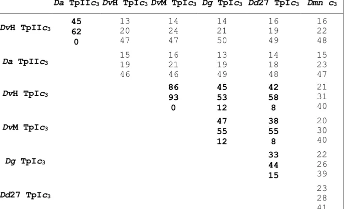
The relative identities and similarities between all the cytochromes obtained from the structure-based alignment (Figure 4) are shown in Table 1. These values support the separation of the Desulfovibrio cytochromes in two types. The classification of the Desulfomicrobium cytochrome c3 is presently not straightforward because it has some features of both types. Nevertheless, it has more similarities to type I cytochromes (Table 1), it also acts as the physiological partner for the Dmn [NiFeSe] hydrogenase, and can mediate the reduction of other multi-heme cytochromes by this hydrogenase.[32] This suggests that the Dmn cytochrome c3 is probably a type I cytochrome c3 more distantly related than the Desulfovibrio TpI-c3s because it originates from a bacterium of a different genus.
In order to better visualise the TpII-c3s features, the surface electrostatic potentials of the fully oxidised DvH TpII-c3, Da TpII-c3 and, as a reference, DvH TpI-c3 were calculated using GRASP[46] (Figure 6). Overall, the DvH TpII-c3 is not as acidic as the Da one, as would be expected since it has 13 acidic residues (D and E) and 14 basic ones (K and R), compared to 19 acidic and 6 basic for Da TpII-c3. However, the similarity between the two TpII-c3s and the contrast with DvH TpI-c3 is readily apparent. Looking at the view from the edge of heme 1 (Figure 6A), it is clear that this heme is indeed
16
surrounded by an acidic surface, more pronounced in the Da TpII-c3[41] than in the DvH one, but which is absent in DvH TpI-c3. Looking at the structures it is obvious that the N-terminal residues covering heme 1 in DvH TpI-c3 are not present in the two TpII-c3s. These features support the suggestion that heme 1 of TpII-c3s plays a role in intermolecular electron transfer,[41] contrary to other cytochromes c3 where this heme is surrounded by a neutral environment, and is protected against solvent by the N-terminus.
The view from the edge of heme 4 (Figure 6B) shows that the basic region that can be found close to this heme in DvH TpI-c3 due to the lysine patch is reduced in DvH TpII-c3 and even more so in Da TpII-c3.[41] This lysine patch, also present in other Desulfovibrio type I cytochromes c3, was proposed to be the site of interaction and electron exchange with a negatively charged region of the redox partner hydrogenase.[45,47,48] These differences in surface charge distribution indicate that TpII-c3s may exchange electrons via a different heme (namely heme 1) from that used by TpI-c3s (probably heme 4).
The structure of the nine-heme cytochrome c (9Hcc) from Dd27k was also analysed to investigate whether each of its cytochrome c3-like domains had any resemblances to type I or type II c3s. Interestingly, several of the TpIIc3s structural features are observed in both 9Hcc domains, although differences are also present not least because 9Hcc contains
17
one extra heme. The main similarities between the 9Hcc cytochrome c3-like domains and the TpII-c3s are: i) all heme binding sites of 9Hcc are of the form CXXCH; ii) the first heme of 9Hcc N-terminal domain is also significantly exposed and surrounded by a negative surface area;[49] iii) in both 9Hcc domains the loop after the second heme-binding site is shifted away from the fourth heme towards the second heme; iv) in the 9Hcc C-terminal domain the loop before the third heme-binding site, which interrupts two -helices in TpI-c3s, is also absent; v) The fourth heme of the 9Hcc C-terminal domain is not surrounded by a positive surface region. This suggests that, overall, 9Hcc has the same fundamental characteristics of TpIIc3s, namely in the N-terminal domain an exposed heme 1 surrounded by an acidic surface region, which is probably the heme involved in intermolecular electron transfer, and in the C-terminal domain a heme 4 (9Hcc heme 9) which lacks the TpI-c3 characteristic positive surface patch. These similarities may indicate that TpII-c3s and 9Hcc have analogous reaction modes and/or physiological partners, which is supported by the evidences that both are part of membrane-bound redox complexes[30] (see below). A similar conclusion may be expected for the 16Hcc since its last two domains have a strong similarity to 9Hcc.[8]
Analysis of the partial sequence obtained from the DvH genome shows that upstream of the gene coding for TpII-c3 an open reading frame is present coding for a protein which has a
18
high similarity (33% identity, 53% similarity) to an FeS protein belonging to the DvH hmc operon, HmcF, and which is predicted to contain two [4Fe-4S]2+/1+ centres[29,50] (Figure 7). The DvH hmc operon codes for a transmembrane redox complex that is proposed to transfer electrons from the periplasm to the cytoplasm.[29] Although HmcF has no transmembrane sequences, it was shown to be present in the membrane fraction of DvH cells.[50] Genes coding for proteins similar to the FeS protein and HmcF can be found in the genomes of several bacteria and they are usually associated with membrane-bound oxido-reductase complexes. The genome of the sulfate-reducing archaeon Archaeoglobus fulgidus contains at least nine genes coding for similar proteins.[51]
The genes coding for TpII-c3 and FeS protein in DvH are located in tandem with an intervening gap of only 25 nucleotides, and the absence of promoter sequences suggests that the two proteins belong to the same operon. The unavailability of nucleotide sequence data upstream of the gene coding for the FeS protein impedes the search for other putative genes and/or promoter sequences. Using BLASTX, no significant sequence similarity to known genes could be found downstream of the DvH TpII-c3 gene and the search for typical bacterial terminator sequences was also unsuccessful. Therefore, it is not possible to exclude the hypothesis that other genes may be part of this operon. Remarkably, a similar situation is observed in Da, where 15 nucleotides upstream of
19
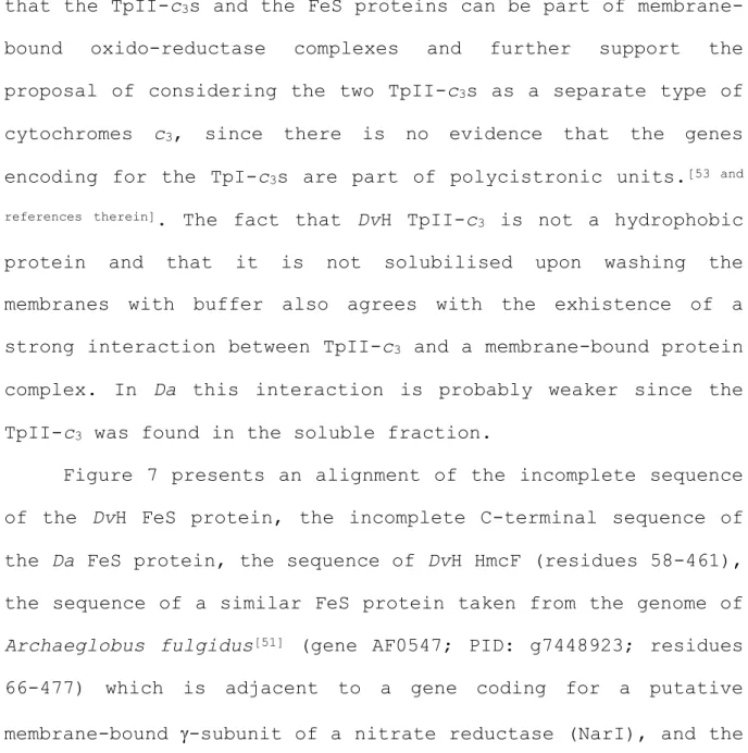
the region encoding Da TpII-c3[52] an incomplete open reading frame also encodes an FeS protein that shares 67% identity and 79% similarity in the C-terminal sequence available (174 amino-acids) to the corresponding sequence of the FeS protein found in the DvH genome (Figure 7). In Da the close proximity between the genes for FeS and TpII-c3 also suggests that they are part of the same operon, and questions the proposal that TpII-c3 is encoded by a monocistronic gene.[34] These observations suggest that the TpII-c3s and the FeS proteins can be part of membrane-bound oxido-reductase complexes and further support the proposal of considering the two TpII-c3s as a separate type of cytochromes c3, since there is no evidence that the genes encoding for the TpI-c3s are part of polycistronic units.[53 and references therein]. The fact that DvH TpII-c3 is not a hydrophobic protein and that it is not solubilised upon washing the membranes with buffer also agrees with the exhistence of a strong interaction between TpII-c3 and a membrane-bound protein complex. In Da this interaction is probably weaker since the TpII-c3 was found in the soluble fraction.
Figure 7 presents an alignment of the incomplete sequence of the DvH FeS protein, the incomplete C-terminal sequence of the Da FeS protein, the sequence of DvH HmcF (residues 58-461), the sequence of a similar FeS protein taken from the genome of Archaeglobus fulgidus[51] (gene AF0547; PID: g7448923; residues 66-477) which is adjacent to a gene coding for a putative membrane-bound -subunit of a nitrate reductase (NarI), and the
20
sequence of a similar FeS protein taken from the genome of Aquifex aeolicus[54] (gene AF0543, PID: g7448926; residues 32-434) which codes for a putative subunit of a heterodisulfide reductase (HrdD). These proteins contain two [4Fe4S]2+/1+ centres and share a considerable similarity indicating they are homologous proteins (see Figure 7). Although the sequence retrieved from the DvH genome data still lacks the N-terminal part of this FeS protein, the last two Cys of the first [4Fe-4S]2+/1+ centre and the cysteine binding motif of the second iron-sulfur cluster are conserved. A cysteine motif formed by CX34CCGX39-41CX2C is also conserved in these proteins, as was observed for other similar FeS proteins from several organisms.[55] All of these FeS proteins are membrane-associated although they all lack obvious transmembrane helices. It was proposed that the membrane binding domain of these FeS proteins could be formed by the hydrophobic face of several amphipatic
-helices,[55] as observed for several other monotopic membrane-proteins (e.g.[56]). The predicted secondary structure of the DvH FeS protein, as well as that of the other proteins aligned in Figure 7, show they also contain several conserved amphipatic -helices in the region of the cysteine motif which could be involved in a similar membrane attachment.
Reduction by DvH hydrogenases. The difference in molecular characteristics between the DvH type I and type II cytochromes c3 suggest they will have different redox partners and physiological roles. The TpI-c3 is generally considered to be
21
the partner for the enzyme hydrogenase, and was shown to mediate the reduction of some other cytochromes by hydrogenases, namely DvH 16Hcc,[31] DvH cytochrome c553,[31] Dd27k 9Hcc,[8] and the Dmn octaheme cytochrome c3 (Mr 26,000).[32] However, for Desulfovibrio desulfuricans Essex 6, it was recently reported that cytochrome c3 does not affect the reduction rate of the 9Hcc by the [NiFe] hydrogenase.[58] In DvH three different hydrogenases were identified, two of which are membrane-bound (the [NiFe][19] and [NiFeSe] hydrogenases[20]). Thus, it was investigated whether the TpII-c3 can interact specifically with one of them. For each DvH hydrogenase a comparison between the rates of the two cytochromes was obtained, and the effect of catalytic amounts of TpI-c3 on the reduction of the TpII-c3 was also observed. A concentration of TpII-c3 lower than the Km was used to ensure that the enzyme is not saturated so that a catalytic effect of TpI-c3 can be observed.
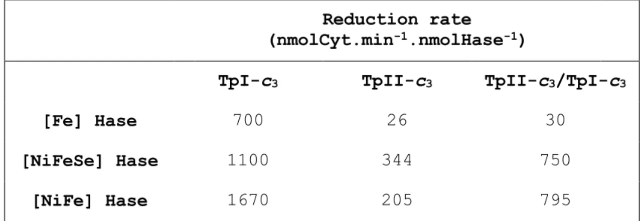
Reduction of the TpII-c3 by the DvH periplasmic [Fe] hydrogenase proceeds at a rate about 30 times slower (26 nmolCyt.min-1.nmolHase-1) than that of the TpI-c3 in the same conditions (700 nmolCyt.min-1.nmolHase-1) (Table 2). When the reduction of the TpII-c3 is performed in the presence of catalytic amounts of TpI-c3 (equimolar with the hydrogenase) the rate is only marginally increased (30 nmolCyt.min -1.nmolHase-1). The behaviour of the TpII-c3 with the [Fe] hydrogenase is very similar to that observed with the monoheme
22
cytochrome c553.[31] The lack of catalytic effect of the TpI-c3 may indicate that TpII-c3 will preferentially interact directly with the hydrogenase. In the case of Da no reduction of TpII-c3 by the Desulfovibrio desulfuricans ATCC 7757 [Fe] hydrogenase was observed, even in the presence of TpI-c3, although this last cytochrome was efficiently reduced.[34] However, since the proteins used belong to different organisms no definite conclusion can be drawn from this result, and it should be pointed out that no [Fe] hydrogenase was ever detected in Da. The absolute reduction rates observed for the DvH [Fe] hydrogenase can not be directly compared to those obtained with the other two hydrogenases because the experimental procedures used in each case were different,[31] and this will affect the activation state of the hydrogenases.
With the membrane-bound [NiFeSe] hydrogenase the reduction of the TpII-c3 is faster (344 nmolCyt.min-1.nmolHase-1), being only three-fold slower than that of the TpI-c3 (1100 nmolCyt.min-1.nmolHase-1). A catalytic amount of TpI-c3 increased the rate of reduction of the TpII-c3 two-fold (750 nmolCyt.min -1.nmolHase-1), which indicates that the electron transfer is more efficient via the TpI-c3, which may be the physiologically preferred electron acceptor of hydrogenase. In the case of reduction of the Da cytochromes c3 by the Da [NiFeSe] hydrogenase, a much larger difference between the TpII-c3 and TpI-c3 was observed, since the latter is reduced with a rate about 60-fold faster than the former.[33] When both cytochromes
23
were present, the rate of reduction was close to that of the TpI-c3, indicating also that this is an efficient intermediary electron carrier between the hydrogenase and the TpII-c3.
The membrane-bound [NiFe] hydrogenase reduces the TpII-c3 with a rate (205 nmolCyt.min-1.nmolHase-1) about 8-fold slower than that of the TpI-c3 (1670 nmolCyt.min-1.nmolHase-1). As with the [NiFeSe] hydrogenase, a catalytic amount of TpI-c3 increased the rate of reduction of the TpII-c3 (795 nmolCyt.min-1.nmolHase-1), indicating that the electron transfer is more efficient when it is carried out via the TpI-c3. Again, in Da the TpI-c3 is reduced 90 times faster by the Dg [NiFe] hydrogenase than the TpII-c3, and the presence of small amounts of the TpI-c3 increases this rate of reduction 5-fold.[34]
Altogether, these results show that the DvH TpII-c3 is less efficient than the TpI-c3 as an electron acceptor for all the three DvH hydrogenases. However, its behaviour is different with each hydrogenase. The [NiFeSe] hydrogenase is that for which less difference between the two cytochromes was observed, indicating that TpII-c3 may preferentially interact with this hydrogenase. However, both for this enzyme and the [NiFe] hydrogenase, the reduction of TpII-c3 is faster with TpI-c3 as a mediator. Nevertheless, the two DvH membrane-bound hydrogenases, and in particular the [NiFeSe], can reduce the TpII-c3 with a considerable rate, raising the question of whether, in vivo, this cytochrome is able to receive electrons from these hydrogenases. It should also be pointed out that the
24
physiological interaction between these proteins will take place in a bidimensional lipidic phase which can significantly alter the properties of this interaction. By contrast, for the [Fe] hydrogenase not only the reduction of the TpII-c3 is much slower than that of TpI-c3, but also it is not affected by the presence of the latter. It is interesting to note that overall the DvH TpII-c3 reacts better with the hydrogenases than the Da one.[33,34] This may be related to the fact that the surface region around heme 4 is more basic in the DvH TpII-c3 than in the Da one, which may permit a better interaction with the hydrogenases.
In relation to the catalytic effect of the TpI-c3 on the reduction of the TpII-c3 by the [NiFe] and [NiFeSe] hydrogenases, it should be noted that for the Dd27k 9Hcc, which presents a similar effect,[8] a high probability specific interaction was observed by modelling studies between the negative heme 1 N-terminal region of 9Hcc and the positive heme 4 region of the TpI-c3.[49] Since heme 1 of the Dd27k 9Hcc has similar characteristics to that of the TpII-c3s (solvent exposed and surrounded by a negative surface charge[49]), it is likely that an analogous situation may be present in the interaction between the DvH TpII-c3 and TpI-c3.
CONCLUSIONS
A new tetraheme cytochrome c3 was isolated from the membranes of DvH. This cytochrome is homologous to the TpII-c3
25
from Da, which was the first sulfate-reducing bacteria reported to contain two tetraheme cytochromes c3.[33,34] The TpII-c3 from this organism was isolated from the soluble fraction, whereas the one from DvH was found exclusively in the membrane extract. This membrane association may be an important feature of the TpII-c3, and may constitute one of the reasons why it has not been found in most other sulfate-reducers studied, since few studies of membrane-bound proteins have been carried out in these bacteria.
The DvH and Da TpII-c3s share structural and genetic characteristics that distinguish them from the other cytochromes c3 and suggest they belong to a different type of cytochromes c3, possibly associated with membrane-bound oxido-reductase complexes, and having different redox partners and physiological function. The considerable amount of TpII-c3 isolated from DvH indicates that it plays an important role in its metabolism.
Experimental Section
Bacterial growth: DvH (DSM 644) cells were grown in lactate/sulfate medium as described.[18] The cells (360 g) were suspended in 350 ml Tris-HCl buffer 10 mM pH 7.6, and ruptured by passing twice through a Manton-Gaulin press. The resulting extract was centrifuged at 10,000 g for 15 min to remove cell debris, and the supernatant at 100,000 g for 2h.
26
Preparation of the membrane extract: The pellet (membrane fraction) was washed twice with 10 mM Tris-HCl pH 7.6, 1 mM EDTA, by suspending the membranes in buffer and re-centrifuging, in order to remove any remaining soluble components (desulfoviridin was used as a marker to ascertain the presence of soluble proteins).
The detergent Zwittergent 3-12 (N-dodecyl-N,N-dimethyl-3-ammonio-1-propansulfonate) (SB12) was used to solubilise the membrane components. The membrane pellet obtained after the final washing was resuspended in Tris-HCl buffer 20 mM pH 7.6, and SB12 added to a final concentration of 2% (w/v). The suspension was stirred for 2 h and then centrifuged at 100,000 g for 40 min. Two extraction steps were performed.
Protein Purification: All purification procedures were performed at pH 7.6 and 4ºC. The solubilised membrane extract was loaded on a DEAE Sepharose Fast Flow (Pharmacia) column (5x40 cm), equilibrated with Tris-HCl 20 mM buffer, 0.2 % SB12 (w/v) (Buffer A). The column was washed with 400 ml Buffer A and a linear gradient from 0-40% Buffer B (100 mM Tris-HCl, 0.2% SB12, 1M NaCl) (2.4 l) was applied. After this a second gradient of 40-100% Buffer B (0.75 l) was utilised.
TpII-c3: The cytochrome-containing fraction that eluted from the DEAE column at around 15%B was pooled, concentrated and dialysed against 20 mM Tris-HCl, 0.2% SB12. This fraction was then passed on a Pharmacia Q-Sepharose High Performance column (Hiload 26/10, flow rate 5 ml/min) equilibrated with 20 mM
27
Tris-HCl, 0.2% SB12 buffer. A stepwise gradient of 0-1 M NaCl was performed. The cytochrome-containing fraction that eluted at 150 mM NaCl was pooled, concentrated and loaded on a Pharmacia S-75 gel filtration column equilibrated with 50 mM Tris-HCl, 100 mM NaCl, 0.2% SB12. The cytochrome fraction from this column was pooled, concentrated and dialysed against 20 mM Tris-HCl, 0.2% SB12. This fraction was then passed again on a Pharmacia Q-Sepharose High Performance column (Hiload 16/10, flow rate 3 ml/min) equilibrated with 20 mM Tris-HCl, 0.2%SB12 buffer. A linear gradient was performed (0-200 mM NaCl), and pure TpII-c3 eluted at 120 mM NaCl. The purity index (A551(red)-A570(red)/A280(ox)) was 2.2 and the amount obtained was 7 mg of TpII-c3.
TpI-c3 from the membrane fraction: The cytochrome-containing fraction that eluted from the DEAE column before the start of the gradient was pooled, concentrated and dialysed against 20 mM Tris-HCl, 0.2% SB12. This fraction was then passed on a Pharmacia Q-Sepharose High Performance column (Hiload 26/10, flow rate 5 ml/min) equilibrated with 20 mM Tris-HCl, 0.2% SB12 buffer. A stepwise gradient of 0-1 M NaCl was performed. The fraction that eluted before the start of the gradient was pooled, concentrated and dialysed against 20 mM Tris-HCl. This fraction was then passed again on a Pharmacia S-Sepharose High Performance column (Hiload 16/10, flow rate 3 ml/min) equilibrated with 20 mM Tris-HCl buffer. A stepwise gradient of 0-1 M NaCl was performed, and pure TpI-c3 eluted at 250 mM
28
NaCl. The purity index (A551(red)-A570(red)/A280(ox)) was 3.8 and the amount obtained was 10 mg of TpI-c3. The identity of this cytochrome was confirmed by determination of its N-terminal. Analytical methods: Protein concentration was determined with the Bicinchoninic Acid assay from Pierce, using soluble TpI-c3 as a standard. The pyridine hemochrome was performed according to Berry and Trumpower[59] using the millimolar absorptivity of
r-o,550-535=23.97 mM-1cm-1 for heme c. SDS-polyacrylamide gel electrophoresis was performed according to Laemmli.[60] Gels were stained with Coomasie Blue for proteins, and tetramethylbenzidine for c-type cytochromes.[61] The isoelectric point was determined by analytical isoelectric focusing using a Bio-Rad Model 111 Mini IEF Cell. A pH gradient of 3.5 to 10 was obtained with carrier ampholites. Protein molecular masses were determined by SDS-PAGE using BioRad low-range protein standards. Molecular mass determination by native size-exclusion chromatography was done on a Pharmacia Superdex 75 HR column, calibrated with Pharmacia low molecular mass calibration kits. For N-terminal sequencing, the protein was adsorbed to a PVDF membrane (ProSorb, Perkin–Elmer) and washed repeatedly to remove salts and detergent. The N-terminal sequence was obtained by the method of Edman and Begg[62] using an Applied Biosystem model 470A sequenator.
Spectroscopic methods: UV-visible spectra were obtained using a Shimadzu (UV 260) spectrophotometer. EPR spectra were recorded using a Brucker ESP 380 spectrometer equipped with an ESR 900
29
continuous-flow helium cryostat from Oxford Instruments, as described earlier.[63] Redox titrations monitored by Visible spectroscopy were performed in an anaerobic chamber in 40 mM Tris-Maleate pH 7.6 buffer, following the changes in absorbance at the band of the reduced hemes, corrected for the corresponding isosbestic points and using buffered sodium dithionite as the reductant and potassium ferricianide as oxidant. The following redox mediators were used (at a final concentration of 3.5 M each): Gallocyanine; Indigo tetrasulfonate; Indigo trisulfonate; Indigo disulfonate; 2-Hydroxy-1,4-naphtoquinone; Antraquinone-2,7-disulfonate; Antraquinone-2-sulfonate; Safranine; Neutral red; Benzyl viologen; Methyl viologen. The reduction potentials are referenced to the standard hydrogen electrode.
Enzymatic activities: Reduction of cytochromes with hydrogenases - All experiments were performed with a Shimadzu UV3100 spectrophotometer, in a stirred cell, with a hydrogen overpressure of 15 kPa flowing through the cell. The buffer used in all cases was 100 mM Tris-HCl pH 7.6. The [NiFe] and [NiFeSe] hydrogenases were activated by flushing the H2ase with hydrogen for about 1h, and then leaving them overnight at 4ºC under a hydrogen atmosphere. The [Fe] hydrogenase was pre-activated by standing 1h inside an anaerobic chamber. The reduction of the cytochromes was measured by following the increase in absorption at 551 nm for TpII-c3, and 553 nm for TpI-c3, using the respective absorption coefficients. The rates
30
were measured from the linear portion of the reduction curves. The concentrations used were chosen so that a reasonable rate could be measured, having assured that the rates were proportional to the hydrogenase concentrations. Each of the experiments, performed as described,[31] was repeated at least three times. The same procedure was used for the [NiFe] and [NiFeSe] hydrogenases, with the following concentrations: TpII-c3/H2ase - 4 M TpII-c3 and 14 nM H2ase; TpI-c3/H2ase - 4 M TpI-c3 and 14 nM H2ase; TpII-TpI-c3/H2ase/TpI-TpI-c3 - 4 M TpII-c3, 14 nM H2ase and 14 nM TpI-c3. The concentrations used in the experiments with the Fe hydrogenase were: TpII-c3/H2ase - 4 M TpII-c3 and 28 nM H2ase; TpI-c3/H2ase - 4 M TpI-c3 and 2.8 nM H2ase; TpII-c3/H2ase/TpI-c3 - 4 M TpII-c3, 28 nM H2ase and 28 nM TpI-c3. At the end of each experiment the cytochromes were reduced with dithionite to check that reduction by hydrogenase had been complete.
Sequence analysis tools and molecular modelling: Preliminary sequence data from the DvH genome was obtained from The Institute for Genomic Research website at http://www.tigr.org. The nucleotide sequence data were analysed using the Genetics Computer Group (Wisconsin) package provided by the Portuguese EMBnet Node (PEN) and Neural Networks for Eukaryotic Promoter Prediction.[64] A three-dimensional model of DvH TpII-c3 was generated using Swiss-Model,[43] with the structures of the oxidised and reduced Da TpII-c3[41] (PDB accession numbers 3CAOA
31
and 3CARA, respectively) as templates. The structures of the several cytochromes c3 were retrieved from the Protein Data Bank, with the following accession numbers: DvH TpI-c3 - 2CTH;[24] DvM - 2CDV;[22] Dg - 1WAD;[26] Dd27k - 3CYR;[24] Dmn - 2CY3.[25] The cytochromes were structurally aligned using the program MODELLER 4.[44] The surface electrostatic potential of the cytochromes was calculated using GRASP.[46] Figure 6 was prepared using GRASP[46] and Raster 3D.[57] Sequence alignments not based on structure were performed using ClustalW version 1.8.[65]
Acknowledgements
We would like to thank Mr. João Carita and the staff of the IBET Fermentation Plant for growing the bacterial cells, and Mrs. M. Regalla for N-terminal sequence determinations. We would also like to thank Dr. Claudio Gomes and Dr. Claudio Soares for help with the surface charge mapping and structural alignment, and Dr. Ricardo Louro for help with the anaerobic redox titration. This work was supported by FCT grants POCTI 36562/ESP/2000 to ICP and POCTI 35021/ BME/2000 to AVX.
32 REFERENCES
[1] I.A.C. Pereira, M. Teixeira, A.V. Xavier Structure and Bonding 1998, 91, 65.
[2] R.O. Louro, T. Catarino, C.A. Salgueiro, J. LeGall, D.L. Turner, A.V. Xavier, In Biological electron transfer chains: Genetics, composition and mode of operation. (Canters, G.W. and Vijgenboom, E., Eds) NATO ASI series vol.512, Kluwer Academic Publishers, 1998, p.209 .
[3] M. Bruschi, E.C. Hatchikian, L. Golovleva, J. LeGall, J. Bacteriol 1977, 129, 30.
[4] E. Samain, G. Albagnac, J. LeGall, FEBS Lett. 1986, 204, 247.
[5] E.C. Hatchikian, P. Papavassiliou, P. Bianco, J. Haladjian, J Bacteriol. 1984, 159 (3), 1040.
[6] P.M. Matias, J. Morais, A.V. Coelho, R. Meijers, A. Gonzalez, A.W. Thompson, L. Sieker, J. LeGall, M.A. Carrondo, J Biol Inorg Chem., 1997, 2, 507.
[7] C. Frazão, L. Sieker, G. Sheldrick, V. Lamzin, J. LeGall, M.A. Carrondo, J Biol Inorg Chem., 1999, 4 (2), 162.
[8] P.M. Matias, R. Coelho, I.A.C. Pereira, A.V. Coelho, A.W. Thompson, L.C. Sieker, J. LeGall, M.A. Carrondo, Structure, 1999, 7 (2) 119.
[9] I.A.C. Pereira, J. LeGall, A.V. Xavier, M. Teixeira, J. Biol. Inorg. Chem., 1997, 2, 23.
33
[10] G. Fauque, M. Bruschi, J. LeGall, Biochem. Biophys. Res. Comm., 1979, 86, 1020.
[11] R.O. Louro, T. Catarino, C.A. Salgueiro, J. LeGall, A.V. Xavier, J. Biol. Inorg. Chem., 1996, 1, 34.
[12] R.O. Louro, T. Catarino, J. LeGall, A.V. Xavier, J. Biol. Inorg. Chem, 1997, 2, 488.
[13] L. Brennan, D.L. Turner, A.C. Messias, M.L. Teodoro, J. LeGall, H. Santos, A.V. Xavier, J Mol Biol., 2000, 298 (1), 61.
[14] J.M. Odom, H.D. Peck Jr., FEMS Microbiol. Lett., 1981, 12, 47.
[15] H. Cypionka, in Sulfate Reducing Bacteria, Barton, L.L. (Ed.), Plenum Press New York, 1995, pp 151-184.
[16] A. Brandis, R.K. Thauer, J. Gen. Microbiol., 1981, 126, 249.
[17] G. Voordouw, V. Niviere, F.G. Ferris, P.M. Fedorak, D.W.S. Westlake, Appl. Environ. Microbiol., 1990, 56, 3748.
[18] B. H. Huynh, M. H. Czechowski, H.-J. Krüger, D.V. DerVartanian, H. D. Peck, J. LeGall, Proc. Nat. Acad. Sci. USA, 1984, 81, 3728.
[19] C. V. Romão, I. A. C. Pereira, A. V. Xavier, J. LeGall, M. Teixeira, Biochem. Biophys. Res. Commun., 1997, 240, 75. [20] F. M. A. Valente, N. Gnadt, J. LeGall, A. V. Xavier, M.
34
[21] R. Haser, M. Pierrot, M. Frey, F. Payan, J.-P. Astier, M. Bruschi, J. LeGall, Nature, 1979, 282, 806.
[22] Y. Higuschi, M. Kusunoki, Y. Matsuura, N. Yasuoka, M. Kakudo, J. Mol. Biol., 1984, 172, 109.
[23] Y. Morimoto, T. Tani, H. Okumura, Y. Higuschi, N. Yasuoka, J. Biochem. (Tokyo), 1991, 110, 532.
[24] P. Simões, P.M. Matias, J. Morais, K. Wilson, Z. Dauter, M.A. Carrondo, Inorganica Chimica Acta, 1998, 273, 213. [25] M. Czjzek, F. Payan, F. Guerlesquin, M. Bruschi, R. Haser,
J. Mol. Biol., 1994, 243, 653.
[26] P.M. Matias, J. Morais, R. Coelho, M.A. Carrondo, K. Wilson, Z. Dauter, L. Sieker, Protein Science, 1996, 5, 1342.
[27] M. Czjzek, F. Guerlesquin, M.Bruschi, R. Haser, Structure, 1996 4, 395.
[28] W. Brent, W.B.R. Pollock, M. Loutfi, M. Bruschi, B.J. Rapp-Giles, J.D. Wall, G. Voordouw, J. Bacteriol., 1991, 173, 220.
[29] M. Rossi, W.B.R. Pollock, M.W. Reij, R.G. Keon, R. Fu, G. Voordouw, J. Bacteriol., 1993, 175, 4699.
[30] L.M. Saraiva, P.N. da Costa, J. LeGall, Biochem Biophys Res Commun., 1999, 262 (3), 629.
[31] I.A.C. Pereira, C.V. Romão, A.V. Xavier, J. LeGall, M. Teixeira, J. Biol. Inorg. Chem., 1998, 3, 494.
35
[32] C. Aubert, M.Brugna, A. Dolla, M.Bruschi, M.-T. Giudici-Orticoni, Biochim. Biophys. Acta, 2000, 1476, 85.
[33] L. Pieulle, J. Haladjian, J. Bonicel, E.C. Hatchikian, Biochim. Biophys. Acta, 1996, 1273, 51.
[34] V. Magro, L. Pieulle, N. Forget, B. Guigliarelli, Y. Petillot, E.C. Hatchikian, Biochim. Biophys. Acta, 1997, 1342, 149.
[35] J. LeGall, G.Mazza, N. Dragoni, Biochem. Biophys. Acta, 1965, 99, 385.
[36] I.A.C. Pereira, J. LeGall, A.V. Xavier, M. Teixeira, Biochem. Biophys Acta, 2000, 1481, 119.
[37] M.D. Roldán, H.J. Sears, M.R. Cheesman, S.J. Ferguson, A.J. Thomson, B.C. Berks, D.J. Richardson, J. Biol. Chem., 1998, 273, 28785.
[38] J. Simon, R. Gross, O. Einsle, P.M.H. Kroneck, A. Kröger, O. Klimmek, Mol. Microbiol., 2000, 35 (3), 686.
[39] A. T’sai, G. Palmer, Biochim. Biophys. Acta, 1982, 681, 484.
[40] F.A. Walker, B.H. Huynh, W.R. Scheidt, S.R. Osvath, J. Am. Chem. Soc., 1986, 108, 5288.
[41] S. Nørager, P. Legrand, L. Pieulle, C. Hatchikian, M. Roth, J. Mol. Biol., 1999, 290, 881.
[42] D.V. Dervartanian, A.V. Xavier, J. LeGall, Biochimie, 1978, 60, 321.
36
[44] A. Sali, T. L. Blundell, J.Mol.Biol., 1993, 234, 779.
[45] D.E. Stewart, J. LeGall, I. Moura, J.J.G. Moura, H.D. Peck, A.V. Xavier, P.K. Weiner, J.E. Wampler, Biochemistry, 1988, 27, 2444.
[46] A. Nicholls, GRASP: Graphical representation and analysis of surface properties. Columbia University, New York, 1992. [47] F. Guerlesquin, A. Dolla, M. Bruschi, Biochimie 1994, 76,
515.
[48] P.M. Matias, C.M. Soares, L.M. Saraiva, R. Coelho, J. Morais, J.LeGall, M.A. Carrondo, J.Biol.Inorg.Chem., 2001, 6 (1) 63.
[49] P.M. Matias, L.M. Saraiva, C.M. Soares, A.V. Coelho, J. LeGall, M.A. Carrondo, J. Biol. Inorg. Chem., 1999, 4, 478. [50] R.G. Keon, G. Voordouw, Anaerobe, 1996, 2, 231.
[51] H.P. Klenk, et al. Nature, 1997, 390 (6658), 364.
[52] V.Magro, L.Pieulle, N. Forget, B. Guigliarelli, Y. Petillot, E.C. Hatchikian, EMBL:DACYTC3A, AC Y09718, GI:1770203.
[53] P.N. da Costa, P. Marujo, W.M.A. van Dongen, C.Arraiano, L. M. Saraiva, Bioch.Biophys. Acta, 2000, 1492, 271.
[54] G. Deckert, P.V. Warren, T. Gaasterland, W.G. Young, A.L. Lenox, D.E. Graham, R. Overbeek, M.A. Snead, M. Keller, M. Aujay, R. Huber, R.A. Feldman, J.M. Short, G.J. Olson, R.V. Swanson, Nature, 1998, 392 (6674), 353.
37
[55] R.S. Lemos, C.M. Gomes, M. Teixeira, Biochem. Biophys. Res. Comm., 2001, 281 (1), 141.
[56] A.G. Spencer, E. Thuresson, J.C. Otto, I. Song, T. Smith, D.L. DeWitt, R.M. Garavito, W.L. Smith, J Biol Chem, 1999, 274 (46), 32936.
[57] E. A. Merritt, D. J. Bacon, Methods in Enzymology, 1997, 277, 505.
[58] G.Fritz, D. Griesshaber, O. Seth, P.M.H. Kroneck, Biochemistry, 2001, 40 (5) 1317.
[59] E.A. Berry, B.L. Trumpower, Anal. Biochem., 1987, 161, 1. [60] U.K. Laemmli, Nature, 1970, 227, 680.
[61] C.F. Goodhew, K.R.Brown, G.W. Pettigrew, Biochem. Biophys. Acta, 1986, 852, 288.
[62] P. Edman, G. Begg, Eur. J. Biochem., 1976, 1, 80.
[63] M. Teixeira, A.P. Campos, A.P. Aguiar, H.S. Costa, H. Santos, D.L. Turner, A.V. Xavier, FEBS Lett., 1993, 317, 233.
[64] M.G. Reese, N.L. Harris, F.H. Eeckman, in Large Scale Sequencing Specific Neural Networks for Promoter and Splice Site Recognition. Hunter, L. and Klein, T.E.( Eds) World Scientific Publishing Co, Singapore, 1996.
[65] J.D. Thompson, D.G. Higgins, T.J. Gibson, Nucleic Acids Research, 1994, 22, 4673.
38
Table 1: Percentage of identity (first line), similarity (second line) and residues aligned with gap characters (third line) between the several cytochromes c3, obtained from the structural alignment of Figure 4. The values for the TpII-c3s and for the TpI-c3s are in bold.
Da TpIIc3 DvH TpIc3 DvM TpIc3 Dg TpIc3 Dd27 TpIc3 Dmn c3
DvH TpIIc3 45 62 0 13 20 47 14 24 47 14 21 50 16 19 49 16 22 48 Da TpIIc3 15 19 46 16 21 46 13 19 49 14 18 48 15 23 47 DvH TpIc3 86 93 0 45 53 12 42 58 8 21 31 40 DvM TpIc3 47 55 12 38 55 8 20 30 40 Dg TpIc3 33 44 15 22 26 39 Dd27 TpIc3 23 28 41
39
Table 2: Rates of reduction of DvH TpII-c3 and TpI-c3 with the three DvH hydrogenases (in nmolCyt.min-1.nmolHase-1).
Reduction rate
(nmolCyt.min-1.nmolHase-1)
TpI-c3 TpII-c3 TpII-c3/TpI-c3
[Fe] Hase 700 26 30
[NiFeSe] Hase 1100 344 750
40 Figure legends
Figure 1: UV-Visible spectra of the DvH TpII-c3 oxidised (full line), and reduced with dithionite (dashed line).
Figure 2: Trace a: EPR spectrum of oxidised DvH TpII-c3. Temperature: 14K; Modulation amplitude: 0.9 mT; Microwave power: 0.75 mW; Microwave frequency: 9.63 GHz. Traces b-e: Simulation of spectrum a obtained by adding three theoretical spectra with g-values of: gz=2.98, gy=2.24 and gz=1.50 (2 hemes; trace c), gz=2.98, gy=2.24 and gz=1.40 (1 heme; trace d) and gz=3.5 and gy=2.0 (1 heme; trace e).
Figure 3: Redox titration of DvH TpII-c3 obtained by following absorption changes at 552 nm (pH 7.6; reduction, oxidation). The experimental points are from three different experiments. The full line corresponds to a theoretical simulation obtained by assuming reduction of four hemes with redox potentials of –170 mV, -235 mV, -260 mV and –325 mV.
Figure 4: A- Sequence alignment of the TpII-c3s from DvH and Da: Identical residues are coloured black, and similar residues are coloured grey.
B- Structure-based alignment of the sequences of DvH TpII-c3, Da TpII-c3, DvH TpI-c3, DvM TpI-c3, Dg TpI-c3, Dd27k TpI-c3 and Dmn cytochrome c3 obtained using MODELLER 4.[44] Fully conserved residues are coloured black, acidic residues are coloured red and basic residues coloured blue. The boxed His (ligand to heme 3) does not align in the TpII-c3s with the corresponding residue in the other cytochromes because their C is quite distant, although the side-chain ring is actually very close to that of the other cytochromes c3. The numbering in both alignments corresponds to the sequence of DvH TpII-c3.
41
Figure 5: Superposition of the DvH TpII-c3 model (pink) and DvH TpI-c3 structure (blue) obtained by superimposing the heme groups (only one set of heme groups is shown: Heme 1 – green; Heme 2 – blue; Heme 3 – yellow; Heme 4 – orange). The N-terminus of the TpI-c3 and C-N-terminus of the TpII-c3 are identified by N and C. The grey arrows point to the features highlighted in each view: A – shorter N-terminal of the TpII-c3; B – the two loops covering heme 2 in the TpII-TpII-c3; C – Loop missing in TpII-c3; D – Shorter C-terminal region of TpII-c3.
Figure 6: Representation of the surface electrostatic potential of the fully oxidised DvH TpII-c3, Da TpII-c3 and DvH TpI-c3. Red coloured zones correspond to negative potentials while blue coloured zones correspond to positive potentials. The range of potentials spans from -20 to 20 kT/e. The structures of Da TpII-c3 and DvH TpI-c3 and the model of DvH TpII-c3 are shown below in the same orientation as the surface representations. A: View from the edge of heme 1 (coloured green); B: View from the edge of heme 4 (coloured red). The figures were prepared using GRASP[46] and Raster 3D.[57]
Figure 7: Alignment of the incomplete sequence of the DvH FeS protein, the incomplete C-terminal sequence of the Da FeS protein, the sequence of DvH HmcF (residues 58-461), the sequence of a similar FeS protein from Archaeglobus fulgidus[47] (gene AF0547; PID: g7448923; residues 66-477), and the sequence of a similar FeS protein from Aquifex aeolicus[49] (gene AF0543, PID: g7448926; residues 32-434). The conserved amphipatic -helices potentially involved in membrane attachment are represented over the sequences. %I = % Identity; %S = % Similarity; *- the values of identity and similarity for Da FeS protein refer to the alignment of the 174 aminoacids available for the sequence of this protein. The Cys binding the two
[4Fe-42
4S]2+/1+ centres are marked by and those belonging to the cysteine motif by .
43
A novel tetraheme cytochrome c3 isolated from the membranes of the sulfate reducer Desulfovibrio vulgaris Hildenborough provides evidence for the separation of cytochromes c3 in two types. Type II cytochromes c3 are most likely associated with membrane-bound respiratory redox complexes.