115 115 115 115 115 Mem Inst Oswaldo Cruz, Rio de Janeiro, Vol. 92, Suppl. II: 115-123, 1997
Signal Transduction and Activation of the NADPH Oxidase
in Eosinophils
Mark A Lindsay
+, Mark A Giembycz
Thoracic Medicine, Imperial College School of Medicine, National Heart and Lung Institute, Dovehouse Street, London SW3 6LY, UK
Activation of the eosinophil NADPH oxidase and the subsequent release of toxic oxygen radicals has been implicated in the mechanism of parasite killing and inflammation. At present, little is known of the signal transduction pathway that govern agonist-induced activation of the respiratory burst and is the subject of this review. In particular, we focus on the ability of leukotrine B4 to activate the NADPH oxidase in guinea-pig peritoneal eosinophils which can be obtained in sufficient number and purity for detailed biochemical experiments to be performed.
Key words: leukotriene B4 - eosinophil - NADPH oxidase - signal transduction
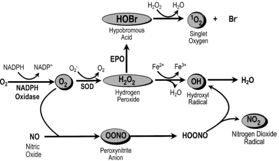
The NADPH oxidase (E.C. 1.23.45.3) cataly-ses the single electron reduction of molecular O2 to superoxide (O2- ), a powerful oxidising and re-ducing agent (Fig. 1) (Babior et al. 1973). In the presence of superoxide dismutase, O2- dismutates to hydrogen peroxide (H2O2) which can be subse-quently converted into hypobromous acid in the presence of eosinophil peroxidase (a highly basic protein stored within specific eosinophil granules) and bromide (Weiss et al. 1986) (Fig. 1). Alterna-tively, in the presence of ferrous ions, O2- and H2O2 interact to form the membrane-perturbing hydroxyl radical (OH.), one of the most unstable oxidising species known (Fig. 1). Other pathways of free radical formation have also been described includ-ing the reaction of O2- with nitric oxide to form peroxynitrite which provides an additional, iron-independent route of OH. formation together with nitrogen dioxide radicals (Fig. 1). Hypobromous acid is able to interact with H2O2 to form singlet oxygen, the biological significance of which is currently unclear (Fig. 1). Activation of the NADPH oxidase and the subsequent production of toxic oxygen radicals is thought to be important to the role of eosinophils during host defence (Butterworth & Thorne 1993). However, it is now appreciated that NADPH oxidase activation maybe cytotoxic to many mammalian cells, particular those of the gut, skin and lung, a finding that has implicated eosinophils in the pathogenesis of a number of non-parasitic inflammatory disorders, including Crohn’s disease, atopic dermatitis and
+Corresponding author. Fax: +44-171-351. 5675. E-mail:
[email protected] Received 3 September 1997 Accepted 30 September 1997
allergic asthma (Butterfield & Leiferman 1993). Indeed, the activity of the NADPH oxidase is sig-nificantly higher in eosinophils that in other ph-agocytes (Yamashita et al. 1985, Petreccia et al. 1987, Sedgwick et al. 1988, Yagisawa et al. 1996). At present, little is known of the intracellular mechanisms responsible for NADPH oxidase ac-tivation in eosinophils. This is in contrast to neu-trophils, where studies of the mechanism of O2 -release by the chemotactic peptide, formyl-methyl-leucyl-phenylalanine (fMLP) have suggested the participation of phospholipase A2- (PLA2), phos-pholipase C- (PLC), phosphos-pholipase D- (PLD) pro-tein kinase C- (PKC), phosphatidylinositol 3-kinnase- (PI-3K) and tyrosine kinase-dependent pathways (possibly those leading to mitogen acti-vated protein kinase stimulation) (Bokoch 1995). This lack of knowledge relates primarily to the dif-ficulty in obtaining sufficient numbers of cells, par-ticular human eosinophils. Thus, we and others have overcome this problem by using guinea-pig eosinophils as a model system, which can be har-vested from the peritoneum in sufficient numbers for detailed biochemical studies.
116 116 116 116
116 Activation of the NADPH Oxidase in Eosinophils • MA Lindsay, MA Giembycz
with sub-threshold concentrations of PAF has been demonstrated to prime the subsequent NADPH oxidase response to opsonized particles (Tool et al. 1992) and fMLP (Zoratti et al. 1992). More re-cent studies have demonstrated a similar priming in human eosinophils adherent to tissue culture plates coated with a range of extracellular matrix proteins (e.g. fibronectin, fibrinogen, collagen, laminin) and fetal calf serum. Under these condi-tions, the cytokines tumor necrosis factor-α
(TNF-α), granulocyte macrophage-colony stimulating factor (GM-CSF), which are unable to stimulate the NADPH oxidase in ‘non-adherent’ cells, pro-duce a slowly developing and sustained genera-tion of O2- (Dri et al. 1991, Horie & Kita 1994). However, since there are no studies concerning the biochemical mechanism of NADPH oxidase acti-vation in adherent eosinophils, this review will focus predominately upon those studies on ‘non-adherent’ cells. In particular, we will concentrated upon recent studies of the mechanism of LTB4 -induced NADPH oxidase activation in guinea-pig eosinophils (Perkins et al. 1995, Lindsay et al. 1995a, b).
STRUCTURE AND ASSEMBLY OF THE NADPH OXI-DASE
In neutrophils, an active NADPH oxidase com-plex assembles at the phagocytic and plasma mem-branes following activation (Segal & Abo 1993) (Fig. 2). At least five proteins are required for the formation of an active oxidase complex: the mem-brane-bound cytochrome b558 (consisting of two subunits, gp91phox and p22phox ) and the cytosolic proteins, p47phox, p67phox and a small
GTP-bind-ing protein, Rac-1 or Rac2 (Casimer & Teahan 1994, Bokoch 1994). Recently, two additional com-ponents have been identified, these being the cy-tosolic protein, p40phox, that appears to be associ-ated with p67phox (Wientjes et al. 1993, Tsunawaki et al. 1994) and the membrane associated small GTP-binding protein, Rap1a (Gabig et al. 1995). Under resting conditions, the cytosolic components exist as a 240-300 kDa oligomer (Park et al. 1992, 1994). Following activation, translocation of these components to the membrane-bound cytochrome b558 and assembly of the active oxidase complex is thought to be mediated by a mechanism involv-ing both protein bindinvolv-ing through Src homology 3 (SH3) domains and phosphorylation of p47phox (Rosrosan & Leto, 1990, McPhail 1994, Park & Ahn, 1995, Demendez et al. 1996).
Fig. 2: structure of the NADPH oxidase. PPP: proline rich re-gions; SH3: src homology domain 3.
Fig. 1: generation of reactive oxygen species in eosinophils.
HOBr
H2O2
O2 OH
1O 2
OONO
NO2
gp22phox gp91phox s
2e
-p67phox
p47phox
p40phox
Rap1a
rac1/2
SH3
SH3
Protein Kinase C MAP Kinases Protein Kinase A Phosphatidynositol 3-kinase regulated
protein kinases Phosphatidic acid regulated protein
117 117117 117117 Mem Inst Oswaldo Cruz, Rio de Janeiro, Vol. 92, Suppl. II, 1997
In eosinophils, evidence for a similar if not identical mechanism of oxidase assembly and ac-tivation is also available. Thus, the cytosolic com-ponents, p47phox, p67phox, p40phox and membrane components, p22phoxand gp91phox have been iden-tified (Segal et al. 1981, Yagisawa et al. 1996, Zhan et al. 1996) whilst p47phox and p67phox have been shown to reconstitute NADPH oxidase activity in cell free systems prepared from both neutrophils and eosinophils fractions (Bolsher et al. 1990).
ROLE OF PHOSPHOLIPASE C, INTRACELLULAR CA2+ AND PROTEIN KINASE C
In neutrophils, stimulation of phospholipase C (PLC) is thought to be central to the activation of the NADPH oxidase. PLC catalyses the hydrolysis of phosphatidylinositol (4,5)-bisphosphate to inositol (1,4,5)-trisphosphate (IP3) and diacylglyc-erol (DAG). IP3 can release Ca2+ from intracellular stores whilst DAG is known to activate protein ki-nase C (PKC). Studies in eosinophils have demon-strated a rapid and transient increase in both IP3 and [Ca2+]i following exposure of guinea-pig and human eosinophils to LTB4, PAF and fMLP (Kroegel et al. 1991, Perkins et al. 1995, Wymann et al. 1995). Furthermore, human eosinophils re-lease DAG following stimulation with opsonized particles (Koenderman et al. 1990). However, the generation of O2-derived free radicals is only mar-ginally suppressed in Ca2+-depleted cells, suggest-ing that neither IP3 nor Ca2+ play a major role in the activation of the NADPH oxidase (Subramanian et al. 1992, Perkins et al. 1995, Wymann et al. 1995). Similarly, whilst the PKC activators, phorbol es-ters, are potent and robust stimulants of oxidase activation in guinea-pig and human eosinophils (Petreccia et al. 1987, Perkins et al. 1995), the PKC inhibitors Ro-31 8220 (Perkins et al. 1995) and 1-O-hexadecyl-2-O-methylglycerol (Rabe et al. 1992) only partially inhibit (by 20 to 30%) agonist-induced H2O2 release in guinea-pig eosinophils, suggest-ing that PKC is not central to this response. Indeed, in human eosinophils exposed to opsonised par-ticles, the rate of oxygen consumption is augmented in the presence of inhibitors of PKC (van der Bruggen et al. 1993) implying that one of more of these enzymes can negatively regulate oxidase ac-tivation. Collectively, therefore, these data provide persuasive evidence that agonist-induced activation of the NADPH oxidase in eosinophils is mediated by mechanisms that are largely independent of in-tracellular Ca2+and PKC.
ROLE OF PHOSPHOLIPASE D AND PHOSPHATIDY-LINOSITOL 3-KINASE
Phospholipase D (PLD) catalyses the hydroly-sis of phosphatidylcholine (PC) to phosphatidic
acid (PA) which can subsequently hydrolysed to diradylglycerol (DRG) by phosphatidic acid phosphohydrolase. Since PLD is generally consid-ered to be the predominate pathway for the pro-duction of DAG, it was originally thought that PLD mediates NADPH oxidase activation following PKC stimulation (Bonser et al. 1989, Thompson et al. 1990, Kessels et al. 1991). However, recent studies in cell free system have suggested the pos-sible involvement of PA-regulated protein kinases in the mechanism of p47phox phosphorylation and NADPH oxidase activation (McPhail et al. 1995). Attempts to measure PLD activation in eosinophils have produced conflicting results which is prob-ably related to differences in the stimuli used. Thus, although C5a stimulated PLD activation in human eosinophils (Minnicozzi et al. 1990) this was not observed in guinea-pig eosinophils exposed to LTB4 (Perkins et al. 1995). Unusually, the latter study found that butan-1-ol, an inhibitor of PLD was able to inhibit NADPH oxidase activation. However, it is likely that the action of butan-1-ol was due to its ability to elevate intracellular cyclic AMP, which is known to inhibit the activation of the NADPH oxidase in eosinophils (see below) (Perkins et al. 1995).
Phosphatidylinositol 3-kinase (PI 3-kinase) catalyses the enzymatic conversion of tidylinositol 4,5-bisphosphate to phospha-tidylinositol 3,4,5-trisphosphate. In neutrophils, this reaction is apparently pre-requisite for the ac-tivation of the NADPH oxidase since selective in-hibitors of PI 3-kinase, such as wortmannin and LY294002, effectively suppress the generation of O2- in response to fMLP (Ding et al. 1995, Vlahos et al. 1995). Furthermore, the use of these inhibi-tors has facilitated the identification and characterisation of PI 3-kinase activated protein kinases that are able to phosphorylate peptides derived from p47phox (Ding et al. 1995, 1996).
Currently, little is known of the role of PI 3-kinase during activation of the eosinophil NADPH oxidase. While wortmannin attenuates eotaxin-in-duced NADPH oxidase activation in human eosi-nophils (Elsner et al. 1996), it has no affect upon LTB4-induced H2O2 generation in guinea-pig eosi-nophils at concentrations that abolish the fMLP evoked respiratory burst in neutrophils (Perkins et al. 1995).
ROLE OF PHOSPHOLIPASE A2 AND ARACHIDONIC ACID
118 118 118 118
118 Activation of the NADPH Oxidase in Eosinophils • MA Lindsay, MA Giembycz
Henderson et al. 1993). The mechanism underly-ing these responses is still unknown although AA has been demonstrated to have a number of intrac-ellular actions in other cell types. These include the inhibition of ras GTPase activating protein (Homayoun & Stacey, 1993, Sermon et al. 1996), activation of PKC (Khan et al. 1995) and MAP kinases (Rao et al. 1994, Hii et al. 1995), increas-ing intracellular Ca2+ concentration (Hardy et al. 1995) and to synergise with GTPγS to cause rac p21 translocation to membrane fractions and the subsequent activation of the NADPH oxidase in cell-free systems (Sawai et al. 1993). We have found that addition of exogenous AA to guinea-pig eosinophils stimulates H2O2 generation in a concentration-dependent manner (Lindsay et al. 1995a). This response was unaffected by inhibi-tors of cyclo-oxygenase and lipoxygenase indicat-ing that is not mediated by its metabolism to pros-taglandins, thromboxane or leukotrienes and may reflect a direct action of AA. However, the role of PLA2 activation and the release of AA during re-ceptor mediated NADPH oxidase activation in eosinophils is virtually unknown. Studies with fMLP- (White et al. 1993) and opsonized zymo-san-stimulated (Shute et al. 1990) eosinophils have
implied a possible role for endogenous PLA2 in the mechanism of O2- generation. However, these conclusions were derived pharmacologically us-ing the non-selective PLA2 inhibitors, mepacrine and 4-bromophenacyl bromide and did not attempt to measure the AA release. In recent experiments, using the release of [3H]AA from pre-loaded cells as a marker of PLA2 activation, we have investi-gated the role of PLA2 during LTB4-induced NADPH oxidase activation. We have found that the liberation of [3H]AA from eosinophils occurs with a time- and concentration-dependence con-sistent with a causal role in the generation of H2O2 (Fig. 3). However, since the non-selective PLA2 inhibitor, mepacrine caused only a small inhibi-tion of H2O2 generation at a concentration (50mM) that completely attenuated [3H]AA release, this suggests that PLA2 activation is not central to the mechanism of LTB4-induced NADPH oxidase ac-tivation (Fig. 3).
ROLE OF MAP KINASES AND TYROSINE KINASES
MAP kinases is the generic term used to de-scribe an ever increasing family of serine/threo-nine kinases. At present, the three most characterised MAP kinases families are the
119 119119 119119 Mem Inst Oswaldo Cruz, Rio de Janeiro, Vol. 92, Suppl. II, 1997
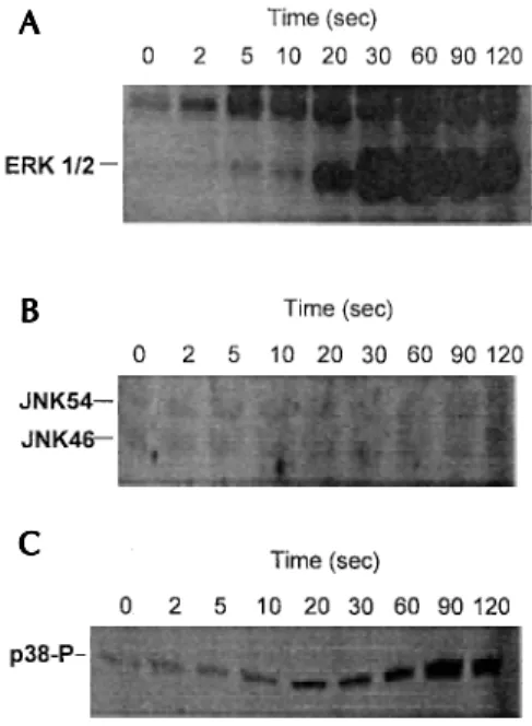
cellular regulated kinases 1 and 2 (ERK1/2), the c-jun N-terminal kinases 46 and 54 (JNK46/JNK54) and the p38 kinases. The upstream mechanisms that regulate the activation of the MAP kinases are pres-ently an area of intense investigation.
The LTB4-, C5a- and fMLP-stimulated re-sponses are thought to activate eosinophils via in-tercalation with receptors linked to the pertussis toxin sensitive G-protein, Gi (Kita et al. 1991, Miyamasu et al. 1995, Wymann et al. 1995, Lind-say et al. 1995b). Recent studies in both neutro-phils and transfected cell lines, have identified some salient aspects of the mechanism of Gi-linked MAP kinase activation (for reviews see Bokoch, 1995, 1996, Denhardt 1996). In the case of ERK1/2 acti-vation, the release of the βγ subunit of Gi results in the phosphorylation of Shc and the subsequent engagement of Grb2-Sos by a mechanism involv-ing phosphatidylinositol 3-kinase (Downey et al. 1996) and the a Src-like tyrosine kinase (Wan et al. 1996). The guanine nucleotide exchanger, Sos stimulates GDP/GTP exchange and activation of p21ras. Activated p21ras recruits the serine/threo-nine kinase Raf-1 to the plasma membrane where it is stimulated by an as yet unidentified mecha-nism. Raf-1 then catalyses the phosphorylation and activation of MAP kinase kinase 1/2 (MEK1/2) which can subsequently phosphorylate and acti-vate the ERK1/2 MAP kinase. At present, much less is known of the pathway responsible for Gi -linked activation of the JNK and p38 MAP kinases. Once again the mechanism is thought to involve the βγ subunit which acts through members of the Rho family of small GTP-binding proteins (rac1 and cdc42). These GTP-binding proteins are be-lieved to stimulate PAK, a p21-activated kinase, which in turn phosphorylates and activates a se-quence containing MEK kinases, then MEKs and finally the JNK and p38 MAP kinases. Since the cytosolic component p47phox has been demon-strated to contain possible MAP kinase phospho-rylation sites whilst another cytosolic component, rac1 is involved in the mechanism of MAP kinase activation, this pathway is potentially important in the mechanism of NADPH oxidase activation.
Although there are no studies demonstrating NADPH oxidase activation by interleukin-5 (IL-5), this cytokine has been reported to cause activa-tion of the lyn-ras-raf1-MEK-ERK pathway in human eosinophils (Pazdrak et al. 1995, Bates et al. 1996). Furthermore, 5-oxo-eicosatetraenoate (5-oxoETE) has been shown to phosphorylate the p42 and p44 MAP kinase (probably ERK1/2) in hu-man eosinophils (O’Flaherty et al. 1996) whilst Araki et al. (1995) have demonstrated PKC-inde-pendent activation of raf1 and ERK following LTB4-activation of guinea-pig eosinophils. We
have extended the later study and shown LTB4 -induced phosphorylation of the p38 MAP kinases although we were unable to demonstrated activa-tion of JNKs (Fig. 4). However, since the selective inhibitors of ERK and p38 MAP kinases, PD098059 (Alessi et al. 1995, Dudley et al. 1995) and SK203580 (Lee et al. 1994) respectively, failed to significantly attenuate H2O2 generation (Fig. 5), this suggested that MAP kinases do not mediate LTB4-induced NADPH oxidase activation.
Fig. 4: LTB4-induced MAP kinase activation in guinea-pig eosi-nophils. Time dependent effect of LTB4 stimulation (1µM) upon ERK1/2 (A) and JNK46/54 (B) activation and p38 MAP kinase phosphorylation (C) in guinea-pig eosinophils. ERK1/2 and JNK46/54 activity were measured using an in-gel renaturation assay employing myelin basic protein and GST-c-jun, respec-tively, as the substrates whilst p38 phosphorylation was deter-mined by western blotting with an anti-phospho-p38 specific antibody (p38-P).
Fig. 5: effect of MAP kinase inhibitors upon LTB4-induced NADPH oxidase activation in guinea-pig eosinophils. Eosino-phils were pre-incubated for 10 min and 30 min with PD098059 (A) and SB203580 (B), respectively, stimulated with 1µM LTB4 and the maximum rate of H2O2 generation determined. Control H2O2 release was essentially zero.
A A A A A
B B B B B
C
CC
120 120 120 120
120 Activation of the NADPH Oxidase in Eosinophils • MA Lindsay, MA Giembycz
A number of inhibitor studies have implicated a possible role for protein tyrosine kinases during NADPH oxidase activation in eosinophils (Nagata et al. 1995, Elsner et al. 1996). Since these inhibi-tors may exert their action through inhibition of the src-related tyrosine kinases, their affects maybe secondary to inhibition of the MAP kinases cas-cade. However, our observation that the tyrosine kinase inhibitors, herbimycin A and lavendustin A, can dose dependently inhibit the MAP kinase-independent LTB4 response in guinea-pig eosino-phil (Fig. 6), suggests the existence of an additional tyrosine kinase dependent pathway(s) responsible for NADPH oxidase activation.
Fig. 6: Effect of tyrosine kinase inhibitors upon LTB4-induced NADPH oxidase activation in guinea-pig eosinophils. Eosino-phils were pre-incubated for 5min with the stated concentration of lavendustin A and herbimycin A. Following 1µM LTB4 stimu-lated, the maximal rate of H2O2 generation was determined. Control H2O2 release was essentially zero.
INHIBITION OF THE NADPH OXIDASE BY CYCLIC AMP
A number of cyclic AMP-elevating drugs in-hibit agonist-induced activation of the NADPH oxidase in eosinophils. Pre-treatment of eosinophils with β2-adrenoceptor agonists such as salbutamol, partially suppress this response but short periods of pre-incubation are necessary if inhibition is to be seen (Yukawa et al. 1990, Rabe et al. 1993). This phenomenon is believed to be due to the rapid development of tachyphylaxis, and may be due to uncoupling of β-adrenoceptors since receptor down-regulation is not observed. Paradoxically, the long-acting β2-agonists salmeterol is inactive on guinea-pig eosinophils and actually behaves as a competitive antagonist. However, this might relate to the very poor efficacy of salmeterol coupling, with a low density of β-adrenoceptors on eosino-phils.
Lipophilic cyclic AMP analogues (Dent et al. 1991) and selective inhibitors of the
phosphodi-esterase (PDE) 4 isoenzymes family also effec-tively prevent activation of the respiratory burst oxidase (Dent et al. 1991, 1994, Souness et al. 1991, Barnette et al. 1995, Hatzelmann et al. 1995).
CONCLUSION
In comparison to neutrophils, little is known of the mechanism of NADPH oxidase activation in eosinophils. As a consequence of the difficul-ties in obtaining sufficient numbers of cells for chemical studies, the majority of the detailed bio-chemical studies have been performed using guinea-pig peritoneal eosinophils. However, where detailed studies have been performed, these results suggest there maybe fundamental difference be-tween the mechanism of NADPH oxidase in eosi-nophils and neutrophils. Thus, increases in intrac-ellular Ca2+ concentration and protein kinase C activation are not required for NADPH oxidase activation in either human or guinea-pig eosino-phils. Furthermore, in contrast to fMLP stimula-tion of neutrophils, LTB4-stimulated NADPH oxi-dase activation in guinea-pig eosinophils appears to be mediated via a tyrosine kinase dependent mechanism that is esssentially independent of PLD, PI 3-kinase, PLA2 and MAP kinases. These dis-parities probably derive from the both the differ-ences in the stimuli and/or the functional roles of these two cell types.
REFERENCES
Aebischer CP, Pasche I, Jorg A 1993. Nanomolar arachi-donic acid influences the respiratory burst in eosi-nophils and neutrophils induced by GTP-binding protein. A comparative study of the respiratory burst in bovine eosinophils and neutrophils. Eur J Biochem 218: 669-677.
Alessi DR, Cuenda A, Cohen P, Dudley DT, Saltiel AR 1995. PD 098059 is a specific inhibitor of the acti-vation of mitogen-activated protein kinase kinase in vitro and in vivo. J Biol Chem270: 27489-27494. Araki R, Komada T, Nakatani K, Naka M, Shima T,
Tanaka T 1995. Protein kinase C-independent acti-vation of Raf-1 and mitogen-activated protein ki-nase by leukotriene B4 in guinea pig eosinophils. Biochem Biophys Res Commun210: 837-843. Babior BM, Kipnes RS, Curnette JT 1973. Biological
defense mechanism: the production by leukocytes of superoxide, a potential bactericidal agent. J Clin Invest 52: 741-744.
Badwey JA, Curnette JT, Robinson JM, Berde CB, Karnovsky MJ, Karnovsky ML 1984. Effects of free fatty acids on release of superoxide and on change of shape by human neutrophils. J Biol Chem 259: 7870-7877.
121 121121 121121 Mem Inst Oswaldo Cruz, Rio de Janeiro, Vol. 92, Suppl. II, 1997
activity. J Pharmacol Exp Ther 273: 674-679. Bates ME, Bertics PJ, Busse WW 1996. IL-5 activates a
45-kilodalton mitogen-activated protein (MAP) ki-nase and Jak-2 tyrosine kiki-nase in human eosinophils. J Immunol156: 711-718.
Bokoch GM 1994. Regulation of the human neutrophil NADPH oxidase by the Rac GTP-binding proteins. Curr Opin Cell Biol 6: 212-218.
Bokoch GM 1995. Chemoattractant signaling and leu-kocyte activation. Blood86: 1649-1660.
Bokoch GM 1996. Interplay between Ras-related and hetertrimeric GTP binding proteins: lifestyles of the BIG and little. FASEB J 10:1290-1295.
Bolsher BGJM, Koenderman L, Tool AJT, Stokman PM, Roos D 1990. NADPH: O2 oxidoreductase of hu-man eosinophils in cell-free system. FEBS Lett268: 269-273.
Bonser RW, Thompson NT, Randall RW, Garland LG 1989. Phospholipase D activation is functionally linked to superoxide generation in human neutro-phils. Biochem J 1989: 617-620.
Butterfield JH, Leiferman KM 1993. In Smith H & Cook RM (eds). The Handbook of Immunopharmacology: Immuopharmacology of Eosinophils: Eosinophil-associated diseases. Academic Press, London. p. 151-192.
Butterworth AE, Thorne KJI 1993. In Smith H & Cook RM (eds). The Handbook of Immunopharmacology: Immunopharmacology of Eosinophils: Eosinophils and parasitic diseases. Academic Press, London. p. 119-150.
Casimar CM, Teahan CG 1994. In Hellewell PG & Wil-liams TJ (eds). The Handbook of Immunopharma-cology: Immunopharmacology of Neutrophils: The respiratory burst of neutrophils and its deficiency. Academic Press, London. p. 27-54.
Curnette JT, Badwey JA, Robinson JM, Karnonsky MJ, Karnovsky ML 1984. Studies on the mechanism of superoxide release from human neutrophils stimu-lated with arachidonate. J Biol Chem 259: 11851-11857.
Demendez I, Adams AG, Sokolic RA, Malech HL, Leto TL 1996. Multiple SH3 domain interactions regu-late NADPH oxidase assembly in whole cells. EMBO Journal15: 1211-1220.
Denhardt DT 1996. Signal-transducing protein phospho-rylation cascades mediated by Ras/Rho proteins in the mammalian cell: the potential for multiplex sig-nalling. Biochem J 318: 729-747.
Dent G, Giembycz MA, Evans PM, Rabe KF, Barnes PJ 1994. Suppression of human eosinophil respiratory burst and cyclic AMP hydrolysis by inhibitors of type IV phosphodiesterase: interaction with the beta adrenoceptor agonist albuterol. J Pharmacol Exp Ther271: 1167-1174.
Dent G, Giembycz MA, Rabe KF, Barnes PJ 1991. Inhi-bition of eosinophil cyclic nucleotide PDE activity and opsonised zymosan-stimulated respiratory burst by ‘type IV’-selective PDE inhibitors. Br J Pharmacol103: 1339-1346.
Ding J, Knaus UG, Lian JP, Bokoch GM, Badwey JA 1996. The renaturable 69- and 63-kDa protein
ki-nases that undergo rapid activation in chemoattratant-stimulated guinea-pig neutrophils are p21-activated kinases. J Biol Chem 271: 24869-24873.
Ding J, Vlahos CJ, Liu R, Brown RF, Badwey JA 1995. Antagonists of phosphatidylinositol 3-kinase block activation of several novel protein kinases in neu-trophils. J Biol Chem 270: 11684-11691.
Downey GP, Butler JR, Brumell J, Borregaard N, Kjeldsen L, Sueaquan AK, Grinstein S 1996. Chemo-tactic peptide-induced activation of MEK-2, the pre-dominate isoform in human neutrophils: inhibition by wortmannin. J Biol Chem 271: 21005-21011. Dri P, Cramer R, Spessotto P, Romano M, Patriarca P
1991. Eosinophil activation on biologic surfaces. Production of O2- in response to physiologic soluble stimuli is differentially modulated by extracellular matrix components and endothelial cells. J Immunol 147: 613-620.
Dudley DT, Pang L, Decker SJ, Bridges AL, Satiel AR 1995. A synthetic inhibitor of the mitogen-activated protein kinase cascade. Proc Natl Acad Sci USA 92: 7686-7689.
Elsner J, Hochstetter R, Kimmig D, Kapp A 1996. Hu-man eotaxin represents a potent activator of the res-piratory burst of human eosinophils. Eur J Immunol 26: 1919-1925.
Gabig TG, Crean CD, Mantel PL, Rosli R 1995. Func-tion of wild-type or mutant Rac2 and Rap1a GTPases in differentiated HL60 cell NADPH oxidase activa-tion. Blood85: 804-811.
Hardy SJ, Robinson BS, Ferrante A, Hii CST, Johnson DW, Poulos A, Murray AW 1995. Polyenoic very-long-chain fatty acids mobilize intracellular calcium from a thapsigargin-insensitive pool in human neu-trophils: The relationship between Ca2+ mobilisation and superoxide production induced by long- and very-long-chain fatty acids. Biochem J 311: 689-697. Hatzelmann A, Tenor H, Schudt C 1995. Differential effects of non-selective and selective phosphodi-esterase inhibitors on human eosinophil functions. Br J Pharmacol114: 821-831.
Henderson LM, Moule SK, Chappell JB 1993. The im-mediate activator of the NADPH oxidase is arachidonate not phosphorylation. Eur J Biochem 211: 157-162.
Hii CST, Ferrante A, Edwards YS, Huang ZH, Hartfield PJ, Rathjen DA, Poulos A, Murray AW 1995. Acti-vation of mitogen-activated protein kinase by arachi-donic acid in rat liver epithelial WB cells by protein kinase C-dependent mechanism. J Biol Chem 270: 4201-4204.
Homayoun P, Stacey DW 1993. Inhibitory effect of arachidonic acid on GTPase activating protein is antagonized by 1-stearoyl, 2-arachidonoyl glycerol. Biochem Biophys Res Commun 194: 1413-1419. Horie S, Kita H 1994. CD11b/CD18 (Mac-1) is required
for degranulation of human eosinophils induced by human recombinant granulocyte-macrophage colony-stimulating factor and platelet-activating fac-tor. J Immunol152: 5457-5467.
hu-122 122 122 122
122 Activation of the NADPH Oxidase in Eosinophils • MA Lindsay, MA Giembycz
man neutrophils. Dependence on changes in cyto-solic free Ca2+ concentration and relation with res-piratory burst activation. J Biol Chem 266: 23152-23156.
Khan WA, Blobe GC, Hannun YA 1995. Arachidonic acid and free fatty acids as secondary messengers and the role of protein kinase C. Cellular Signalling 7: 171-184.
Kita H, Abu Ghazaleh RI, Gleich GJ, Abraham RT 1991. Role of pertussis toxin-sensitive G proteins in stimu-lus-dependent human eosinophil degranulation. J Immunol147: 3466-3473.
Koenderman L, Tool AT, Roos D, Verhoeven AJ 1990. Priming of the respiratory burst in human eosino-phils is accompanied by changes in signal transduc-tion. J Immunol145: 3883-3888.
Kroegel C, Chilvers ER, Giembycz MA, Challiss RA, Barnes PJ 1991. Platelet-activating factor stimulates a rapid accumulation of inositol (1,4,5) trisphosphate in guinea pig eosinophils: relationship to calcium mobilization and degranulation. J Allergy Clin Immunol88: 114-124.
Kroegel C, Giembycz MA, Barnes PJ 1990. Character-ization of eosinophil cell activation by peptides. Differential effects of substance P, melittin, and FMET-Leu-Phe. J Immunol145: 2581-2587. Lee JC, Laydon JT, McDonnell PC, Gallagher TF, Kumar
S, Green D, McNulty D, Blumenthal MJ, Heys JR, Lanvatter SW, Strickler JE, McLaughlin MM, Si-emens IR, Fisher SM, Livi GP, White JR, Adams JL, Young PR 1994. A protein kinase involved in the regulation of inflammatory cytokine biosynthe-sis. Nature372: 739-746.
Lindsay MA, Perkins RS, Barnes PJ, Giembycz MA 1995a. Role of phospholipase A2 in LTB4-induced activation of the NADPH oxidase in guinea pig eosi-nophils. Res Crit Care Med 151: A678.
Lindsay MA, Perkins RS, Barnes PJ, Giembycz MA 1995b. Evidence that leukotriene B4 receptors on guinea pig eosinophils can couple independently to phospholipase A2 and C: Relationship to activation of the NADPH oxidase. Res Crit Care Med 151: A680.
Maghni K, de Brum Fernandes AJ, Foldes Filep E, Gaudry M, Borgeat P, Sirois P 1991. Leukotriene B4 receptors on guinea pig alveolar eosinophils. J Pharmacol Exp Ther258: 784-789.
McPhail LC 1994. SH3-dependent assembly of the ph-agocytic NADPH oxidase. J Exp Med180: 2011-2015.
McPhail LC, Qualliotine-Mann D, Waite KA 1995. Cell-free activation of neutrophil NADPH oxidase by a phosphatidic acid-regulated protein kinase. Proc Natl Acad Sci USA 92: 7931-7935.
Minnicozzi M, Anthes JC, Siegel MI, Billah MM, Egan RW 1990. Activation of phospholipase D in normodense human eosinophils. Biochem Biophys Res Commun170: 540-547.
Miyamasu M, Hirai K, Takahashi Y, Lida M, Yamaguchi, M, Koshino T, Takaishi T, Morita Y, Ohta K, Kasahara T, Ito K 1995. Chemotactic agonists in-duce cytokine generation in eosinophils. J Immunol
154: 1339-1349.
Nagata M, Sedgwick JB, Bates ME, Kita H, Busse WW 1995. Eosinophil adhesion to vascular cell adhesion molecule-1 activates superoxide anion generation. J Immunol155: 2194-2202.
O’Flaherty JT, Kuroki M, Nixon AB, Wijkander J, Yee E, Lee SL, Smitherman PK, Wykle WK, Daniel LW 1996. 5-oxo-eicosanoids and hematopoietic cytokines cooperate in stimulating neutrophil func-tion and mitogen-activated protein kinase pathway. J Biol Chem 271: 17821-17828.
Palmblad J, Gyllenhammar H, Lindgren JA, Malmsten CL 1984. Effects of leukotrienes and f-Met-Leu-Phe on oxidative metabolism of neutrophils and eosino-phils. J Immunol132: 3041-3045.
Park J-W, Ahn SM 1995. Translocation of recominant p47phox cytosolic component of the phagocytic oxi-dase by in vitro phosphorylation. Biochem Biophys Res Commun211: 410-416.
Park J-W, Benna JE, Scott KE, Christensen BL, Chanock, SJ Babior BM 1994. Isolation of a complex of res-piratory burst oxidase components from resting neu-trophil cytosol. Biochemistry33: 2907-2911. Park J-W, Ma M, Ruedi JM, Smith RM, Babior BM 1992.
The cytosolic components of the respiratory burst oxidase as a M(r) approximately 240,000 complex that acquires a membrane-binding site during acti-vation of the oxidase in a cell-free system. J Biol Chem267: 17327-17332.
Pazdrak K, Justement L, Alam R 1995. Mechanism of inhibition of eosinophil activation by transforming growth factor-beta. Inhibition of Lyn, MAP, Jak2 kinases and STAT1 nuclear factor. J Immunol155: 4454-4458.
Perkins RS, Lindsay MA, Barnes PJ, Giembycz MA 1995. Early signalling events implicated in leukotriene B4-induced activation of the NADPH oxidase in eosinophils: role of Ca2+, protein kinase C and phospholipases C and D. Biochem J310: 795-806.
Petreccia DC, Nauseef WM, Clark RA 1987. Respira-tory burst of normal human eosinophils. J Leukoc Biol41: 283-288.
Rabe KF, Giembycz MA, Dent G, Barnes PJ 1992. Ac-tivation of guinea pig eosinophil respiratory burst by leukotriene B4: role of protein kinase C. Fundam Clin Pharmacol6: 353-358.
Rabe KF, Giembycz MA, Dent G, Perkins RS, Evans P, Barnes PJ 1993. Salmeterol is a competitive antago-nist at beta-adrenoceptors mediating inhibition of respiratory burst in guinea-pig eosinophils. Eur J Pharmocol 231: 305-308.
Rao GN, Baas AS, Glasgow WC, Eling TE, Runge MS, Alexander RW 1994. Activation of mitogen-acti-vated protein kinases by arachidonic acid and its me-tabolites in vascular smooth muscle cells. J Biol Chem 269: 32586-32591.
123 123123 123123 Mem Inst Oswaldo Cruz, Rio de Janeiro, Vol. 92, Suppl. II, 1997
T, Kaibuchi K, Takai Y, Katayama K. Combination of arachidonic acid and guanosine 5’-O-(3-thiotriphosphate) induce translocation of rac p21s to membrane and activation of NADPH oxidase in a cell-free system. Biochem Biophys Res Commun 195: 264-269.
Sedgwick JB, Vrtis RF, Gourley MF, Busse WW 1988. Stimulus-dependent differences in superoxide anion generation by normal human eosinophils and neu-trophils. J Allergy Clin Immunol81: 875-883. Segal AW, Abo A 1993. The biochemical basis of the
NADPH oxidase of phagocytes. Trends Biochem Sci 18: 43-47.
Segal AW, Garcia R, Goldstone H, Cross AR, Jones OT 1981. Cytochrome b-245 of neutrophils is also present in human monocytes, macrophages and eosi-nophils. Biochem J196: 363-367.
Sermon BA, Eccleston JF, Skinner RH, Lowe PN 1996. Mechanism of inhibition by arachidonic acid of the catalytic activity of ras GTPase-activating proteins. J Biol Chem 271: 1566-1572.
Shute JK, Rimmer SJ, Akerman CL, Church MK, Holgate ST 1990. Studies of cellular mechanisms for the generation of superoxide by guinea-pig eosi-nophils and its dissociation from granule peroxidase release. Biochem Pharmacol40: 2013-2021. Souness JE, Carter CM, Diocee BK, Hassall GA, Wood
LI, Turner NC 1991. Characterization of guinea-pig eosinophil phosphodiesterase activity. Assessment of its involvement in regulating superoxide genera-tion. Biochem Pharmacol42: 937-945.
Souness J, Maslen C, Webber S, Foster M, Raeburn D, Palfreyman MN, Ashton MJ, Karlsson JA 1995. Sup-pression of eosinophil function by RP 73401, a po-tent and selective inhibitor of cyclic AMP-specific phosphodiesterase: comparison with rolipram. Br J Pharmacol115: 39-46.
Subramanian N 1992. Leukotriene B4 induced steady state calcium rise and superoxide anion generation in guinea pig eosinophils are not related events. Biochem Biophys Res Commun187: 670-676. Tenscher K, Metzner B, Schopf E, Norgauer J, Czech
W 1996. Recombinant human eotaxin induces oxy-gen radical production, Ca2+-mobilization, actin re-organization, and CD11b upregulation in human eosinophils via a pertussis toxin-sensitive heterotrimeric guanine nucleotide-binding protein. Blood88: 3195-3199.
Thompson NT, Tateson JE, Randall RW, Spacey GD, Bonser RW, Garland LG 1990. The temperol rela-tionship between phospholipase activation, diradylglycerol formation and superoxide produc-tion in the human neutrophil. Biochem J 271: 209-213.
Tool AT, Koenderman L, Kok PT, Blom M, Roos D, Verhoeven AJ 1992. Release of platelet-activating factor is important for the respiratory burst induced in human eosinophils by opsonized particles. Blood 79: 2729-2732.
Tsunawaki S, Mizunari H, Nagata M, Tatsuzawa O, Kuratsuji T 1994. A novel cytosolic component, p40phox, of respiratory burst oxidase associates with
p67phox and is absent in patients with chronic granu-lomatous disease who lack p67phox. Biochem Biophys Res Commun199: 1378-1387.
Van der Bruggen T, Kok PTM, Blom M, Verhoeven AJ, Raaijmakers JAM, Lammers J-MJ, Koenderman L 1993. Transient exposure of human eosinophils to the protein kinase C inhibitors CGP39-360, CGP41-251, and CGP44-800 leads to priming of the respi-ratory burst induced by opsonized particles. J Leuk Biol54: 552-557.
Vlahos CJ, Matter WF, Brown RF, Traylor Kaplan AE, Heyworth PG, Prossnitz ER, Ye RD, Marder P, Schelm JA, Rothfuss KJ, Rothfuss KJ, Serlin BS, Simpson PJ 1995. Investigation of neutrophil signal transduction using a specific inhibitor of phosphatidylinositol 3-kinase. J Immunol 154: 2413-2422.
Wan Y, Kurosaki T, Huang X-Y 1996. Tyrosine kinases in activation of the MAP kinase cascade by G-pro-tein-coupled receptors. Nature380: 541-544. Weiss SJ, Test ST, Eckmann CM, Roos D, Regiani S
1986. Brominating oxidants generated by human eosinophils. Science234: 200-203.
White SR, Strek ME, Kulp GVP, Spaethe SM, Burch RA, Neeley SP, Leff AR 1993. Regulation of hu-man eosinophil degranulation and activation by en-dogenous phospholipase A2. J Clin Invest 91: 2118-2125.
Wientjes FB, Hsuan JJ, Totty NF, Segal AW 1993. p40phox, a third cytosolic component of the activa-tion complex of the NADPH oxidase to contain src homology 3 domains. Biochem J296: 557-561. Wymann MP, Kernen P, Von Tscharner V, Tai PC, Spry
CJ, Baggiolini M 1995. Activation of the respira-tory burst in eosinophil leucocytes—a transduction sequence decoupled from cytosolic Ca2+ rise. Eur J Clin Invest25: 25-31.
Yagisawa M, You A, Yonemaru M, Imajoh-Ohmi S, Kanegasaki S, Yazaki Y, Takaku F 1996. Superox-ide release and NADPH oxidase components in mature human phagocytes: Correlation between functional capacity and amount of functional pro-teins. Biochem Biophys Res Commun228: 510-516. Yamashita T, Someya A, Hara E 1985. Response of su-peroxide anion production by guinea-pig eosino-phils to various soluble stimuli: comparison to neu-trophils. Arch BiochemBiophys 241: 447-452. Yukawa T, Ukena D, Kroegel C, Chanez P, Dent G,
Chung KF, Barnes PF 1990. Beta 2-adrenergic re-ceptors on eosinophils. Binding and functional stud-ies. Am Rev Respir Dis 141: 1446-1452.
Zhan SX, Vazquez N, Zhan SL, Wientjes FB, Budarf ML, Schrock E, Ried T, Green ED, Chanock SJ 1996. Genomic structure, chromosomal localization, start of transcription, and tissue expression of the human p40-phox, a new component of the nicotinamide adenine dinucleotide phosphate-oxidase complex. Blood88: 2714-2721.