i n t e r o b s e r v e r r e l i a b i l i t y i n u l t r a s o u n d
a s s e s s m e n t o f r h e u m a t o i d w r i s t j o i n t s
Karine R. Luz
*, Rita N.V. Furtado
*, Sonia V. Mitraud
**, Jorge Porglhof
**,
Conceição Nunes
*, Artur R. C. Fernandes
**, Jamil Natour
**Rheumatology Division, Universidade Federal de São Paulo – Escola Paulista de Medicina, São Paulo, Brazil.
** Department of Radiology, Universidade Federal de São Paulo – Escola Paulista de Medicina, São Paulo, Brazil.
Introduction
In recent years, musculoskeletal ultrasound has be-come an important diagnostic tool for rheumatic disease, as it allows the detection of the inflamma-tory process in intra-articular and periarticular structures as well as the identification of bone ero-sion1,2. This exam has a number of advantages over
other imaging methods, including its non-invasive nature, good visualization of the joint cavity, ab-sence of radiation, and wide acceptance by pa-tients. The exam’s dynamic and rapid execution ena bles it to assess multiple joints at low cost, thereby making it is a “bedside exam”3,4. Despite
these significant advantages, ultrasound findings remain highly dependent on the individual exami -ner’s findings. This occurs partially due to the sub-jective assessment of the images and the low degree of standardization of the technique due to the small number of multi-center studies involving the method5-9.
In cases of rheumatoid arthritis (RA), the wrist is affected in 90% of patients in the first 10 years of the disease10. The wrist is an anatomical complex made
up of various articular recesses and interbone liga -ments. The three main recesses in the wrist are the radiocarpal (RC), midcarpal (MC) and ulnocarpal (UC)11,12. Ultrasound has proven useful in the
as-sessment of these articular recesses as well as in the distinction between healthy individuals and pa-tients with chronic inflammatory arthropathy of the wrist13-16. It is a helpful tool for guiding
proce-dures, assessing sub-clinical findings and moni-toring treatment16,17.
There are few studies investigating interobser ver reliability in the ultrasound assessment of muscu-loskeletal conditions6-9,18-21. The majority of these
studies have analyzed ultrasound reliability for the joints of the hands and feet, knees and periarticular structures, such as in cases of rotary cuff inju -ry7-9,14-16. The reliability of ultrasound assessment of
Abstract
Objective: To evaluate interobserver reliability in
the ultrasound assessment of synovitis in the ra-diocarpal (RC), midcarpal (MC) and ulnocarpal (UC) joints in RA.
Methods: Ultrasound examinations of 295
rheuma-toid wrist joints were performed over a three month period. The RC, MC and UC joints were examined using dorsal longitudinal ultrasound scans. Syno -vial thickening was assessed by quantitative mea-surement and a previously established semiquantitative scoring system (Grades 0 to 3). Interobser -ver reliability was determined by the comparing the findings of two radiologists who were unaware of each other findings.
Results: The intraclass correlation coefficient (ICC)
between examiners for the quantitative measure-ment of synovitis in the RC, MC and UC recesses were 0.508, 0.346 and 0.240 (p<0.001), respectively. Weighted kappa values using the semi-quantita-tive scoring system were 0.308, 0.312 and 0.153 for the RC, MC and UC joints, respectively.
Conclusion: Interobserver reliability of the
ultra-sound assessment in rheumatoid wrists proved good for the quantitative measurement of synovi-tis in the RC joint, but poor agreement was found for the MC and UC joints. Using the semi-quanti-tative scoring system, interobserver agreement was poor for all three joints (RC, MC and UC).
Keywords: Ultrasonography; Reliability; Wrist;
were performed from the radial and ulnar sides as well as midline to assess the RC, MC and UC recesses in accordance with the standards establi shed by the European League Against Rheumato -logy24.
Both quantitative and semi-quantitative measurements were performed in each recess for syno -vial thickening13,19. The quantitative measurement
was obtained from the distance between the joint capsule and subchondral bone (Figure 1). For the semi-quantitative assessment, a modified version of a previously established semiquantitative sco -ring system to evaluate metacarpophalangeal (MCP), proximal interphalangeal and metatar-sophalangeal joints were used19. A single score was
used for effusion and synovitis, ranging from 0 to 3: 0- no synovial thickening; 1- minimal synovial thickening up to the joint capsule; 2- synovial thickening causing curvature of the joint capsule, but without extending to the bone diaphysis; 3- synovial thickening with curvature of the joint capsule and extending to at least one bone diaphy -sis. Figure 2 displays the semi-quantitative mea-surement at radiocarpal joint and respective scores.
Interobserver Reliability
Interobserver reliability was determined by com-paring the mean quantitative and semi-quantita-tive scores obtained by two radiologists who were unaware of clinical assessments. Each operator performed the ultrasound exams sequentially and independently. The assessments were performed in different rooms, using the same machine and settings, and the measurements were recorded on separate charts. Therefore, each evaluator was blinded to the measurements of the other. the wrist has only been evaluated regarding the
presence or absence of synovitis in a small num-ber of patients with different chronic inflammato-ry conditions8-9,22. There is no evidence of any study
investigating interobserver reliability in the ultra-sound assessment of the synovium in different ar-ticular recesses of the wrist.
The aim of the present study was to determine interobserver reliability in the ultrasound assess-ment of the radiocarpal (RC), midcarpal (MC) and ulnocarpal (UC) recesses of the wrist in patients with RA and clinical synovitis.
Methods
A cross-sectional study was carried out involving patients with RA based on the classification crite-ria of the American College of Rheumatology23
pre-senting clinical synovitis in at least one of the wrists. The patients included had no diagnostic criteria for any other collagen disease
Ultrasound Assessment
Assessments were carried out by two radiologists with experience in musculoskeletal ultrasound. Two hundred and ninety five wrists of RA patients with clinical synovitis were examined by ultra-sound over a three-month period. The ultraultra-sound examinations were performed using a Sonosite 180 Plus (SonoSite. Inc – United States) device equip -ped with a linear probe (5 to 10 MHz).
All patients were instructed to stay seated in a comfortable position in front of the examiner with their hand in a pronated position on top of the desk to take dorsal scans in neutral position of the wrist.
The ultrasound examinations were performed from the dorsal aspect of the wrist with the trans-ducer in a longitudinal position. The examinations
Figure1.B-mode US synovial thickness measurements
in the wrist joint, scanned in a longitudinal plane of the dorsal central and ulnar surface of radiocarpal (RC), midcarpal (MC) and ulnocarpal (UC) recess. Synovial measurements were performed perpendicular to the great axis and at the point of greatest thickness
Figure2.Ilustration of semi-quantitative scoring system
at radiocarpal joint: L – lunate; R- radius; RCJ – radiocarpal joint; JC – joint capsule; BD – bone diaphysis; * – synovial thickening
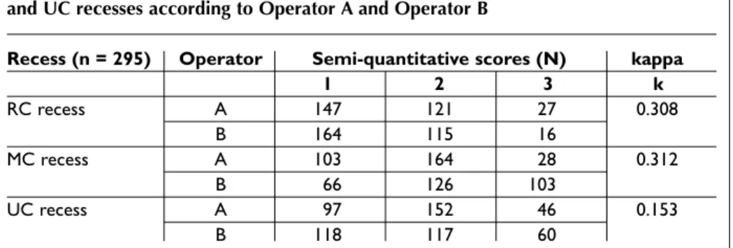
of 1 in 147 and 164 assessments, a score of 2 in 121 and 115 measurements, and a score of 3 in 27 and 16 measurements. In the semi-quantitative mea-surements of the MC recess, Operators A and B de-termined a score of 1 in 28 and 103 assessments, respectively; a score of 2 in 164 and 126 measure-ments, respectively; and a score of 3 in 27 and 16 measurements, respectively. In the semi-quanti-tative measurements of the UC recess, Operators A and B determined a score of 1 in 97 and 118 as-sessments, respectively; a score of 2 in 152 and 117 measurements, respectively; and a score of 3 in 46 and 60 measurements, respectively (Table III).
The absolute agreement for semiquantitative scoring for both observers was 58,3% for RC, 47,5% for MC and 46,4% for UC recess.
Interobserver reliability
The ICC between the two evaluators for the quan-titative measurements of the RC, MC and UC recesses was 0.508, 0.3463 and 0.240 (p<0.001), res -pectively. Weighted Weighed kappa values for semi-quantitative assessments of the RC, MC and UC recesses were 0.308, 0.312 and 0.153, respec-tively (Tables II and III).
Discussion
The present study assessed the interobserver relia -bility ultrasonography for quantitative and semi--quantitative measurements of wrist in patients with long-standing RA. The wrist is one of the most affected joints in RA and is a complex anatomical structure made up of various joint recesses and
pe-Statistical Analysis
The intra-class correlation coefficient (ICC) was used for the quantitative measurements and weighted Kappa test (κ) was used for the semiquan titative measurements. For the ICC, inte -robser ver reliability was considered excellent if R > 0.75, good to optimal if R was 0.4 to 0.75 and poor if R < 0.425. With the Kappa test, interobser ver
relia bility was considered excellent if κ > 0.81, subs -tantial when values were 0.61 to 0.80, moderate when values were 0.41 to 0.60, good when values were 0.21 to 0.40, minimal when values were 0.20 to 0 and poor when the value was 026.
Results
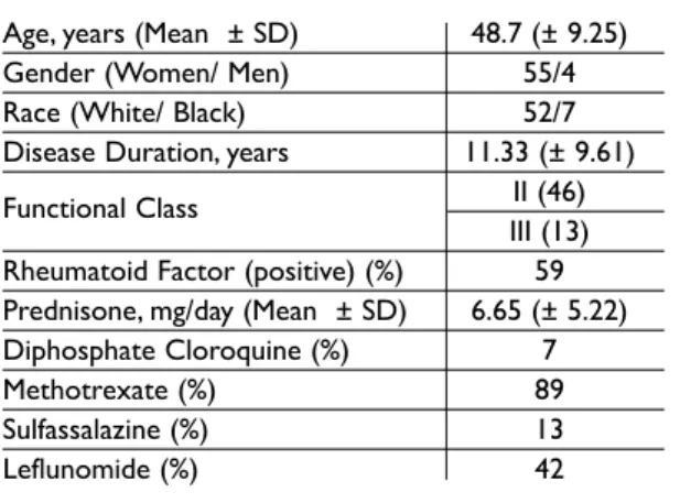
Fifty-nine patients with RA were analyzed. Table I dis-plays the demographic data and clinical parameters.
Ultrasound Assessment
A total of 295 assessments were performed on the rheumatoid wrists over a three-month period. The mean quantitative measurement of synovium in the RC, MC and UC recesses according to Opera-tor A was 5.09 ± 1.83 (1.2 – 10.12) mm, 4.82 ±1.83 (0 –11.66) mm and 5.34 ± 1.68 (1.16 – 12.23) mm, respectively. According to Operator B, these mea-surements were 4.53 ± 1.41 (1.7 – 9.9) mm, 4.40 ±1.34 (0.66 – 8.3) mm and 7.03 ± 1.74 (1.16 – 11.56) mm, respectively (Table II).
In the semi-quantitative measurements of the RC recess, Operators A and B determined a score
Table I. Demographic parameters, disease related variables in 59 RA patients
Age, years (Mean ± SD) 48.7 (± 9.25)
Gender (Women/ Men) 55/4
Race (White/ Black) 52/7
Disease Duration, years 11.33 (± 9.61)
Functional Class II (46)
III (13)
Rheumatoid Factor (positive) (%) 59
Prednisone, mg/day (Mean ± SD) 6.65 (± 5.22)
Diphosphate Cloroquine (%) 7
Methotrexate (%) 89
Sulfassalazine (%) 13
Leflunomide (%) 42
SD – standard desviation
Table II. The mean quantitative measurement (mm) of synovial in the RC, MC and UC recess according to Operator A and Operator B
Recesses Operator A Operator B ICC (Mean ± SD) (n-295) (n-295) (R) RC recess 5.09 mm 4.82 mm 0.482 (Mean ± SD) (± 1.83) (± 1.41) MC recess 4.53 mm 4.40 mm 0.509 (Mean ± SD) (±1.83) (±1.34) UC recess 5.34 mm 7.03 mm 0.240 (Mean ± SD) (± 1.68) (± 1.74)
RC – radiocarpal; MC – mediocarpal; UC – ulnocarpal; SD – standard desviation; ICC – intra-class correlation coefficient
moderate reliability (k = 0.49) for the presence of synovitis in the RC recess of the wrist27.
Few studies on ultrasound have employed a quantitative measurement of the synovia as an as-sessment instrument. Schmidt et al., determined reference values for the measurement of the syno -vium in different joints in healthy individuals; in the wrist, the mean distance between the joint capsu le and scaphoid bone profile was < 1.5 mm28.
Koski (2003) established the measurement of the synovium in the recesses of rheumatoid joints and considered unequivocal synovitis to be a mea-surement greater than 2 mm in the RC recess and the presence of any area of synovial proliferation in the MC recess13. However, interobserver reliabi
-lity was not determined in either of these studies. The poor interobserver reliability (ICC = 0.346) for the quantitative measurement of synovitis in the MC recess in the present study may have occurred due to the fact that the patients had long--standing RA, in which erosion is common and the possible destruction of the carpal bones, such as the lunate and capitate, has occurred, which would hamper the visualization of the subchondral bone profile for the quantitative mea-surement. In the UC recess, there is a presence of the triangular fibrocartilage and the styloid pro-cess, which may have impaired the exact posi-tioning of the transdu cer, thereby causing an anisotropic effect and lea ding to the poor reliabili -ty in the measurement of the synovium in this re-cess. Moreover, a portable ultrasound device of lesser resolution was used in the present study, which may have compromised the adequate lo-calization of the joint capsule and hampered the quantitative measurement.
A semi-qualitative assessment of synovitis in the riarticular structures, which makes the physical
exam of this joint a difficult task that requires the use of imaging methods11,14,15. A number of studies
have demonstrated the ultrasound is capable of revealing inflammatory alterations in this joint. However, there are no previous studies that have evaluated the interobserver reliability of this method for joint recesses of the wrist1-4.
In the present study, good reliability (ICC = 0.5081) was found for the RC recess, whereas poor correlations were found for the MC (ICC = 0.3463) and UC (ICC = 0.240) recesses. Likewise, poor inter-observer reliability was found for the semi-quanti-tative assessment for all three recesses ana lyzed (RC, MC and UC), with Kappa values of k = 0.308, k = 0.312 and k = 0.153, respectively. Two previous studies in-volving experts in musculoskeletal ultrasound and the assessment of different joints found moderate in-terobserver reliability (k = 0.59 to 0.61) regarding the presence or absence of synovitis in the wrist; these studies report interobserver reliability similar to that found in the present study in the quantitative mea-surement of synovia in the RC recess8,9. Unlike the
present study, however, these studies only performed a qualitative assessment (presence or absence of syn-ovitis) and did not perform a quantitative measure-ment of synovitis. Moreover, no systematic exami-nations of rheumatoid wrists were carried out, but rather the evaluation of different joints and degene -rative inflammatory conditions8,9.
Iagnocco et al., investigated the presence or absen ce of synovitis in the wrists of patients with systemic lupus erythematosus and found optimal interobserver reliability for the RC recess (k = 0.73 to 0.89)22. A recent study investigated interobser
ver reliability in the ultrasound assessment of syno -vitis in 28 joints in patients with RA and found
Table III. Interobserver agreement for the semi-quantitative scores of the RC, MC and UC recesses according to Operator A and Operator B
Recess (n = 295) Operator Semi-quantitative scores (N) kappa
1 2 3 k RC recess A 147 121 27 0.308 B 164 115 16 MC recess A 103 164 28 0.312 B 66 126 103 UC recess A 97 152 46 0.153 B 118 117 60
RC, MC and UC recesses was employed in the pre-sent study, as this method is the most common form of measuring synovial thickening. For this assess-ment, a semi-quantitative scoring system which was previously estabilished by Szkularek et al., for small joints of the hand and feet (proximal metacar-pophalangeal, interphalangeal and metatarsopha-langeal) was used19. In this study the sco ring system
for synovitis and joint effusion showed moderate to optimal interobserver reliability (ICC of 0.61 and 0.78, respectively) for all evaluations19. In the present
study, the scoring system was mo dified to deter-mine the presence of synovitis and joint effusion in the same assessment. This decision was made due to the fact that both alterations occur simultaneously in the chronic inflammatory process. Howe -ver, there was poor interobserver reliability in the as-sessment of the recesses. The MCP joints used by Skzudlarek et al., are consi dered joints with a sim-ple anatomical model, in which the subchondral bone and cartilage may be assessed and detection of synovitis is easy26. An explanation for different
levels of agreement between studies may be that the wrist is a more elaborate joint with diverse re-cesses and multiple ligament structures11,12.
The assessment of synovial proliferation in the present study was not carried out with the aid of a power Doppler signal. This decision was made due to the low resolution of the ultrasound device in the assessment of a power Doppler, which could have compromised the results.
In conclusion, there was moderate interobser ver reliability for the quantitative measurement of the synovium in the RC recess and poor reliability re-garding the MC and UC recesses. The semiquantitative assessment of the synovium using a pre -viously established scoring system for small joints demonstrated poor interobserver correlations for the RC, MC and UC recesses of rheumatoid wrists. Further studies are needed for the standar dization of a quantitative measurement of the synovium in joint recesses of the wrist as well as the va lidation of semi-quantitative scoring systems for this fre-quently affected joint in patients with RA.
Correspondence to
Jamil Natour
Disciplina de Reumatologia, Universidade Federal de São Paulo Rua Botucatu, 740 São Paulo, SP – Brazil 04023-900 Phone/Fax: 55 11 5576 4239 E-mail: jnatour@unifesp.br
References
1. Canoso JJ. Ultrasound Imaging: A Rheumatologist’s Dream. J Rheumatoi 2000; 27:2063-2064.
2. Kane D, Ballin PV, Sturrock R, Grassi W. Muscu-loskeletal Ultrasound – a state of the art review of musculoskeletal ultrasound in rheumatology. Rheumatology (Oxford) 2004; 43:823028.
3. Manger B, Kalden JR. Joint and Connective Tissue Ul-trasonography – a Rheumatologic Bedside Proce-dure? A German Experience. Arthritis and Rheuma-tism 1995; 38:736-742.
4. Grassi W, Cervini C. Ultrasonography in Rheumatolo-gy: an evolving technique. Ann Rheum Dis 1998;57:268-271.
5. Joshua F, Lassere M, Bruyn GA, et al. Summary find-ings of a systematic review of the ultrasound assess-ment of synovitis: proceedings of OMERACT 8. J Rheumatol 2007;34:839–847.
6. Naredo E, Rodriguez M, Campos C, et al, and the Ul-trasound Group of The Spanish Society of Rheuma-tology. Validity, reproducibility and responsiveness of a twelvejoint simplified power Doppler ultraso -nographic assessment of joint inflammation in rheumatoid arthritis. Arthritis Rheum 2008;59: 515–522.
7. D’Agostino MA, Maillefert JF, Said-Nahal R, Breban M, Ravaud P, Dougados M. Detection of small joint synovitis by ultrasonography: the learning curve of rheumatologists. Ann Rheum Dis 2004;63:1284–1287. 8. Scheel AK, Schmidt WA, Hermann K-GA, et al. Inter-observer reliability of rheumatologistis performing musculoskeletal ultrasonography; results from a EU-LAR “Train the trainers” course. Ann Rheum Dis 2005;64:1043-1049.
9. Naredo E, Moller I, Moragues C, de Agustin JJ, Scheel AK, Grassi W. Interobserver reliability in muscu-loskeletal ultrasonography: results from a “Teach and Teachers” rheumatologist course. Ann Rheum Dis 2006;65:14-19.
10. Hamalainen M, Kamoonen M, Lethimaki M. Epi-demiology of wrist involvement of rheumatoid arthritis. Rheumatol 1992;17:1-7.
11. Harrison MO, Freiberger RH, Ranawat CS. Arthrogra-phy of the wrist joint. Am J Roentgenol Radium The Nucl Med 1971;112:480-486.
12. Hastings DE, Evans JA. Rheumatoid wrist deformities and their relation to ulnar drift. J Bone Joint Surg 1975;57:930-934.
13. Koski JM. Ultrasonography in detection of effusion in the radiocarpal and midcarpal joints. Scand J Rheumatol 1992;21:79-81.
14. van Vugt RM, van Dallen A, Bijlsma JW. The current role of high-resolution ultrasonography of the hand and wrist in rheumatic disases. Clin Exp Rheumatol 1998;16:454-458.
15. van Vugt RM, van Jaarsveld CH, Hofman DM, Helders PJ, Bijlsman JW. Patterns of disease progression in the rheumatoid wrist; a long term followup. J Rheumatol 1992;26:1467-1473.
16. Koski JM, Hermunen H. Intra-articular glucocorticoid treatment of the rheumatoid wrist. An ultrasono-graphic study. Scan J Rheumatol2001;30:268-270. 17. Luz KR, Furtado RN, Nunes CC, Rosenfeld A,
Fernan-der AR, Natour J. Ultrasound guided intra-articular injections in the wrist in patients with rheumatoid arthritis: a Double –blind, randomized controlled study. Ann Rheum Dis 2008;67:1198-2000.
18. Balint OV, Sturrock RD. Intraobserver repeatability and interobserver reproductibility in musculoskeletal ultrasound imaging measurements. Clin Exp Rheu -matol 2001;19:89-92.
19. Szkudlarek M, Court-Payen M, Jacobsen S, Klarlund M, Thomsen HS, Ostergaard M. Interobserver agree-ment in ultrasonography of the fingers and toe joints in rheumatoid arthritis. Arhritis and Rheum 2003; 48:995-962.
20. Middleton WD, Teefey SA, Yamaguchi K. Sonography of the rotator cuff: analysis of interobserver variabili-ty. AJR Am J Roentgenol 2004;183:1465-1458.
21. O Connor PJ, Rankine J, Gibbon WW, Richardson A, Winter F, Miller JH. Interobserver variation in sonog-raphy of the painful shoulder. J Clin Ultrasound 2005;33:53-56.
22. Iagnocco A, Ossandon A, Coari G, et al. Wrist joint in-volvement in systemic lúpus erythematosus. An ul-trasonographic study. Clin Exp Rheumatol 2004;22:621-624.
23. Arnett FC, Edworthy SM, Bloch DA, et al. The Ameri-can Rheumatism Association 1987 revised criteria for the classification of rheumatoid arthritis. Arthritis Rheum 1988;31:315-324.
24. Backhaus M, Burmester GR, Gerber T, et al. Guide-lines for musculoskeletal ultrasound in rheumatolo-gy. Ann Rheum Dis 2001;60:641-649.
25. Shourt PE, Fleiss JL. Intraclass correlation: use in as-sessing rater reliability. Psychol Bull 1979;86:420-428. 26. Landis JR, Koch GG. The measurement of observer
agreement for categorical data. Biometrics 1977;33: 159-174.
27. Salaffi F, Filippucci E, Carotti M, et al. Interobserver agreement of standard joint couts in early rheuma-toid arthritis; a comparasion with grey scale ultra-sonography – a preliminary study. Rheumatology (Oxford) 2008;47:54-58.
28. Schmidt WA, Schmidt H, Schicke B, Gromnica-lhle E. Standard reference values for musculoskeletal ultra-sonography. Ann Rheum Dis 2004;63:988-994. 29. Conaghan PG, Green M. Emery P. Established rheu
matoid arthritis. Bailliere’s Best Prac Res Clin Rheu -matol 1999;13:561-575.