208 209
208 209
Tomographic evaluation of TMJ in adolescents with
temporomandibular disorders
Avaliação tomográfica da ATM em adolescentes com
disfunção temporomandibular
Luciano José Pereira*
Maria Beatriz Duarte Gavião**
ABSTRACT: This study evaluated the TMJ images of normal subjects and of those who presented TMD (temporo-mandibular disorders) using linear tomography at rest position. Cranio(temporo-mandibular Index (CMI) and a question-naire on subjective symptoms were used to assess 217 subjects aged 12 to 18 years. Those with the highest and the lowest scores were divided into Control (n = 20) and TMD groups (n = 20), respectively. Corrected tomography was used to measure the narrowest anterior, superior and posterior joint spaces, to determine the condyle position in all 40 subjects. The distance means did not correlate with CMI scores (p > 0.05). The number of posteriorly po-sitioned condyles was significantly higher in TMD patients (p = 0.05), especially in females. It was concluded that condyle position in linear tomography at rest position cannot yield TMD diagnosis.
DESCRIPTORS: Tomography; Temporomandibular joint disorders; Teen health.
RESUMO: O objetivo deste estudo foi avaliar imagens da ATM de indivíduos portadores e não portadores de distúr-bios temporomandibulares (DTM), através da tomografia linear corrigida na posição de repouso. Foram avaliados 217 adolescentes com idade entre 12 e 18 anos através do Índice Craniomandibular (CMI) e de questionário de sintomas subjetivos. Foram selecionados 40 indivíduos com escores extremos, 20 sem DTM e 20 com DTM, para a formação dos grupos controle e DTM, respectivamente. Os espaços articulares anterior, superior e posterior foram mensurados pela tomografia corrigida, tendo como referência a região de menor comprimento subjetivo, determi-nando a posição condilar. As médias das distâncias não se correlacionaram com os escores do CMI (p > 0,05). O número de côndilos posteriorizados foi significativamente maior nos pacientes com DTM (p = 0,05), principalmen-te no sexo feminino. Concluímos que a posição condilar na posição de repouso não pode predizer diagnóstico de DTM.
DESCRITORES: Tomografia; Transtornos da articulação temporomandibular; Saúde do adolescente.
INTRODUCTION
The term temporomandibular disorders (TMD) is defined as “collective term embracing a number of clinical problems that involve the masticatory musculature, the temporomandibular joint (TMJ) and associated structures, or both”16.
Temporo-mandibular disorders are considered to be a sub-classification of musculoskeletal disorders and are a major source of orofacial pain of non-dental origin18.
TMD in children and adolescents is consid-ered to have a multifactorial etiology8,17,28.Because
of the difficulty in establishing a precise etiology, TMD is often defined on the basis of signs and symptoms, the most common of which are TMJ and muscle pain, mouth opening limitation, click and crepitation5,12.
Whenever a patient presents a suspected TMJ problem, the radiographic examination is part of the patient’s complete evaluation1. Plain-film
radi-ography and tomradi-ography are basic imaging tech-niques for the assessment of the TMJ.
Observations on condyle position are now being studied and related to TMD; however, its therapeutic implications still remain unclear in the related literature, as well as its importance in the TMD etiology. Previous studies have evalu-ated the prevalence of anterior, concentric, and posterior condyle position in asymptomatic vol-unteers2,3,19,22, and in symptomatic patients3,11,21,25.
Research has suggested that the measurement of the joint space and the determination of condylar position is of questionable value, given the high
208 209
208 209
variability of condylar position within the fossa in the adult population6,23.Although the variability
in condylar position in adults may minimize the diagnostic value of the measurement, the assess-ment of the joint space and condylar position in adolescents may be of value13.
The purpose of this study was to determine joint space and condylar position in adolescents with and without TMD viewed in axial linear cor-rected tomography at mandibular rest position and to correlate these findings to the clinical diagnosis of TMD.
MATERIAL AND METHODS
The subjects were selected from public schools in the city of Piracicaba/Brazil. The parents and the adolescents were informed of the purpose of the study and they gave their written and verbal consent. The Ethics Committee of the School of Dentistry of Piracicaba approved the research. Adolescents who received any type of orthodon-tic treatment prior to or during the research ex-amination period were excluded from the study. Initially, the signs of TMD were assessed in 217 adolescents (120 female/97 male) aged 12 to 18 years, according to the Craniomandibular Index (CMI), as described by Fricton, Schiffman9 (1986),
by two calibrated examiners (kappa = 0.936). The CMI has a 0 to 1 scale that measures tenderness and dysfunction in the stomatognathic system and includes all currently recognized signs of TMJ dis-orders9,10. There are 2 subscales: the Dysfunction
Index (DI) and the Palpation Index (PI). The DI is designed to measure limitation in mandibular movement, pain and deviation in movement, TMJ noise, and TMJ tenderness. The PI measures the prevalence of muscle tenderness in the stomato-gnathic system. Thus, this index separates joint problems from muscle problems.
A self-report questionnaire was used to assess subjective symptoms according to Riolo et al.24
(1987), regarding pain in the jaws when in func-tion, unusually frequent headaches (more than once a week), stiffness/tiredness in the jaws, dif-ficulty in opening the mouth wide, grinding teeth, and TMJ sounds. Each question should be an-swered with “yes” or “no”.
After all parameters had been checked, the median score for the CMI was determined. Forty adolescents out of the 217 subjects were select-ed to dichotomize the data in order to compare “extreme” groups, the lower and upper extremity
values being used to compose the control group (10 males/10 females) and the TMD group (10 males/10 females), respectively. In addition, sub-jects from the TMD group should present at least one subjective symptom of TMD. The linear tomog-raphy was performed in both groups.
Linear tomography
Corrected linear tomograms (Quint Secto-graph, Quint Co., Los Angeles, USA) were ob-tained with teeth at rest position. The center cut film was used for analysis. Linear measurements, as described by Pullinger, Hollender20 (1986), were
taken twice by a single examiner of the subjective closest posterior (P), superior (S) and anterior (A) radiological intra-articular spaces. The mean for each space (P, S and A) was used for statistical analysis. The position of the mandibular condyle was described as anterior, concentric, or poste-rior according to the following equation: linear ratio = (P – A)/(P + A) × 100, where P = the closest posterior linear measurement and A = the closest anterior linear measurement. If the linear ratio was less than –12, the condyle was considered posterior. If the ratio was between –12 and +12, the condyle position was considered concentric. If the ratio was greater than +12, the condyle posi-tion was considered anterior.
Statistical analysis
The values of the distance for anterior, supe-rior and postesupe-rior joint spaces measured by to-mography were analyzed using the paired t-test in both control and TMD groups to verify differences between right and left sides and the non-paired t -test to compare distances between groups and gen-ders. Pearson’s correlation coefficient and linear regression were determined among the distances obtained in the exams and the CMI, DI and PI. The significance level was set at p ≤ 0.05. Fisher’s exact test (2-tail) was used to analyze the association between condyle position, groups and gender.
RESULTS
The mean values obtained for control and TMD groups regarding CMI, DI and PI scores are shown in Table 1.
con-210 211
210 211
trol and TMD groups for both sides. There was no statistical difference between right and left sides in both groups. Data were pooled and an individual mean was obtained for each patient to perform the correlation tests. In relation to gender, there was no significant difference in the studied sample.
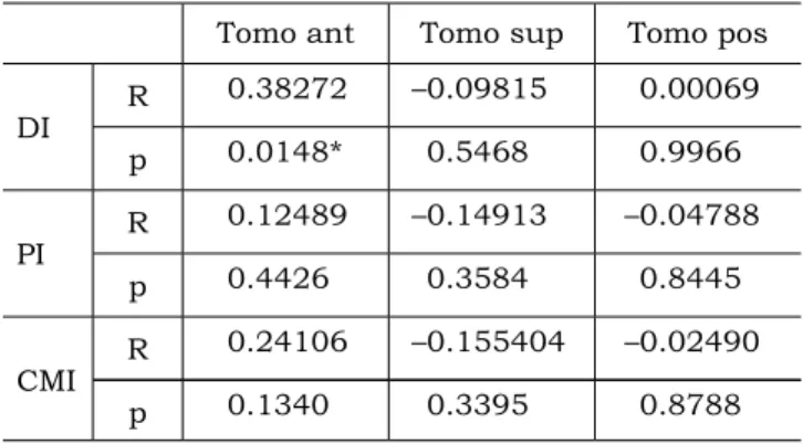
Table 3 and Figures 1, 2 and 3 show the cor-relation between the obtained values in the images and the indexes. There was a significant positive correlation between the anterior joint space and the DI.
The linear regression showed that signs and symptoms of TMD that determined the index val-ues were not responsible for the variability of to-mographic measurements, represented by the R2
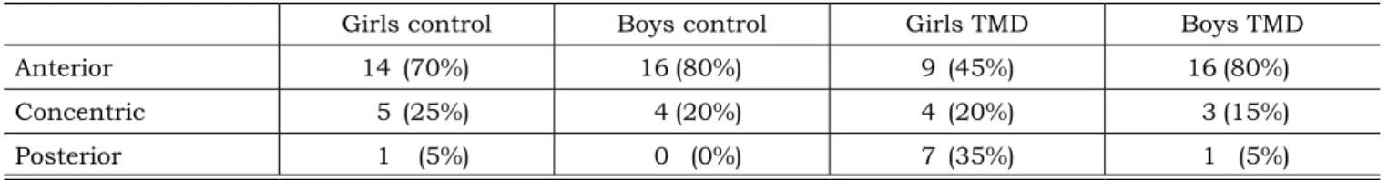
of the equations. Despite these coefficients being low, the scatter plots exhibited slightly positive relationships between the anterior joint space and DI, PI and CMI, and a negative relationship be-tween the superior joint space and the indexes. Tables 4 and 5 represent the condylar positions in both groups and the gender distribution of these positions. Each condyle position was determined in isolation. There was a significant difference in the condyle position between control and TMD groups (p = 0.05). Data were analyzed with gender dif-ferentiation and there was a significant difference between genders for the total sample (p = 0.03). When the condyle position of females was evalu-ated independently, the p value was 0.065, which may suggest a tendency towards a significant dif-ference in the condyle position for girls between both groups; for boys the p value was 1.0.
DISCUSSION
Epidemiologic studies have shown that signs and symptoms of TMD can be found in all age TABLE 1 - Mean values relative to the Dysfunction Index (DI), Palpation Index (PI) e Craniomandibular Index (CMI) scores for control and TMD groups.
Groups DI PI CMI
Control group 0.056 ± 0.03a 0.008 ± 0.02a 0.033 ± 0.01a
TMD group 0.150 ± 0.10b 0.321 ± 0.17b 0.233 ± 0.09b
Values with different superscript letters differ significantly (p < 0.05). TMD: temporomandibular disorders.
TABLE 2 - Mean (mm) and standard deviation for the articular space measurements in linear tomograms using digital caliper.
Groups Tomo ant/r Tomo ant/l Tomo sup/r Tomo sup/l Tomo pos/r Tomo pos/l Control group 1.90 ± 0.62 1.58 ± 0.52 3.89 ± 1.17 3.71 ± 1.29 3.66 ± 1.16 3.23 ± 1.38 TMD group 2.23 ± 0.85 1.81 ± 0.69 3.50 ± 1.55 3.23 ± 1.45 3.13 ± 1.56 3.01 ± 1.08 Tomo: tomography; ant: anterior; sup: superior; pos: posterior; r: right side; l: left side (p > 0.05); TMD: temporomandibular disorders.
TABLE 3 - Correlation coefficients (R) and p values be-tween the indexes and the articular space measure-ments obtained by tomograms.
Tomo ant Tomo sup Tomo pos
DI R 0.38272 –0.09815 0.00069
p 0.0148* 0.5468 0.9966
PI R 0.12489 –0.14913 –0.04788
p 0.4426 0.3584 0.8445
CMI R 0.24106 –0.155404 –0.02490
p 0.1340 0.3395 0.8788
Tomo: tomography; ant: anterior; sup: superior; pos: posterior; *p < 0.05. DI: Dysfunction Index; PI: Palpation Index; CMI: Craniomandibular Index.
TABLE 4 - Number of condyles in each position in con-trol and TMD groups.
Anterior Concentric Posterior*
Control group 30 9 1
TMD group 25 7 8
210 211
210 211
groups26,27,29. According to Egermark et al.7 (2001)
the prevalence is low in young children but in-creases with age in adolescence up to young adult-hood. Since signs and symptoms of TMD involve the muscle of mastication, TMJ and associated structures, it was considered important to deter-mine these variables associated with imaging ex-ams in young subjects, allowing the early diagnosis of structural alterations that could influence the normal growth and of the stomatognathic develop-ment system.
The decision to implement a dysfunction in-dex in the present study (CMI) was based on the possibility of objectively measuring the severity of
problems in mandibular movements, joint noise, and muscle and joint tenderness, using clearly defined criteria, simple clinical methods and easy scoring. In addition, this index has a good intra- and inter-examiner agreement10.
With regard to the condyle position in the lin-ear tomograms, it is important to blin-ear in mind that all studies in the related literature refer to the maximal intercuspal measurements. However, most of the time, people generally maintain the mandible at rest position. Thus, the condyle at rest position could cause more cumulative effects on TMJs than the maximal intercuspal position. TABLE 5 - Condyle position gender distribution in control and TMD (temporomandibular disorders) groups (p = 0.03).
Girls control Boys control Girls TMD Boys TMD
Anterior 14 (70%) 16 (80%) 9 (45%) 16 (80%)
Concentric 5 (25%) 4 (20%) 4 (20%) 3 (15%)
Posterior 1 (5%) 0 (0%) 7 (35%) 1 (5%)
FIGURE 1 - Linear regression between the mean values of the articular anterior, superior and posterior spaces obtained by tomography measurements and the Dysfunction Index (DI) (p < 0.05 for tomography anterior versus DI).
y = �0.0013 x + 0.1042 R2 = 0.0004
y = 0.0086 x + 0.0715 R2 = 0.0149
Tomography superior � DI Tomography anterior � DI
Tomography posterior � DI 0
0.1 0.2 0.3 0.4 0.5
Articular space measurements (mm)
D
ysf
un
ct
io
n
In
de
x
1 2 3 4 5 6 7
0
212 213
212 213
Bonilla-Aragon et al.4 (1999) measured joint
spaces in linear tomograms for condyle position determination. Even though there was a significant distal positioning of the condyle in patients with disk displacement, the authors suggested that the position of the condyle was a poor predictor of the presence of internal derangement. In this study, the TMD group also presented a significantly high-er numbhigh-er of distally positioned condyles when compared to the control group, corroborating the findings of the above-mentioned authors. However, a higher number of anteriorly positioned condyles were found due to the rest position image used in this study. According to Kordass14 (1999) the
occlusal pressure transmitted into the joint com-partments makes the condyle slide more or less retrocranially whereas the articular disk takes up a more displaced position and a deformed shape, which may change the width of joint spaces in those images in which the soft tissues cannot be well defined.
The joint spaces were also measured in adoles-cents by Major et al.15 (2002)and the values found
for the anterior, superior and posterior jointspaces were similar. The minor differences could be ex-plained by the mandible position since the mean age of the adolescents was the same. The authors concluded that as disc displacement increases, the superior joint space decreases, and anterior and posterior joint spaces increase significantly. In this study, such relationships could also be seen as shown in Figures 1 to 3. As the index values (CMI, DI and PI) increase, the anterior joint space also increases and the superior joint space decreases.
In the current study, there were no signifi-cant differences between groups and genders regarding the joint space measurements. How-ever, there was a significant difference in condyle position (p = 0.05) between groups, indicating a high number of distally positioned condyles in the TMD group. Kinniburgh et al.13 (2000) conducted
a study considering the condyle position and the joint spaces in adolescents with and without an-terior disk displacement. The combined and fe-male samples showed a statistically significant
0 0.1 0.2 0.3 0.4 0.5 0.6 0.7 0.8
7 6
5 4
3 2
1 0
Articular space measurements (mm)
P
al
pa
tio
n
In
de
x
y = 0.0436 x + 0.0747 R2 = 0.0143
y = �0.0207 x + 0.2309 R2 = 0.0195
y = �0.0031x + 0.1667 R2 = 0.0004
Tomography superior � PI Tomography anterior � PI
Tomography posterior � PI
212 213
212 213
difference between joints with normal and anterior disk position for all joint-space measurements and condylar position. Similarly, male joints displayed significantly different values for anterior and su-perior joint spaces and condylar position. In this study, when the condyle position was evaluated in differing genders, there was a significant difference (p = 0.03) in the total sample. When female joints were analyzed alone, this difference did not occur, with a p value of 0.065; and for the male joints the p was 1.0, which may suggest that the difference found in the total sample was due to the greater evidence of distally positioned condyles in females. The differences found between the studies may be corroborated by the fact that those authors had a magnetic resonance diagnosis of internal de-rangement for the experimental group. However, in this study, the TMD group had only a clinical diagnosis, which may suggest that some patients might have had muscle alterations without inter-nal derangement.
CONCLUSIONS
Even though there was a significant difference for condyle position determined by tomograms, it cannot be inferred that distally positioned condyles can predict TMD. As there was a great variation in condylar position in both groups, this evaluation was considered inconclusive for TMD diagnosis. This finding implies that changing the position of the condyle based on tomography is not justified as a therapeutic procedure. However, the diagnostic validity of tomography can not be discarded, since it is a reliable exam for osseous diseases.
ACKNOWLEDGMENTS
This work was supported by the State Uni-versity of Campinas (UNICAMP) and the Research Foundation of the State of São Paulo (FAPESP - #01/10442-3). We are grateful to Dr. Gláucia Maria Bovi Ambrosano for her advice on the sta-tistical analysis and to Waldeck Ribeiro Moreira and Antonio Custódio Camargo for their advice on tomography.
0 0.1 0.2 0.3 0.4 0.5 0.6
7 6
5 4
3 2
1 0
C
ra
ni
om
an
di
bu
la
r
In
de
x
Articular space measurements (mm)
Tomography superior � CMI Tomography anterior � CMI
Tomography posterior � CMI
y = 0.0547 x + 0.0295 R2 = 0.0581
y = �0.0024 x + 0.1403 R2 = 0.0006
y = �0.0143 x + 0.1837 R2 = 0.024
214 PB
REFERENCES
1. Berrett A. Radiology of the temporomandibular joint. Dent Clin North Am 1983;27:527-40.
2. Blaschke D, Chase DC. Differences in TMJ condyle-tem-poral relationships in normal men and women [Abstract 863]. J Dent Res 1984;63:266.
3. Blaschke DD, Blaschke TJ. Normal TMJ bony relationships in centric occlusion. J Dent Res 1981;60:98-104.
4. Bonilla-Aragon H, Tallents RH, Katzberg RW, Kyrkanides S, Moss ME. Condyle position as a predictor of temporo-mandibular joint internal derangement. J Prosthet Dent 1999;82:205-8.
5. Bronstein SL, Tomasetti BJ, Ryan DE. Internal derange-ments of the temporomandibular joint: correlation of ar-thrography with surgical findings. J Oral Surg 1981;39:572-84.
6. Dumas AL, Moaddab MB, Willis HB, Homayoun NM. A tomographic study of the condyle/fossa relationship in patients with TMJ dysfunction. J Craniomandibular Pract 1984;2:315-25.
7. Egermark I, Carlsson GE, Magnusson T. A 20-year longitu-dinal study of subjective symptoms of temporomandibular disorders from childhood to adulthood. Acta Odontol Scand 2001;59:40-8.
8. Egermark-Eriksson I, Carlsson GE, Magnusson T. A long term epidemiologic study of the relationship between oc-clusal factors and mandibular dysfunction in children and adolescents. J Dent Res 1987;66:67-71.
9. Fricton JR, Schiffman EL. Reliability of a craniomandibular index. J Dent Res 1986;65:1359-64.
10. Fricton JR, Schiffman EL. The craniomandibular in-dex: validity. J Prosthet Dent 1987;58:222-8.
11. Katzberg RW, Keith DA, Ten Eick WR, Guralnick WC. Internal derangements of the temporomandibular joint: an assessment of condylar position in centric occlusion. J Prosthet Dent 1983;49:250-4.
12. Katzberg RW, Westesson PL, Tallents RH, Drake CM. Anatomic disorders of the temporomandibular joint disc in asymptomatic subjects. J Oral Maxillofac Surg 1996;54:147-53.
13. Kinniburgh RD, Major PW, Nebbe B, Glover KE. Osse-ous morphology and spatial relationships of the temporo-mandibular joint: comparisons of normal and anterior disc position. Angle Orthod 2000;70:70-80.
14. Kordass B. The temporomandibular joint in video motion – noninvasive image techniques to present the func-tional anatomy. Anat Anz 1999;181:33-6.
15. Major PW, Kinniburgh RD, Nebbe B, Prasad NG, Glov-er KE. Tomographic assessment of temporomandibular joint osseous articular surface contour and spatial relation-ships associated with disc displacement and disc length. Am J Orthod Dentofacial Orthop 2002;121:152-61. 16. McNeill C, editor. Temporomandibular disorders:
guide-lines for classification, assessment, and management. The
American Academy of Orofacial Pain. Chicago: Quintes-sence; 1993.
17. Miller VJ. The effect of age on condylar asymmetry in patients with craniomandibular disorders of arthrogenous origin. J Prosthet Dent 1992;67:845-6.
18. Mohl ND, Dixon C. Current status of diagnostic proce-dures for temporomandibular disorders. J Am Dent Assoc 1994;125:56-64.
19. Pullinger A. The significance of condyle position in normal and abnormal temporomandibular joint function. In: Clark GT, Solberg W, editors. Perspectives in temporo-mandibular disorders. Chicago: Quintessence; 1987. p. 89-103.
20. Pullinger A, Hollender L. Variation in condyle-fossa re-lationships according to different methods of evaluation in tomograms. Oral Surg Oral Med Oral Pathol 1986;62:719-27.
21. Pullinger AG, Solberg WK, Hollander L, Guichet D. Tomographic analysis of mandibular condyle position in diagnostic subgroups of temporomandibular disorders. J Prosthet Dent 1986;55:723-9.
22. Pullinger AG, Solberg WK, Hollander L, Petersson A. Relationship of mandibular condylar position to dental occlusion factors in an asymptomatic population. Am J Orthod Dentofacial Orthop 1987;91:200-6.
23. Ren YF, Isberg A, Westesson PL. Condyle position in the temporomandibular joint. Comparison between asymp-tomatic volunteers with normal disk position and patients with disk displacement. Oral Surg Oral Med Oral Pathol Oral Radiol Endod 1995;80:101-7.
24. Riolo ML, Brandt D, TenHave TR. Associations be-tween occlusal characteristics and signs and symptoms of TMJ dysfunction in children and young adults. Am J Orthod Dentofacial Orthop 1987;92:467-77.
25. Ronquillo HI, Guay J, Tallents RH, Katzberg RW, Murphy W. Tomographic analysis of mandibular condyle position as compared to arthrographic findings of the tem-poromandibular joint. J Craniomand Disord 1988;2:59-64.
26. Sonmez H, Sari S, Oksak Oray G, Camdeviren H. Prevalence of temporomandibular dysfunction in Turk-ish children with mixed and permanent dentition. J Oral Rehabil 2001;28:280-5.
27. Thilander B, Rubio G, Pena L, de Mayorga C. Preva-lence of temporomandibular dysfunction and its associa-tion with malocclusion in children and adolescents: an epidemiologic study related to specified stages of dental development. Angle Orthod 2002;72:146-54.
28. Vanderas AP. Relationship between oral parafunctions and craniomandibular dysfunction in children and adoles-cents: a review. ASDC J Dent Child 1994;61:378-81. 29. Wahlund K. Temporomandibular disorders in
adoles-cents. Epidemiological and methodological studies and a randomized controlled trial. Swed Dent J Suppl 2003;inside front cover:2-64.