491 1 . Master degree; Coordinator of the Heart Surgery Service at
Hospital Regional Hans Dieter Schmidt de Joinville-SC e do Centro Hospitalar UNIMED de Joinville-SC.
2 . Heart Surgeron
3 . General and Abdominal Surgeon.
4 . Resident Physician – General Surgery - Hospital Municipal São José de Joinville-SC.
This study was carried out at Hospital Regional Hans Dieter Schmidt – Joinville, SC, e Centro Hospitalar Unimed - Joinville, SC. Correspondance address:
Robinson Poffo. Rua Mário Lobo, 61 sala 1314. CEP 89201-330 Joinville, SC, Brasil. Telefone: (47) 3423-0310.
E-mail address: drpoffo@hotmail.com
Robinson POFFO1, Marcos BONIN2,Rafael Armínio SELBACH3, Murilo PILATTI4
Rev Bras Cir Cardiovasc 2007; 22(4): 491-494 CASE REPORT
Article received in 28 Feb 2007 Article accepted in 20 Jul 2007
RBCCV 44205-933
Troca valvar mitral minimamente invasiva videoassistida
Vídeo-assisted minimally invasive mitral valve
replacement
Abstract
The use of minimally invasive techniques in cardiac surgery has become widely discussed, aiming improvements not only in the aesthetics aspects, but also better functional results. In this report, we present a case of a pacient with severe mitral stenosis that underwent succesful video-assisted minimally invasive mitral valve replacement.
Descriptors: Mitral valve, surgery. Minimally invasive
surgical procedures, methods. Video-assisted thoracic surgery.
Resumo
A utilização de técnicas minimamente invasivas em cirurgia cardíaca vem sendo amplamente discutida em nosso meio, visando melhorias não só no aspecto estético, mas também funcional. Neste relato, apresentamos o caso de um paciente portador de estenose valvar mitral grave, submetido à troca valvar mitral minimamente invasiva videoassistida com sucesso.
Descritores: Valva mitral, cirurgia. Procedimentos
492
POFFO, R ET AL - Vídeo-assisted minimally invasive mitral valve replacement
Rev Bras Cir Cardiovasc 2007; 22(4): 491-494
INTRODUCTION
The Constant search for better functional and aesthetic outcomes in thoracic surgery has led surgeons to develop new techniques, among which is the minimally invasive surgery [1-3]. The main goal is to achieve minor surgical trauma and eventually low postoperative pain and recovery time. We cannot reveal the aesthetic aspect, which results in greater satisfaction to the patient [4].
Associated to minithoracotomy, the development of new technologies in cardiopulmonary bypass (CPB), specific surgical instruments, and the use of videothoracocospy has advanced this kind of procedure [5].
The video-assisted minimally invasive mitral valve surgery is already widely accepted and performed in some surgical services with outcomes that are similar to the conventional techniques [5,6].
CASE REPORT
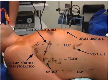
We report a case of a 39-year-old male patient with a history of rheumatic process in childhood progressing to mitral murmur. In 2006, when he was referred to thoracic surgery, he was in New York Heart Association (NYHA) functional class III, recent onset of atrial fibrillation, and a recent history of previous stroke. On physical examination, it was noted a significant holodiastolic murmur (++++/6+) in the mitral valve area and a mild motor deficit on the right. The rhythm was of atrial fibrillation. Echocardiogram highlighted an augmented left atrium (63 mm), a left ventricle end-systolic diameter of 37 mm, a left ventricle end-diastolic diameter of 58 mm, and an ejection fraction of 65%. On Doppler, the mitral regurgitation was graded mild and the stenosis as severe (area = 0.5 cm2) presenting a transvalvar gradient of 14 mmHg with significant thickening and calcification of the cusps and subvalvar system. It was also noted that the presence of an organized thrombus in the left atrium. Cardiac catheterism showed pulmonary capillary pressure of 20 mmHg and normal coronary arteries. The surgical possibilities regarding the procedure were informed to the patient and the minimally invasive surgical procedure was preferred. Surgery was performed through right anterolateral minithoracotomy with video assistance. Patient was intubated using the Carlen orotracheal tube for selective ventilation and placed with the right side of the thorax elevated by 30° (Figure 1), the arm lying alongside the body. Disposable pads for external cardiac defibrillation were placed in the region of the right scapula and anterolateral left hemithorax. A 5-cm incision was made in the 4th intercostals space between the anterior axillary line (AAL) and midaxillary line (MAL).
A rib retractor (Estech, Ino) specific for minimally invasive surgery was used to withdraw the ribs. The right lung was “selected”. In the same intercostal space, in the
midaxillary line, a trocar for a 10-mm lens at 30° with a line for CO2 insufflator was inserted. The surgical instrument used was specific for this kind of procedure. The kit was composed of clamps, needle-holder, and long scissors (length approximately 35 cm) for videothoracoscopy. Under the vision of this optical instrument, the pericardium was opened 2 cm anteriorly to the phrenic nerve, using the specific scissor for thoracoscopy. The incision was made from the inferior vena cava up to the superior vena cava. The pericardium was pulled by four points, which were exteriorized through the thoracic wall using a specific retractor/hook for this purpose.
Fig. 1 – Patient positioning and the site marks for minithoracotomy incision, transthoracic aortic clamping, trocars for optical device, left atrial aspirator, and left atrial retractor. (LA = left atrium; AAL = anterior axillary line; MAL = midaxillary line; PAL = posterior axillary line)
Cardiopulmonary bypass was established by means of femoral cannulation. The skin was incised longitudinally to the vessels in the right inguinal region. A 17 Fr wire-bound cannula (Medtronic BioMedicus, Inc., Edren Prairie, MN,. USA) was inserted into the artery and a venous line was established using Carpentier Bi-Caval Femoral Venous Cannula 24/29 Fr (Medtronic BioMedicus, Inc., Edren Prairie, MN,. USA). A venous drainage was improved connecting a centrifuge pump to the venous line. Patient was maintained at 30°C.
493 coated with Teflon passed through a tourniquet.
Cardioplegic line was exteriorized through the incision. This same line was used to further aspirate the aortic root. Low-volume blood cardioplegia was used at the same patient’s temperature (30°C), repeated each 20 minutes.
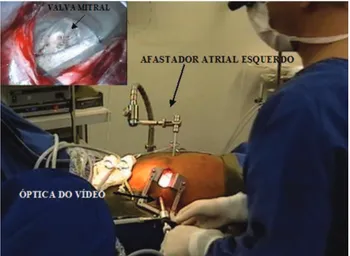
The left atrium was opened anteriorly to the right pulmonary veins and the atrial withdrawal was performed with a specific transthoracic retractor (Estech, Inco.) through the fifth intercostal space, laterally to the region of the right internal thoracic artery. By introducing an optical device into the left atrium, the mitral valve was examined (Figure 2), which was found to be severely stenosed and calcified, especially in the posterior mitral valve ring region. Thrombus was found in the region of left atrium posterior wall, which has been removed.
Once the left atrium was opened, CO2 injection through the optical trocar was maintained at 2 Liters per minute aiming at reducing the possibility of an airway embolism.
Left atriorrhaphy was performed using a 3-0 polypropylene thread through which an aspiration catheter was left in place to remove residual air.
Left chambers deareation maneuvers were performed by moving the operating table to the Trendelenburg and anti-Trendelenburg positions and alternating right-to-left. The aspiration was performed through the aortic root and left atrium. The aorta was then de-clamped e the patient re-warmed. Fibrillation was required to return the heart to normal sinus rhythm.
After CPB was removed, femoral vessels were decannulated and the heparin reversed. The minithoracotomy was closed through the conventional fashion. Left hemithorax was drained through the trocar incision to aspirate the left atrium.
Climping time was 190 minutes. Patient was extubated 8 hours after being admitted to the ICU and was referred to patient’s room on postoperative day 2. The total bleeding volume through the drain was 250 mL. No blood component was used. Patient’s recovery was uneventful.
Echocardiogram showed normofunctioning mitral valve prosthesis and sinus rhythm. Patient was discharged from hospital on postoperative day 7.
DICUSSION
Since the middle 1990s, several studies have shown the usage of the minithoracotomy associated to videothoracoscopy as a safe and efficient method to approach mitral valve [6].
The goals are a better patient’s recovery with less pain and postoperative complications, resulting in less length of hospital stay and consequently cost savings. Another point is the esthetic aspect and the patient’s satisfaction.
Several studies [3,5] have showed similar surgical outcomes to the conventional technique, that is, presenting low morbidity and mortality rates. Aubek et al. [3] reported a thirty-day mortality rate of 3.3% (8/240).
The restrained access to the mitral valve, due to the limited length of incision, is compensated by the use of thoracoscopy. Patients with a small anteroposterior diameter have the mitral valve exposure hampered, once the sternum limits the anterior retraction of left atrium. Thus, patients with pectus excavatum should not undergo this surgical procedure. Other counterindications would be obese patients or with big breasts because the access to the fourth intercostals space is more difficult, or patients with aortic regurgitation, once cardioplegic solution is administered in an anterograde way and can compromise the myocardial protection.
Fig. 2 – Operative Field aspect, showing the left atrial retractor, minithoracotomy, and the site of the optical vídeo system. In detail, left superior corner, internal view of the left atrium though videothoracoscopy
To assist in aspirating the blood from the pulmonary veins, a 7-mm trocar was inserted in the seventh intercostal space in the anterior axillary line (AAL) through which a malleable left atrium aspirator (Medtronic DLP) was passed. Mitral valve was widely resected and a 29-mm St Jude mechanical bileaflet prosthesis (St. Jude Medical, Minneapolis, MN) was implanted with 20 braided stitches Teflon-coated in a “U” pattern. The knots were made using a specific clamp for this type of surgery (Knot-pusher Estech Inc.).
Aortic clamping, left atriotomy, valve excision, passage of the stitches through valve ring, prosthesis placement and knots were exclusively preformed using video monitor visualization.
POFFO, R ET AL - Vídeo-assisted minimally invasive mitral valve replacement
494
The most feared complication is the retrograde aortic dissection, once the cannulation is performed in the femoral artery. This technique is contraindicated for patients with peripheral vascular disease or aorta with significant atheromatosis. Aybek et al. [3] reported only one case of retrograde aortic dissection in a group of 240 patients.
Another important aspect is regarding the preventive care to airway embolization. Due to the restrained access, the direct manipulation of the heart is virtually impossible, thus the constant CO2 insufflation in the operative field and the transesophageal echocardiogram are of great assistance in withdrawing the air from the left chambers.
The surgeon’s suitability and the adequate adaptation of the surgical setting are critical, because there is an alteration in the way we are used to work with the mitral valve. The handling of long instruments and the indirect visualization of the operative field are some of the difficulties carried by the method. These probably were held responsible for the aortic clamping time of 190 minutes. Other studies showed clamping times similar to the conventional technique [3,5]. We believe that with more practice and an accurate control of the technique, similar results to those found in the literature will be achieved.
The patient’s progress was uneventful and the patient was discharged from hospital on postoperative day 7. It is expected that with this less invasive technique there will be a decrease in the length of ICU stay and consequently lower hospital total length of stay, resulting in cost savings. Grossi et al. [5] reported an intensive care unit time of 19 hours, and total hospital stay of 6 days.
Initial surgical cost rises, once there is a requirement to assemble new technologies (thoracoscopy equipment, specific surgical equipment, inservice training, and especial cannulas). A further prospective study will be necessary to highlight the potential economical advantages of this method.
The abovementioned case shows the reproducibility of this technique in Brazil. We believe that despite of being a more complex surgery, it should be part of the therapeutic armamentarium, once the outcomes are highly encouraging [5].
REFERENCES
1. Mulinari LA, Tyszka AL, Costa FDA, Carvalho RG, Silva Jr. AZ, Giublin R, et al. Miniesternotomia: um acesso seguro para a cirurgia cardíaca. Rev Bras Cir Cardiovasc. 1997;12(4):335-9.
2. Pereira MB, Barcellos CS, Kalil RAK, Santana JR, Prates PR, Nesralla IA. Toracotomia minimamente invasiva nas intervenções cirúrgicas valvares. Rev Bras Cir Cardiovasc. 1998;13(3):229-33.
3. Aybek T, Dogan S, Risteski PS, Zierer A, Wittlinger T, Wimmer-Greinecker G, et al. Two hundred forty minimally invasive mitral operations through right minithoracotomy. Ann Thorac Surg. 2006;81(5):1618-24.
4. Chitwood WR Jr. Current status of endoscopic and robotic mitral valve surgery. Ann Thorac Surg. 2005;79(6):S2248-53.
5. Grossi EA, Galloway AC, LaPietra A, Ribakove GH, Ursomanno P, Delianides J, et al. Minimally invasive mitral valve surgery: a 6-year experience with 714 patients. Ann Thorac Surg. 2002;74(3):660-3.
6. Mohr FW, Falk V, Diegeler A, Walther T, van Son JA, Autschbach R, et al. Minimally invasive port-acess mitral valve surgery. J Thorac Cardiovasc Surg. 1998;115(3):567-74. ACKNOWLEDGEMENT
We want to express our profound gratitude to Prof. Friedrich W. Mohr for his encouragement and strength of purpose with which he gently taught us the ways to use this technique.
POFFO, R ET AL - Vídeo-assisted minimally invasive mitral valve replacement